This video demonstrates the concentration of viral particles from environmental water samples using centrifugal ultrafiltration. The method enriches intact viruses by size-based retention and buffer-assisted recovery for downstream molecular applications.
مقالة منهجية
Concentrating Virus-Enriched Water Samples by Centrifugal Ultrafiltration
26 مشاهدة
⸱
أكتوبر 29, 2026
في هذه المقالة
الملخص
البروتوكول
1. Tertiary Concentration
- Prepare a centrifugal concentrator (30,000 molecular weight cutoff) for each sample collected by adding at least 10 mL of 1x PBS (phosphate-buffered saline) and 0.2% bovine serum albumin (BSA) to the upper sample chamber. Ensure that the solution has filled the thin-channel concentration chamber, and then hold overnight (O/N) at 4 °C.
- Discard the fluid. Rinse the concentrator one time with at least 10 mL of sterile reagent-grade water to remove excess BSA and then discard the water.
- Add an amount of secondary water concentrate from each test sample equal to S, the Assay Sample Volume, into separate centrifugal concentrators.
Calculate S using Equation 1,
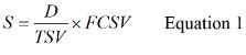
Where D is the Volume of Original Water Sample Assayed, TSV is the Total Sample Volume, and FCSV is the Final Concentrated Sample Volume.
- Centrifuge each test sample at 3,000–6,000 x g at 4 °C in a swinging bucket rotor for 20 – 30 min. Check the volume in the thin-channel concentration chamber.
- If the volume is greater than 400 µl, centrifuge again for 20 min or longer. Continue centrifuging until the sample in the thin-channel concentration chamber has been reduced to less than 400 µl. Do not remove the supernatant.
- Wash the sides of the centrifugal concentrator with 1 mL of sterile 0.15 M sodium phosphate, pH 7–7.5 to increase virus recovery. Centrifuge again at 3,000–6,000 x g and 4 °C until the sample has been reduced to less than 400 µl. Repeat this wash step one additional time.
- Using a 100-200 µl micropipette, carefully measure and transfer each concentrated sample to a 1.5 ml microcentrifuge tube (i.e., transfer 200 µl to the microcentrifuge tube and then measure the remaining concentrate by adjusting the micropipette until the remaining fluid can be completely drawn up into the pipettor tip). Add 0.15 M sodium phosphate, pH 7–7.5, to adjust the final volume to 400 ± 2 µl.
- Extract nucleic acid immediately. Hold any concentrated samples that cannot be processed immediately at 4 °C for no more than 24 hr.
المواد
| الاسم | الشركة | رقم فهرسي | التعليقات |
|---|---|---|---|
| Bovine serum albumin (BSA) | Affymetrix | 10856 | Crystalline grade or better |
| Phosphate buffered saline (PBS) | U.S. Biological | D9820 | |
| Refrigerated centrifuge | Beckman Coulter | 367501 | |
| Vivaspin 20 centrifugal concentrator units | Sartorius-Stedim | VS2022 | |
| Microcentrifuge tubes | Fisher Scientific | 02-682-550 | Use ribonuclease- and deoxyribonuclease-free tubes with snap caps |
