This video demonstrates a method using a human intestinal epithelial organoid monolayer expressing a fluorescent calcium indicator to visualize calcium signaling during rotavirus infection. This enables real-time detection of virus-induced calcium dynamics in the epithelial cells.
方法文章
Visualization of Rotavirus-Induced Calcium Signaling in a Human Intestinal Organoid Monolayer
29 次观看
⸱
2026年10月29日
本文内容
摘要
方案
Viral infection of HIO monolayers
- Prepare the virus inoculum. If necessary, trypsin activates the virus stock. For rotavirus, add 10 µg/mL Worthington's Trypsin to the virus stocks and incubate at 37 °C for 1 h.
- Dilute the activated virus stock using one of the two following methods.
- If aiming for the maximum number of infected cells using only one viral strain, mix 50 μL of the activated viral stock with 50 μL Complete media without growth factors (CMGF-).
- If comparing multiple strains, prepare the inoculums to achieve equivalent multiplicities of infection (MOIs). Use the following formula to determine the volume of virus stock per well necessary for the desired MOI. Add CMGF- to the volume of virus stock calculated to achieve a final volume of 100 μL per well.
- Dilute the activated virus stock using one of the two following methods.

NOTE: A single human intestinal organoid (HIO) monolayer plated in a well with a diameter of 5.46 mm (96-well plate) contains about 200,000 cells. Because of the poor efficiency of infection in monolayers, a high MOI (100-1 million viral particles per cell) is preferred. The optimal MOI must be determined empirically.
- Infect the monolayers by gently removing the media from the HIO monolayer using a 200 mL pipette. Replace with 100 mL of virus inoculum or mock inoculum.
- Since most rotavirus stocks are propagated in MA104 cells, use an uninfected MA104 cell lysate for the mock inoculum. Use a volume equivalent to the volume of virus stock used for the infected wells and add CMGF- for a final volume of 100 mL per well.
- For viruses that infect cells basolaterally, such as rotavirus, use a 25G needle to score the monolayer. It is easiest to make a single score across the length of the monolayer, from the bottom to the top of the well. Gently press the needle into the monolayer at an angle with the bevel side up, opposite the direction of motion, and drag it across the length of the monolayer to create a scar (Figure 1A).
- Incubate in a 37 °C incubator for 2 h. Remove the virus and mock inoculum. Wash monolayers once with 1x phosphate buffered saline.
- Add 100 μL of phenol red-free differentiation media. Place the slide in a 37 °C incubator until ready to image. The optimal imaging window will vary based on the kinetics of the viral infection. For rotavirus, begin imaging at 6-8 h post infection.
结果
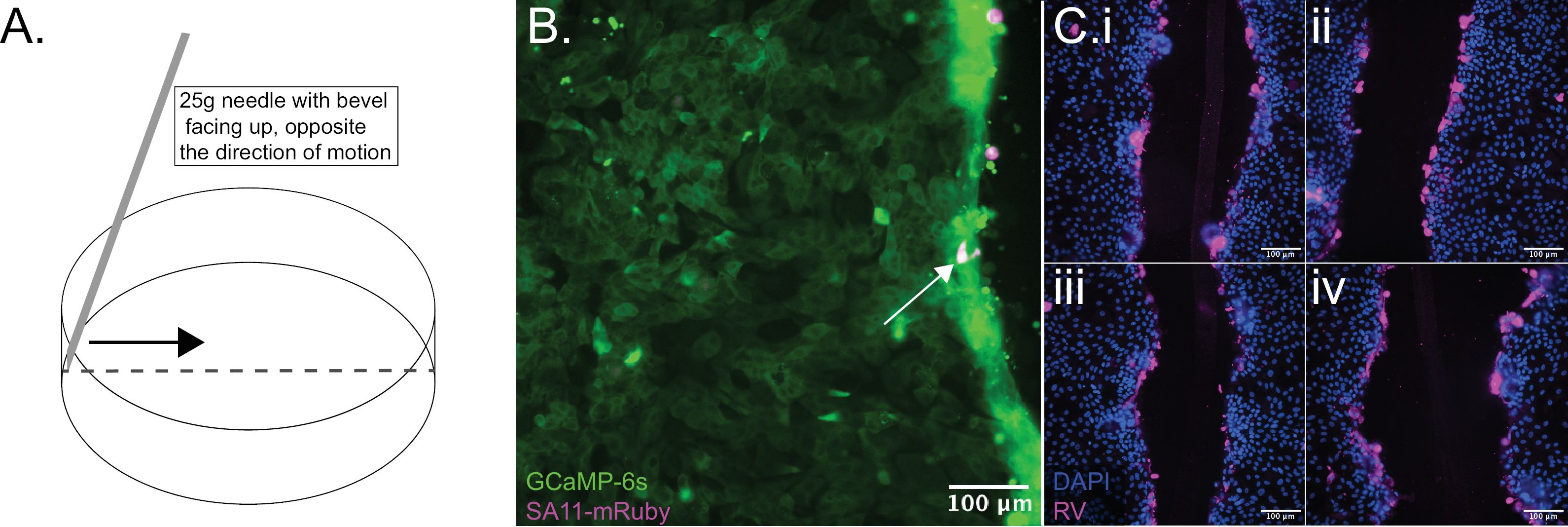
Figure 1: Scoring an HIO monolayer to facilitate RV infection. (A) The figure illustrates a suitable technique for scoring a monolayer using a 25G needle. (B) An HIO monolayer was infected with a recombinant strain of RV that expresses the mRuby fluorescent protein (magenta, arrow), allowing for the identification of infected cells. Alternatively, when using a naturally occurring strain of RV, infected cells can be identified using immunofluorescence. (C) The images shown are from 4 different HIO monolayers after fixation, DAPI staining (blue), and antibody labeling of rotavirus antigen (magenta). Both techniques illustrate the preferential RV infection near the scored portion of the monolayer.
材料
| 姓名 | 公司 | 目录编号 | 评论 |
|---|---|---|---|
| Advanced DMEM F12 | Gibco | 12634028 | |
| 1X PBS | Corning | 21-040-CV | |
| 25 gauge needle | Thermofisher Scientific | 1482113D | |
| Antibiotic-antimycocytic | Gibco | 15240062 | |
| Antibiotic-antimycotic | Gibco | 15240062 | |
| CellView Cell Culture Slide, PS, 75/25 MM, Glass Bottom, 10 compartments | Greiner | 543979 | |
| Fetal bovine serum | Corning | 35010CV |
