癌症发生是一个涉及癌细胞与肿瘤微环境之间相互作用的过程。为了剖析其中的分子事件,需要在癌症发展的不同阶段分离出不同的细胞群体。利用基底细胞癌的小鼠模型,我们描述了一种在癌症发生过程中进行细胞分析的实验方案。
本文内容
摘要
摘要
癌症的发生是一个多步骤过程,涉及多种细胞类型,包括癌前体细胞、免疫细胞、成纤维细胞和内皮细胞。在癌变过程中,每种细胞类型都会经历信号传导和功能上的改变。目前,许多癌症研究者面临的主要挑战是解析在多步骤过程中每种细胞类型所发生的这些变化 在体近年来,作者们建立了一套利用 K14creER/R26-SmoM2 分离皮肤癌发展过程中不同细胞群体的实验方法。YFP 小鼠。该实验流程分为6个部分:1)制备适用于本研究的小鼠(K14creER+ 和 R26-SmoM2YFP+ 小鼠在此方案中);2)诱导 SmoM2YFP 小鼠皮肤中的表达;3)制备小鼠皮肤活检样本;4)从皮肤中分离表皮;5)从表皮制备单细胞悬液;6)对单细胞群体进行标记以用于流式细胞术分析。本方案中限制性步骤是获得足够数量且基因型正确的小鼠,可能需要长达两个月时间。其余步骤耗时数小时至数天。在本方案中,我们还包含了一个故障排除部分。尽管本研究重点是皮肤癌,但该方案可经修改后适用于其他人源疾病动物模型。
引言
作为最常见的人类癌症类型,基底细胞癌(BCC)每年影响约200万美国人1。越来越多的证据表明,Hedgehog(Hh)信号通路的异常激活是驱动BCC发生发展的主要因素(详见综述2,3)。BCC中Hh信号通路的改变包括PTCH1的失活突变4-6、SMO的功能获得性突变7-9、Hh通路转录因子GLI110和GLI211的异常表达,以及负向调控因子Su(Fu)罕见的失活突变12。基于这些及其他相关研究,美国食品药品监督管理局(FDA)已批准使用Hh信号通路抑制剂Vismodegib治疗转移性和局部晚期BCC13-15。
尽管取得了所有这些成就 16我们仍然不了解Hh信号通路驱动致癌作用的分子和细胞机制。建立通过组织特异性激活Hh信号通路的动物模型,对于这些研究以及理解药物耐药性仍然具有重要意义。在小鼠中,即使暴露于高剂量的致癌物、紫外线或电离辐射,野生型小鼠也不会发生基底细胞癌(BCCs)。相比之下, Ptch1+/- 小鼠易发生基底细胞癌(BCC) 17,18. BCC 发生的外显率在 Ptch1+/- 小鼠 超过 50% 18,19 尽管 Ptch1+/- 小鼠在正常条件下很少发育出完全成熟的基底细胞癌(BCCs)。由于胚胎致死性 Ptch1-/-,通常采用组织特异性敲除PTCH1的方法进行研究 20此外,皮肤特异性条件性表达致癌基因SmoM2YFP Krt14-creER:R26-SmoM2YFP 或 Krt14-cre:R26-SmoM2YFP) 会导致在很早的年龄形成多个显微镜下可见的基底细胞癌(BCC),从而为SMO下游的Hh信号通路提供了一种简便的遗传检测方法 21.
目前对基底细胞癌(BCC)生物学的认识存在三个主要问题。首先,BCC的细胞起源尚不完全明确。尽管一些研究支持毛囊隆突区作为干细胞来源22-24,Youssef 等25通过使用细胞特异性Cre激活ROSA26驱动的转基因突变型SMO表达,将小鼠皮肤Hh通路驱动肿瘤的起源细胞定位于表皮间区而非毛囊中。其次,BCC发生过程中的细胞相互作用尚不清楚。已知Hh信号通路激活的角质形成细胞可导致BCC的形成,但其他伴随的细胞变化仍不明确。此外,在小鼠中使用Smo拮抗剂治疗可导致药物耐药性26-28,因此迫切需要发现新的BCC治疗靶点。对上述三个问题的深入理解,依赖于在小鼠癌变过程中对细胞动态变化的系统分析。尽管已有多种关于角质形成细胞培养、流式细胞术及皮肤干细胞分离的方法被报道29-31,但目前尚无针对小鼠BCC细胞分析的系统性操作流程。本文将整合小鼠BCC模型构建、表皮分离、组织单细胞制备及细胞分析等方法,为读者提供一套用于研究BCC发展过程中细胞信号通路、细胞间及分子间相互作用的完整实验方案。我们相信,本实验方案将帮助读者清晰掌握每一步操作的具体实施方式。此外,我们还提供了一些代表性结果数据,以展示预期的实验结果,并附有常见问题的解决方案,以协助读者克服实验过程中可能遇到的困难。
方案
本研究已获得印第安纳大学医学院动物护理与使用委员会(IACUC)的批准。
1. 获得正确基因型的小鼠
- 建立基底细胞癌(BCC)的小鼠模型。将 K14-CreER 小鼠(Jackson 实验室货号 005107)与 R26-SmoM2YFP 小鼠(Jackson 实验室货号 005130)杂交(18),以获得 K14-creER+ 和 R26-SmoM2YFP+ 小鼠。
- 进行基因分型。将尾部组织样本(0.5 cm 长)浸入含 1 mg/ml 蛋白酶 K 的 directPCR 尾部裂解液中,于 55 °C 摇床孵育过夜。次日,通过微型离心机高速离心去除组织碎片(保留上清液用于基因分型)。使用供应商推荐的引物和反应条件,每份 PCR 反应加入 1 μl 上清液。筛选出基因型正确的小鼠(在 3–6 周龄时,剪尾后需皮下注射布托啡诺,剂量为 0.05–0.1 mg/kg,每 8–12 小时一次,持续 2–3 天),用于下一步实验。
2. 在皮肤角质形成细胞中诱导 SmoM2 表达
通过饲喂针(弯曲的20号针头)经口灌胃给予他莫昔芬(每只5 μg/kg体重,溶于50 ml植物油中),连续5天,以在角质形成细胞中诱导SmoM2表达。
3. 准备皮肤样本
通常,K14creER/R26SmoM2GFP 小鼠的耳朵和尾巴表型更为严重。为了进行细胞分析,我们在动物安乐死后(采用机构批准的程序:CO2 加颈椎脱位)首先采集小鼠皮肤。
4. 分离表皮
- 按照批准的程序处死小鼠(参见步骤3)后,使用修剪器去除毛发。将皮肤样本浸入dispase溶液(5 mg/ml,溶于含10%胎牛血清的DMEM中),在37 °C下孵育1–2小时(对于年龄小于8周的小鼠为1小时,年龄大于8周的小鼠为2小时)。
- 经dispase处理后,用镊子将表皮与真皮分离。
5. 生成单细胞
- 为获得单细胞群体,先用剪刀将表皮组织剪碎,然后置于含有胶原酶IV的溶液(1 mg/ml,溶于含10%胎牛血清的DMEM中)中,在37 °C条件下于50 ml离心管中消化1–2小时。每20分钟轻轻摇动离心管,以促进细胞解离。
- 在显微镜下观察细胞解离情况。当在胶原酶溶液中可观察到单个细胞时,继续孵育样本30分钟,以提高细胞得率。通常情况下,一只小鼠的表皮组织可获得高达107个细胞。
- 将组织悬液通过细胞滤网(孔径70 μm)过滤,使用台式离心机以1,500 rpm离心收集细胞,并用PBS洗涤一次。
6. 标记细胞并进行流式细胞术分析
- 将细胞重悬于含10% FBS的PBS中,细胞浓度为2.5 × 10⁶ 个细胞/ml,用于阻断非特异性结合(冰上孵育30分钟)
- 取等量细胞分装至若干1.5 ml微量离心管中,用于特异性抗体标记。向每管中加入不同的抗体,终浓度为2 μg/ml。轻轻涡旋混匀数秒,冰上孵育30分钟,然后用1 ml PBS洗涤。对于BCC样本,我们使用以下抗体标记相应生物标志物:CD11b-PE联合Gr1-APC(髓系细胞或髓系来源的抑制性细胞);vimentin-FITC(成纤维细胞)(详见试剂部分)。
- 对于细胞内染色,我们使用试剂盒并按照厂商说明书操作(详见试剂部分)。我们使用新鲜细胞分析细胞表面标志物。完成表面标志物染色后,洗涤细胞并重悬于PBS中。向细胞悬液中加入DAPI,终浓度为1 μg/ml。DAPI可标记死细胞,以便在后续分析中设门排除。
- 使用特定软件分析数据。我们首先根据FSC与SSC散点图选取单细胞群体,并以DAPI阴性群体(活细胞)进行后续分析。
结果
已知小鼠除非在Hh信号通路激活的情况下,否则不会发生基底细胞癌。图1展示了诱导表达致癌性平滑受体SmoM2YFP后的皮肤表型。图2展示了正常小鼠与荷瘤小鼠中髓系细胞的分析结果。CD11b+ Gr1+细胞来源于髓系细胞,其中部分为髓源性抑制细胞。在正常情况下,CD11b+Gr1+细胞群体几乎无法检测到,但在炎症或肿瘤发生时其数量会增加。我们发现,在皮肤中诱导表达SmoM2YFP会导致CD11b+Gr1+细胞增加。目前,基底细胞癌中这一变化背后的分子机制尚不明确。
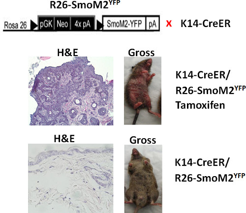
图1. 小鼠皮肤中诱导 SmoM2YFP 后的形态学变化。 上图显示了小鼠基因型的示意图。中排图像:经他莫昔芬处理的 K14creER+ 和 R26-SmoM2YFP+ 小鼠表现出明显的皮肤异常(右侧),H&E 染色切片中可见皮肤肿瘤(左侧)。下排图像:未诱导 SmoM2YFP 的小鼠皮肤外观正常(右侧),H&E 染色切片中无异常形态(左侧)。
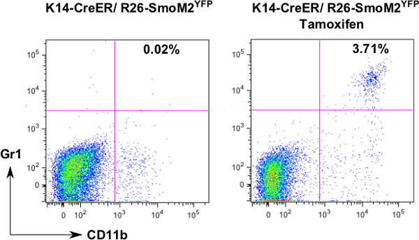
图 2. CD11b+Gr1+ 细胞的流式分析。 为检测皮肤癌发展过程中 CD11b+Gr1+ 细胞的变化,我们分离了单细胞,并使用荧光标记的抗 CD11b 和抗 Gr1 抗体进行染色。通过 FlowJo 分析后,我们发现经他莫昔芬处理的小鼠皮肤组织中该细胞群体有所增加。
讨论
我们已描述了一种用于基底细胞癌(BCCs)细胞群体分析的方法。肿瘤组织由多种细胞类型组成,每种细胞类型在癌变过程的特定时间点表现出不同的行为,这种异质性即使在最佳条件下也极难完全重现。 体外 条件。利用本方案,我们证明了基底细胞癌的发展伴随着表皮干细胞的扩增以及髓系细胞的募集。与免疫组织化学或免疫荧光分析相比,由于目前已存在针对多种细胞类型的特异性抗体,细胞群体分析能够更定量地评估细胞群体的变化。
本方案需要10周时间完成研究,因为基因诱导引起的表型变化需要一定时间。因此,提前规划实验非常重要。细胞分离与分析可能需要整整两天时间。由于分析过程中使用的是活细胞,因此必须提前预约FACSCalibur流式细胞仪。我们还列出了使用本方案时常见的问题(见下文)。
根据我们的经验,以下是我们遇到的常见问题:
- 表皮分离效果差:可能由于dispase处理不充分(请延长dispase处理时间)或dispase溶液不足(皮肤需完全浸没在dispase溶液中)。我们每只小鼠至少使用5 ml dispase溶液,具体体积可根据皮肤大小调整。
- 细胞数量得率低:可能由于胶原酶IV消化不完全。请首先检查胶原酶的浓度或有效期。此外,也可能因使用的表皮组织量不足所致。同时,酶消化应在37 °C条件下伴随震荡进行(消化前请调节好温度)。
- 细胞团块较多:皮肤细胞中此现象非常常见(可通过细胞筛网后用移液器吹打数次,再进行离心;或因溶液中含有Mg2+和Ca2+所致(请使用无Mg2+和Ca2+的PBS进行细胞洗涤)。
- 死细胞过多:可能由于组织降解(请使用新鲜组织)或消化过度(避免dispase和胶原酶IV孵育时间过长)所致。
该方案可与骨髓移植或组织移植相结合,以研究细胞来源。例如,将表达GFP的骨髓细胞移植到致死剂量照射的小鼠体内,可使我们研究骨髓细胞对肿瘤相关成纤维细胞和髓系细胞的贡献。类似地,可将皮肤肿瘤移植到新的小鼠宿主体内,以研究肿瘤与宿主之间的相互作用。
除了研究癌变过程中细胞群体的变化外,本方案还可使研究人员检测药物处理对肿瘤微环境中细胞的影响。尽管本方案旨在用于皮肤癌变过程中的细胞群体分析,但稍加修改后也可适用于其他类型的癌症。
总之,在小鼠皮肤癌模型中进行细胞群体分析,能够揭示癌变过程中的动态细胞变化,有助于更好地理解肿瘤转化过程中的细胞生物学特性以及转化过程中不同的细胞事件。
披露
作者声明不存在任何竞争性经济利益。
致谢
本研究由美国国家癌症研究所(NCI)(R01-94160 和 R01-155086)、印第安纳大学西蒙癌症中心以及威尔斯儿童研究中⼼资助。
材料
| 姓名 | 公司 | 目录编号 | 评论 |
|---|---|---|---|
| directPCR 裂解试剂(尾部) | Viagen Biotech | 102-T | 储存于 4 °C |
| 蛋白酶 K | Sigma | P2308 | 储存于 -20 °C |
| ApliTag 360 taq 聚合酶 | Applied biosystems | N8080155 | 储存于 -20 °C |
| 他莫昔芬 | Sigma | T5648 | 储存于 4 °C |
| 饲喂针头(20 号) | Fisher scientific | 01-290-9B | |
| 分散酶 | Roche Diagnostic | 04942078001 | 储存于 4 °C |
| IV 型胶原酶 | Worthington | LS004189 | 储存于 -20 °C |
| 细胞滤筛 | Fisher scientific | 08-771-2 | |
| 抗 CD11b-PE 抗体 | eBioscience | 12-0112-81 | 储存于 4 °C |
| 抗 Gr1-APC 抗体 | eBioscience | 17-5931-81 | 储存于 4 °C |
| 抗波形蛋白-FITC 抗体 | eBioscience | 11-9897-80 | 储存于 4 °C |
参考文献
- Rogers, H. W., et al. Incidence estimate of nonmelanoma skin cancer in the United States. Arch. Dermatol. 146, 283-287 (2006).
- Epstein, E. H. Basal cell carcinomas: attack of the hedgehog. Nat. Rev. Cancer. 8, 743-754 (2008).
- Yang, L., Xie, G., Fan, Q., Xie, J. Activation of the hedgehog-signaling pathway in human cancer and the clinical implications. Oncogene. 29, 469-481 (2010).
- Johnson, R. L., et al. Human homolog of patched, a candidate gene for the basal cell nevus syndrome. Science. 272, 1668-1671 (1996).
- Hahn, H., et al. A mammalian patched homolog is expressed in target tissues of sonic hedgehog and maps to a region associated with developmental abnormalities. J. Biol. Chem. 271, 12125-12128 (1996).
- Hahn, H., et al. Mutations of the human homolog of Drosophila patched in the nevoid basal cell carcinoma syndrome. Cell. 85, 841-851 (1996).
- Xie, J., et al. Activating Smoothened mutations in sporadic basal-cell carcinoma. Nature. 391, 90-92 (1998).
- Reifenberger, J., et al. Missense mutations in SMOH in sporadic basal cell carcinomas of the skin and primitive neuroectodermal tumors of the central nervous system. Cancer Res. 58, 1798-1803 (1998).
- Lam, C. W., et al. A frequent activated smoothened mutation in sporadic basal cell carcinomas. Oncogene. 18, 833-836 (1999).
- Nilsson, M., et al. Induction of basal cell carcinomas and trichoepitheliomas in mice overexpressing GLI-1. Proc. Natl. Acad. Sci. U.S.A. 97, 3438-3443 (2000).
- Sheng, H., et al. Dissecting the oncogenic potential of Gli2: deletion of an NH(2)-terminal fragment alters skin tumor phenotype. Cancer Res. 62 (2), 5308-5316 (2002).
- Reifenberger, J., et al. Somatic mutations in the PTCH, SMOH, SUFUH and TP53 genes in sporadic basal cell carcinomas. Br. J. Dermatol. 152, 43-51 (2005).
- Guha, M. Hedgehog inhibitor gets landmark skin cancer approval, but questions remain for wider potential. Nat. Rev. Drug Discov. 11, 257-258 (2012).
- Sekulic, A., et al. Efficacy and safety of vismodegib in advanced basal-cell carcinoma. N. Engl. J. Med. 366, 2171-2179 (2012).
- Tang, J. Y., et al. Inhibiting the hedgehog pathway in patients with the basal-cell nevus syndrome. N. Engl. J. Med. 366, 2180-2188 (2012).
- Aszterbaum, M., et al. Ultraviolet and ionizing radiation enhance the growth of BCCs and trichoblastomas in patched heterozygous knockout mice. Nat. Med. 5, 1285-1291 (1999).
- Pazzaglia, S., et al. Modulation of patched-associated susceptibility to radiation induced tumorigenesis by genetic background. Cancer Res. 64, 3798-3806 (2004).
- Athar, M., et al. Inhibition of smoothened signaling prevents ultraviolet B-induced basal cell carcinomas through regulation of Fas expression and apoptosis. Cancer Res. 64, 7545-7552 (2004).
- Mancuso, M., et al. Basal cell carcinoma and its development: insights from radiation-induced tumors in Ptch1-deficient mice. Cancer Res. 64, 934-941 (2004).
- Ellis, T., et al. Patched 1 conditional null allele in mice. Genesis. 36, 158-161 (2003).
- Mao, J., et al. A novel somatic mouse model to survey tumorigenic potential applied to the Hedgehog pathway. Cancer Res. 66, 10171-10178 (2006).
- Wang, G. Y., Wang, J., Mancianti, M. L., Epstein, E. H. Basal cell carcinomas arise from hair follicle stem cells in Ptch1(+/-) mice. Cancer Cell. 19, 114-124 (2011).
- Grachtchouk, M., et al. Basal cell carcinomas in mice arise from hair follicle stem cells and multiple epithelial progenitor populations. J. Clin. Invest. 121, 1768-1781 (2011).
- Kasper, M., et al. Wounding enhances epidermal tumorigenesis by recruiting hair follicle keratinocytes. Proc. Natl. Acad. Sci. U.S.A. 108, 4099-4104 (2011).
- Youssef, K. K., et al. Identification of the cell lineage at the origin of basal cell carcinoma. Nat Cell Biol. 12, 299-305 (2010).
- Yauch, R. L., et al. Smoothened mutation confers resistance to a Hedgehog pathway inhibitor in medulloblastoma. Science. 326, 572-574 (2009).
- Buonamici, S., et al. Interfering with resistance to smoothened antagonists by inhibition of the PI3K pathway in medulloblastoma. Sci. Transl. Med. 2, 51ra70(2010).
- Dijkgraaf, G. J., et al. Small molecule inhibition of GDC-0449 refractory smoothened mutants and downstream mechanisms of drug resistance. Cancer Res. 71, 435-444 (2011).
- Guo, Z., Draheim, K., Lyle, S. Isolation and culture of adult epithelial stem cells from human skin. J. Vis. Exp. (49), e2561(2011).
- Li, L., Fukunaga-Kalabis, M., Herlyn, M. The three-dimensional human skin reconstruct model: a tool to study normal skin and melanoma progression. J. Vis. Exp. (54), e2937(2011).
- Anacker, D., Moody, C. Generation of organotypic raft cultures from primary human keratinocytes. J. Vis. Exp. (60), e3668(2012).
重印与许可
