一种微通道芯片平台通过结合光刻可重铸光刻胶技术、软光刻技术和微流控技术而开发。该内皮化微通道平台可模拟三维(3D)几何结构 体内 微血管,可在受控的连续灌注流下运行,支持高质量的实时成像 并可应用于微血管研究。
需要JoVE订阅才能观看此内容。 请登录或开始免费试用
本文内容
摘要
摘要
研究重点一直集中在开发 体外 用于微血管研究的检测方法,因为 体内 动物实验耗时更长、成本更高,且观察与定量分析极为困难。然而,传统的 体外 微血管实验在表征方面存在局限性 体内 微血管的三维(3D)结构并实现连续流体流动。结合光刻可再流光刻胶技术、软光刻技术和微流控技术,我们开发出一种具有多深度圆形横截面的内皮化微通道芯片,可模拟微血管的三维(3D)结构 在体 微血管在受控的连续灌注流下运行。采用正性可流动光刻胶制作具有半圆形横截面微通道网络的母模。通过将从母模复制的两条聚二甲基硅氧烷(PDMS)微通道对准并键合,形成圆柱形微通道网络。微通道的直径可得到良好控制。此外,在芯片内接种的原代人脐静脉内皮细胞(HUVECs)显示,在持续可控灌注条件下,细胞可在4天至2周的时间内贴附于微通道内表面。
引言
微血管作为循环系统的一部分,介导血液与组织之间的相互作用,支持代谢活动,决定组织微环境,并在多种生理和病理条件下发挥关键作用。体外重建功能性微血管可为研究复杂的血管现象提供平台。然而,传统的体外微血管检测方法,如内皮细胞迁移实验、内皮细胞成管实验以及大鼠和小鼠主动脉环实验,无法重现体内微血管的三维(3D)结构和连续流动控制1-8。利用动物模型和体内实验(如角膜血管生成实验、鸡胚尿囊膜血管生成实验和Matrigel栓塞实验)进行微血管研究则更加耗时、成本高昂,在观察和定量分析方面存在挑战,并引发伦理问题1, 9-13。
微加工和微流控芯片技术的进步为生物医学研究提供了多种新见解,同时降低了与动物实验和体内研究相关的高昂实验成本和复杂性14,例如可轻松且精确地控制生物条件和动态流体环境,而这些在传统宏观尺度技术下是无法实现的。
在此,我们介绍一种构建内皮化微通道芯片的方法,该芯片可模拟三维几何结构 体内 微血管在受控的连续灌注流条件下运行,通过结合光刻可再流光刻胶技术、软光刻技术和微流控技术实现。
访问受限。请登录或开始试用以查看此内容。
方案
1. 光刻法制备光刻胶原模
以下方案展示了制备直径在30-60 μm之间的微通道的过程。若要获得更小直径(小于30 μm)的微通道,需进行一次光刻胶旋涂。
- 将回流光刻胶从4 °C冰箱转移至洁净室,使用前24小时使其升至室温。
- 清洁硅片并在150 °C下烘烤1小时,使其脱水。脱水有助于光刻胶在硅基底上的附着。
- 旋涂第一层 使用以下配方涂布光刻胶层:
| 步骤 | 时间(秒) | 转速(rpm) | 加速度 |
| 1 | 2 | 300 | 最高 |
| 2 | 10 | 0 | |
| 3 | 3 | 300 | 最高 |
| 4 | 60 | 600 | 最高 |
| 5 | 10 | 500 | 最高 |
| 6 | 10 | 600 | 最高 |
- 然后,将晶圆置于温度为110 °C的热板上烘烤90秒。软烘后,光刻胶的厚度为20–30 μm。
- 旋涂第二层 与第一次相同的光刻胶涂覆工艺后的光刻胶层 层
- 将晶圆再次置于温度为110 °C的热板上进行前烘,时间为90秒。此次前烘后,光刻胶的厚度将为40–60 μm。
- 通过紫外光照射光刻胶,曝光剂量为 14,500 mJ/cm²,生成正性母版图案2 通过薄膜掩模
- 将显影液用去离子水(DI水)稀释(体积比1:2)。将硅片在该溶液中反复冲洗,直至图案完全显影。然后 用去离子水清洗晶圆,并用氮气吹干。
- 回流:将晶圆置于温度为120 °C的热板上加热4分钟,并用玻璃培养皿覆盖,以防止溶剂蒸发。从热板上取下晶圆,使其冷却至室温。回流后的光刻胶厚度为50–60 μm。
2. PDMS 微通道网络的软光刻制作
- 按质量比10:1(基料:固化剂)配制聚二甲基硅氧烷(PDMS)溶液,并使用行星式离心混合器充分混合。
- 将PDMS溶液浇注到回流后的光刻胶母模上。将浇注好的PDMS放入真空干燥器中脱气15分钟。如有需要,使用氮气去除残留气泡。
- 将PDMS置于60 °C烘箱中加热3小时,使其固化。随后将固化的PDMS层从母模上剥离。
- 使用锋利的打孔器在通道网络处打孔,以形成进样口和出样口。用氮气清洁PDMS表面。
- 将两层PDMS置于等离子清洗仪中,在4.5 × 10⁻² Torr的工作压力和3.5 ft3/min的氧气流速条件下进行30秒的氧气等离子体处理。然后在光学显微镜下手动对齐PDMS表面。如有需要,可滴加一滴水以更好地控制对准。
- 将器件在60 °C烘箱中加热30分钟,以实现永久键合。
3. 芯片中 HUVEC 的培养与接种
- 使用含L-谷氨酰胺并补充10%胎牛血清(FBS)的培养基培养HUVECs。实验中使用第2至第5代的细胞。
- 当HUVECs达到融合状态时,用HEPES缓冲盐溶液(HEPES-BSS)冲洗细胞,然后用胰蛋白酶/EDTA处理,并在37 °C下孵育2-6分钟以消化细胞。消化完成后,加入胰蛋白酶中和液中和胰蛋白酶/EDTA活性。离心后,将细胞重悬于含8%葡聚糖的培养基中。葡聚糖用于增加培养基黏度,以促进细胞更均匀地接种和贴壁。
- 将装置置于4.5 × 10⁻² Torr的操作压力和3.5 ft3/min的氧气流速下,用氧等离子体处理5分钟。然后 向装置中加入去离子水,并在层流通风生物安全柜中用紫外光照射8小时以进行灭菌。
- 在细胞准备就绪前一天,用1×磷酸盐缓冲液(1× PBS)清洗装置,然后加入纤连蛋白(100 μg/ml,用1× PBS稀释)进行包被,并在4 °C冰箱中孵育过夜。
- 完成纤连蛋白包被后,用1× PBS清洗装置,然后加入培养基。将装置在37 °C下孵育15分钟。
- 将HUVEC细胞以3-4 × 10⁶ cells/ml的浓度悬浮于含8%葡聚糖的培养基中,取20 μl细胞悬液滴加至装置的一个入口处,并倾斜装置使细胞进入微流控通道。15-20分钟后,细胞将开始贴附于通道侧壁。每隔15分钟旋转一次装置,以实现更均匀的细胞分布。如有必要,可进行额外的加样操作。
4. 长期灌注装置
静置培养5-6小时后,贴壁的HUVECs将开始充分铺展。使用可远程控制的注射泵系统建立灌注,保持10 μl/hr的稳定流速。灌注流速可调至更高,并可持续4天至 2周不等。
5. 细胞染色与显微镜表征
- 当细胞在装置内达到汇合状态时,首先用1×PBS清洗装置,彻底去除培养基。然后将稀释后的红色染料(4 μl-1 ml)加入装置中,染料的加载方式与细胞加载步骤类似。室温下避光孵育5分钟,随后用培养基清洗装置以终止染色。染料长时间孵育可能导致细胞毒性及细胞脱落。
- 将蓝色染料用1×PBS稀释后加入装置(每毫升2滴)。室温下避光孵育5分钟,然后用1×PBS彻底清洗装置。
- 在倒置光学显微镜下观察细胞染色情况。若染色效果良好,向微通道中加入固定液(3.5%多聚甲醛用1×PBS稀释),随后将整个装置浸入固定液中,并用铝箔完全包裹。将装置置于4 °C冰箱中保存,以防止装置干燥和荧光漂白。固定后的装置即可用于共聚焦成像,可使用激光扫描共聚焦显微镜进行观察。
访问受限。请登录或开始试用以查看此内容。
结果
我们构建多深度微通道网络的方法模仿了复杂的三维几何结构 体内 微血管,其中微通道具有圆形横截面15此外,母代分支通道与子代通道的直径大致遵循 Murray 定律,以维持流体流动在所需水平,从而使整个网络的通道阻力较低,且流速更加均匀16-18. 展示了半圆形光刻胶母模和圆形横截面PDMS微通道网络的制备过程与结果 视频 1, 图2,以及 视频 2,分别。PDMS 微通道网络的几何特征经过表征,结果如图所示 图2我们的结果表明,利用光刻胶回流技术可通过更为便捷的方法构建多深度分支通道网络,并能够设计出近似遵循 Murray 定律的微血管仿生系统。
在许多体外(in vitro)模型中,血管细胞通常培养在平面培养板、滤膜或涂有水凝胶的这些基质上。在这些条件下,微血管通过细胞自组装随机形成。此外,传统检测方法在实现内皮细胞上持续稳定灌注方面存在固有困难。缺乏长期且连续的培养基...
访问受限。请登录或开始试用以查看此内容。
讨论
1. 主模具制备
血管形态计量学设计与指导原则之一是著名的 Murray 定律16,该定律指出,血管网络中血管直径的分布遵循最小能量消耗原则。此外,该定律还表明,在分叉处,母血管直径的立方等于其子代血管直径立方之和(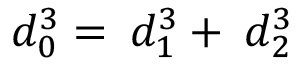 )19。此外,Poiseuille 定律已被用于估算大多数血管系统中剪切应力的大小,即
访问受限。请登录或开始试用以查看此内容。
披露
作者声明不存在任何竞争性经济利益。
致谢
本研究部分得到了美国国家科学基金会(NSF 1227359)、由国家科学基金会资助的西弗吉尼亚大学EPSCoR项目(EPS-1003907)、由国家科学基金会资助的西弗吉尼亚大学ADVANCE办公室(1007978)以及西弗吉尼亚大学PSCoR项目的支持。微加工工作在西弗吉尼亚大学共享研究设施(洁净室设施)和芯片微流控集成细胞研究实验室(MICRoChip Lab)完成。共聚焦成像在西弗吉尼亚大学显微镜成像设施完成。
访问受限。请登录或开始试用以查看此内容。
材料
| 姓名 | 公司 | 目录编号 | 评论 |
|---|---|---|---|
| <强>试剂/材料 | |||
| 回流光刻胶 | AZ电子材料 | AZP4620 | |
| 开发者 | AZ电子材料 | AZ 400K | |
| PDMS | 道康宁公司 | Sylgard 184 | |
| MCDB 131 培养基 | Invitrogen | 10372-019 | |
| NacBlue 细胞核染色 | Invitrogen | H1399 | |
| PKH 红色染料 | Sigma | MINI26 和 PKH26GL | |
| 纤连蛋白 | Gibco | PHE0023 | |
| L-谷氨酰胺 | Sigma | G7513 | |
| 磷酸盐缓冲液 | Invitrogen | 14040-133 | |
| HEPES 缓冲盐溶液 | Lonza | CC-5024 | |
| 胰蛋白酶/EDTA | Invitrogen | 25300-062 | |
| 胰蛋白酶中和液 | Lonza | CC-5002 | |
| PDMS 固化剂 | 道康宁公司 | Sylgard 184 | |
| 原代人脐静脉内皮细胞 | Lonza | CC-2517 | |
| 胎牛血清 | Lonza | 14-501F | |
| 稀释液C | Sigma | CGLDIL | |
| Hoechst33342 | Invitrogen,Molecular Probes | R37605 | |
| 葡聚糖 | Sigma | 95771 | |
| 3.5% 甲醛 | 电子显微镜科学 | 15710-S | |
| <强>仪器设备 | |||
| 旋转器 | Laurell Technologies Corporation | WS-400BZ-6NPP/LITE | |
| 干燥器 | BelArt 产品 | 999320237 | |
| 倒置显微镜 | 尼康 | Eclipse Ti | |
| 注射泵系统 | 哈佛仪器 | PHD Ultra | |
| 层流生物安全柜 | 赛默飞世尔科技 | 1300 系列 A2 | |
| 行星式离心混合器 | Thinky | ARE-310 | |
| Isotemp 培养箱 | Fisher Scientific | 13-246-516GAQ | |
| 光学显微镜 | Zeiss | Invertoskop 40C | |
| 等离子清洗机 | Harrick Plasma | PDC-32G | |
| 加热板 | Barnstead/Thermolyne Cimarec | SP131635 | |
| 激光扫描共聚焦显微镜 | Zeiss | LSM 510 |
参考文献
- Adair, T. H. Angiogenesis: Integrated systems physiology: from molecule to function to disease. , 1st edition, Biota Publishing. (2011).
- Goodwin, A. M. In vitro assays of angiogenesis for assessment of angiogenic and anti-angiogenic agents. Microvasc. Res. 74, 172-183 (2007).
- Smith, E. J., Staton, C. A. Tubule formation assays. Angiogenesis assays: A critical appraisal of current techniques. , John Wiley & Sons Ltd. West Sussex, UK. 65-87 (2006).
- Nakatsu, M. N., Davis, J. J., Hughes, C. C. W. Optimized fibrin gel bead assay for the study of angiogenesis. J. Vis. Exp. (3), e186(2007).
- Nicosia, R. F., Ottinetti, A. Growth of microvessels in serum-free matrix culture of rat aorta. A quantitative assay of angiogenesis in vitro. Lab. Invest. 63, 115-122 (1990).
- Aplin, A. C., Fogel, E., Zorzi, P., Nicosia, R. F. The aortic ring model of angiogenesis. Methods Enzymol. 443, 119-136 (2008).
- Nicosia, R. F. The aortic ring model of angiogenesis: A quarter century of search and discovery. J. Cell. Mol. Med. 13, 4113-4136 (2009).
- Griffith, L. G., Swart, M. A. Capturing complex 3D tissue physiology in vitro. Nat. Rev. Mol. Cell Biol. 7, 211-224 (2006).
- Folkman, J. History of angiogenesis. Angiogenesis: An integrative approach from science to medicine. , Springer. (2008).
- Auerbach, R., Lewis, R., Shinners, B., Kubai, L., Akhtar, N. Angiogenesis assays: A critical overview. Clin. Chem. 49, 32-40 (2003).
- Auerbach, R., Akhtar, N., Lewis, R. L., Shinners, B. L. Angiogenesis assays: Problems and pitfalls. Cancer Metastasis Rev. 19, 167-172 (2000).
- Staton, C. A., Reed, M. W., Brown, N. J. A critical analysis of current in vitro and in vivo angiogenesis assays. Int. J. Exp. Pathol. 90, 195-221 (2009).
- Staton, C. A., Stribbling, S. M., et al. Current methods for assaying angiogenesis in vitro and in vivo. Int. J. Exp. Pathol. 85, 233-248 (2004).
- Moraes, C., Mehta, G., Lesher-Perez, S. C., Takayama, S. Organs-on-a-Chip: A focus on compartmentalized microdevices. Ann. Biomed. Eng. 40 (6), 1211-1227 (2012).
- Huang, Z., Li, X., Martins-Green, M., Liu, Y. Microfabrication cylindrical microfluidic channel networks for microvascular research. Biomedical Microdevices. 14 (5), 873-883 (2012).
- Murray, C. D. The physiological principle of minimum work applied to the angle of branching of arteries. J. Gen. Physiol. 9 (6), 835-841 (1926).
- Zamir, M., Medeiros, J. A. Arterial branching in man and monkey. J. Gen. Physiol. 79, 353-360 (1982).
- Gafiychuk, V. V., Lubashevsky, I. A. On the principles of the vascular network branching. J. Theor. Biol. 212, 1-9 (2001).
- Sherman, T. F. On connecting large vessels to small. The meaning of Murray's law. J. Gen. Physiol. 78 (4), 431-453 (1981).
- Kamiya, A., Bukhari, R., Togawa, T. Adaptive regulation of wall shear stress optimizing vascular tree function. Bull Math Biol. 46 (1), 127-137 (1984).
- LaBarbera, M. Principles of design of fluid transport systems in zoology. Science. 249, 992-1000 (1990).
- Emerson, D. R., Cieslicki, K., Gu, X., Barber, R. W. Biomimetic design of microfluidic manifolds based on a generalized Murray's law. Lab Chip. 6, 447-454 (2006).
- Lu, H., Koo, L. Y., et al. Microfluidic shear devices for quantitative analysis of cell adhesion. Anal. Chem. 76, 5257-5264 (2004).
- Shevkoplyas, S. S., Gifford, S. C., Yoshida, T., Bitensky, M. W. Prototype of an in vitro model of the microcirculation. Microvasc. Res. 65, 132-136 (2003).
- Kaihara, S., Borenstein, J., et al. Silicon micromachining to tissue engineer branched vascular channels for liver fabrication. Tissue Eng. 6, 105-117 (2000).
- Fisher, A. B., Chien, S., Barakat, A. I., Nerem, R. M. Endothelial cellular response to altered shear stress. Am. J. Physiol. Lung Cell. Mol. Physiol. 281 (3), L529-L533 (2001).
- Nerem, R. M., Alexander, R. W., et al. The study of the influence of flow on vascular endothelial biology. Am. J. Med. Sci. 316 (3), 169-175 (1998).
- Daly, D., Stevens, R. F., Hutley, M. C., Davies, N. The manufacture of microlenses by melting photoresist. Meas. Sci. Technol. 1 (8), 759-766 (1990).
- Schilling, A., Merz, R., Ossmann, C., Herzig, H. P. Surface profiles of reflow microlenses under the influence of surface tension and gravity. Opt. Eng. 39 (8), 2171-2176 (2000).
- Young, B., Heath, J. W. Wheater's functional histology: A text and colour atlas. , 4th edition, Churchill Livingstone. (2000).
- O'Neill, F. T., Sheridan, J. T. Photoresist reflow method of microlens production. Part 1: Background and experiments. Optik Int. J. Light Electron Opt. 113, 391-404 (2002).
- de Gennes, P. G. Wetting: statics and dynamics. Rev. Mod. Phys. 57, 827-863 (1985).
- Elias, H. G. An Introduction to Polymer Science. , VCH Publishers. New York. (1997).
- Voinov, O. V. Dynamic edge angles of wetting upon spreading of a drop over a solid surface. J. Appl. Mech. Tech. Phys. 40, 86-92 (1999).
- Daly, D., Stevens, R. F., Hutley, M. C., Davies, N. The manufacture of microlenses by melting photoresist. Meas. Sci. Technol. 1, 759(1990).
- Jay, T. R., Stern, M. B. Preshaping photoresist for refractive microlens fabrication. Opt. Eng. 33, 3552-3555 (1994).
- Nerem, R. M., Alexander, R. W., et al. The Study of the influence of flow on vascular endothelial biology. Am. J. Med. Sci. 316 (3), 169-175 (1998).
- Chien, S., Li, S., Shyy, Y. J. Effects of mechanical forces on signal transduction and gene expression in endothelial cells. Hypertension. 31, 162-169 (1998).
- Li, Y. S., Haga, J. H., Chien, S. Molecular basis of the effects of shear stress on vascular endothelial cells. J. Biomech. 38, 1949-1971 (2005).
- Lee, E. J., Vunjak-Novakovic, G., Wang, Y., Niklason, L. E. A biocompatible endothelial cell delivery system for in vitro tissue engineering. Cell Transplant. 18, 731-743 (2009).
- Lee, E. J., Niklason, L. E. A novel flow bioreactor for in vitro microvascularization. Tissue Eng. Part C Methods. 16, 1191-1200 (2010).
- Chau, L., Doran, M., Cooper-White, J. A novel multishear microdevice for studying cell mechanics. Lab Chip. 9, 1897-1902 (2009).
- Meeson, A., Palmer, M., Calfon, M., Lang, R. A relationship between apoptosis and flow during programmed capillary regression is revealed by vital analysis. Development. 122, 3929-3938 (1996).
- Van Royen, N. J., Piek, J., Schaper, W., Bode, C., Buschmann, I. Arteriogenesis: mechanisms and modulation of collateral artery development. J. Nucl. Cardiol. 8, 687-693 (2001).
- Schaper, W. Therapeutic arteriogenesis has arrived. Circulation. 104 (17), 1994-1995 (2001).
- Tarbell, J. M. Shear stress and the endothelial transport barrier. Cardiovas. Res. 87 (2), 320-330 (2010).
- Potter, C. M., Lundberg, M. H., et al. Role of shear stress in endothelial cell morphology and expression of cyclooxygenase isoforms. Arterioscler. Thromb. Vasc Biol. 31, 384-391 (2011).
- Montesano, R. In vitro rapid organization of endothelial cells into capillary-like networks is promoted by collagen matrices. J. Cell Biol. 97, 1648-1652 (1983).
- Darland, D. C., D'Amore, P. A. TGF beta is required for the formation of capillary-like structures in three-dimensional cocultures of 10T1/2 and endothelial cells. Angiogenesis. 4 (1), 11-20 (2001).
- Lawley, T. J., Kubota, Y. Induction to morphologic differentiation of endothelial cells in culture. J. Invest. Dermatol. 93, 59S-61S (1989).
- Kanzawa, S., Endo, H., Shioya, N. Improved in vitro angiogenesis model by collagen density reduction and the use of type III collagen. Ann. Plast. Surg. 30, 244-251 (1993).
- Davis, G. E., Bayless, K. J., Mavila, A. Molecular basis of endothelial cell morphogenesis in three-dimensional extracellular matrices. Anat. Rec. 268, 252-275 (2002).
- Velazquez, O. C., Snyder, R., Liu, Z., Fairman, R. M., Herlyn, M. Fibroblast-dependent differentiation of human microvascular endothelial cells into capillary-like 3-dimensional networks. FASEB J. 16, 1316-1318 (2002).
- Donovan, D., Brown, N. J., Bishop, E. T. Comparison of three in vitro human "angiogenesis" assays with capillaries formed in vivo. Angiogenesis. 4, 113-121 (2001).
- Tang, D. G., Conti, C. J. Endothelial cell development, vasculogenesis, angiogenesis, and tumor neovascularization: an update. Semin. Thromb. Hemost. 30, 109-117 (2004).
- Takayama, S., McDonald, J. C., et al. Patterning cells and their environments using multiple laminar fluid flows in capillary networks. Proc. Natl. Acad. Sci. U.S.A. 96, 5545-5548 (1999).
- Cho, B. S., Schuster, T. G., et al. Passively driven integrated microfluidic system for separation of motile sperm. Anal. Chem. 75, 1671-1675 (2003).
- Parsa, H., Upadhyay, R., Sia, S. K. Uncovering the behaviors of individual cells within a multicellular microvascular community. Proc. Natl. Acad. Sci. U.S.A. 108 (12), 5133-5138 (2011).
访问受限。请登录或开始试用以查看此内容。
重印与许可
