本研究的目的是通过建立一种可靠的免疫组织化学技术,利用细胞c-fos激活来检测大鼠首次摄入脂肪和糖后多巴胺通路及其终末位点的同步变化,从而鉴定与奖赏相关的分布式脑网络。
需要JoVE订阅才能观看此内容。 请登录或开始免费试用
方法文章
大鼠初次摄入糖和脂肪后从中脑边缘和中脑皮层多巴胺奖赏区域同时检测c-Fos激活
* These authors contributed equally
本文内容
摘要
摘要
本研究利用细胞c-fos激活来评估大鼠初次摄入脂肪和糖类对脑内多巴胺(DA)通路的影响。糖类和脂肪的摄取行为既受其固有吸引力的驱动,也受到后天习得偏好的调节。脑内多巴胺,特别是源自腹侧被盖区(VTA)的中脑边缘和中脑皮层投射通路,已被证实参与介导这两种先天及后天的反应。尽管已有研究提出“分布式脑网络”的概念,认为多个脑区及神经递质/肽类系统相互作用以调节可口食物的摄入,但目前尚缺乏足够的实验证据来明确支持这一机制。因此,摄入糖类可引起多巴胺释放,并增加VTA多巴胺投射区域的c-fos样免疫反应性(FLI),这些区域包括伏隔核(NAC)、杏仁核(AMY)、内侧前额叶皮层(mPFC)以及背侧纹状体。此外,向上述脑区中枢给予特异性多巴胺受体拮抗剂,可差异性地降低由糖类或脂肪诱导的条件性风味偏好的获得与表达。为确定这些脑区是否在摄入糖类或脂肪后以分布式脑网络的形式协同作用,一种可行的方法是同步评估VTA及其主要中脑端脑多巴胺投射区(前边缘和下边缘mPFC、伏隔核核心与壳部、基底外侧及中央-皮质-内侧杏仁核)以及背侧纹状体,在口服摄入无条件刺激的玉米油(3.5%)、葡萄糖(8%)、果糖(8%)和糖精(0.2%)溶液后,是否表现出协调且同步的FLI激活。该方法是成功识别利用细胞c-fos激活在多个相关脑区同步研究啮齿类动物摄食奖赏相关学习可行性的第一步。
引言
脑内多巴胺(DA)被认为通过享乐机制1,2、与努力相关的机制3以及基于习惯的作用机制4,5 ,参与对可口糖类摄入的中枢反应。介导这些效应的主要DA通路起源于腹侧被盖区(VTA),并投射至伏隔核(NAC)的核心与壳部、基底外侧及中央皮质内侧杏仁核(AMY),以及前边缘和下边缘内侧前额叶皮层(mPFC)(参见综述6,7)。VTA已被证实参与蔗糖摄入过程8,9,且在糖摄入后可在NAC10–15、AMY16,17和mPFC18–20中观察到DA的释放。脂肪摄入同样可刺激NAC中DA的释放21,而另一富含DA的投射区域——背侧纹状体(尾状壳核)——也与DA介导的摄食行为相关22,23。Kelley24–27提出,该DA介导系统的多个投射区域通过广泛而紧密的相互连接,构成了一个整合性、交互性的分布式脑网络28–34。
除了多巴胺D1和D2受体拮抗剂能够减少糖类35-37和脂肪38-40的摄入外,多巴胺(DA)信号传导还被认为参与介导糖类和脂肪引发条件性风味偏好的能力(CFP)41-46。将多巴胺D1受体拮抗剂微量注射至伏隔核(NAC)、杏仁核(AMY)或内侧前额叶皮层(mPFC)可消除由胃内葡萄糖引发的条件性风味偏好的获得47-49。将多巴胺D1或D2受体拮抗剂微量注射至mPFC可消除果糖引发的条件性风味偏好的获得50,而在伏隔核和杏仁核中,多巴胺拮抗剂对果糖引发的条件性风味偏好的获得和表达则表现出差异性阻断作用51,52。
c-fos 技术53,54已被用于研究可口食物摄入所诱导的神经激活。本文通篇使用术语"c-fos 激活",其操作性定义为神经元去极化过程中 c-Fos 转录水平的升高。蔗糖摄入可增加杏仁核中央核(AMY)、腹侧被盖区(VTA)以及伏隔核(NAC)壳部而非核部的 fos 样免疫反应性(FLI)55-57。在假饲大鼠中,蔗糖摄入显著增加了杏仁核和伏隔核的 FLI,但未增加腹侧被盖区的 FLI58;而经胃内灌注蔗糖或葡萄糖可显著增加伏隔核以及杏仁核中央核和基底外侧核的 FLI59,60。在定时给予饲料的基础上反复添加蔗糖,可增加前额叶内侧皮质(mPFC)以及伏隔核壳部和核部的 FLI61。采用蔗糖浓度下调范式发现,基底外侧杏仁核和伏隔核的 FLI 增加最为显著,而腹侧被盖区则无明显变化62。在条件反射建立后,糖相关自然奖赏行为的消退过程可增加基底外侧杏仁核和伏隔核的 FLI63。此外,将糖的出现与声音配对后,该声音随后可引起基底外侧杏仁核 FLI 水平的升高64。高脂饮食摄入同样可增加伏隔核和前额叶内侧皮质区域的 FLI65-67。
先前引用的大多数研究仅考察了糖和脂肪对单一脑区c-fos激活的影响,无法提供有关奖赏相关分布式脑网络识别的信息24-27。此外,许多研究也未明确界定伏隔核(NAC,包括核心区和壳区)、杏仁核(AMY,包括基底外侧核和中央-皮质-髓质区)以及内侧前额叶皮层(mPFC,包括前肢核和下肢核)内部亚区的相对作用,而这些亚区的差异本可通过c-Fos图谱所具备的优异空间分辨率和单细胞分辨率加以分析68。本实验室69近期利用c-fos激活技术,同时检测了大鼠初次摄入脂肪和糖类后,腹侧被盖区多巴胺(VTA DA)通路及其投射区域(NAC、AMY和mPFC)发生的变化。本研究详细描述了实验操作与方法步骤,旨在同时分析急性暴露于六种不同溶液(玉米油、葡萄糖、果糖、糖精、水以及脂肪乳剂对照)是否会在NAC、AMY、mPFC以及背侧纹状体的各个亚区中差异性地激活即刻早期基因标记(FLI)。这种对差异的同步检测,使得我们能够确认各脑区FLI的显著变化,并判断某一特定脑区的变化是否与其他相关脑区的变化存在相关性,从而为分布式脑网络的存在提供支持证据24-27。这些实验程序用于检验在口服、非条件性摄入葡萄糖(8%)、果糖(8%)、玉米油(3.5%)和糖精(0.2%)溶液后,VTA、前肢核和下肢核mPFC、NAC的核心区与壳区、AMY的基底外侧核与中央-皮质-髓质区,以及背侧纹状体是否表现出协调且同步的FLI激活。
访问受限。请登录或开始试用以查看此内容。
方案
这些实验方案已获得机构动物照护与使用委员会的批准,确认所有实验对象和操作均符合美国国立卫生研究院《实验动物照护与使用指南》的要求。
1. 受试者
- 购买和/或繁育雄性Sprague-Dawley大鼠(260 - 300 g)。
- 将大鼠单只饲养在金属丝网笼中。保持12小时光照/12小时黑暗循环,自由供应大鼠饲料和水 随意摄取.
- 分配适当的样本量 (例如,n ≈ 6 - 8)随机分组。
2. 测试装置与进样程序
- 使用带有橡胶塞和45°角金属吸管的校准离心管,以准确测量(± 0.1 ml)所提供的溶液。用一根拉紧的金属弹簧将其固定在动物饲养笼上,以便观察刻度。
- 限制大鼠的食物供给量(约15 g/天),使其体重减少至原始体重的85%,以增强其摄入溶液的动机。注意:体重下降过程应在3至5天内完成。
- 在训练前连续四天,每天提供一次1小时的预训练溶液(10 ml,0.2%糖精),以最大化大鼠在后续测试中快速(潜伏期小于1分钟)采样测试溶液的可能性。
- 通过滴出少量液体来确认离心管中的液体流动通畅。
- 每次实验前后均称量离心管重量,以获得摄入量数据。
- 在第五天对接受以下六种溶液之一的亚组进行摄入测试(每种溶液10 ml,测试1小时):a) 水,b) 新口味(0.05%樱桃香精)的0.2%糖精溶液,c) 8%果糖,d) 8%葡萄糖,e) 3.5%玉米油悬浮于0.3%黄原胶中,f) 0.3%黄原胶。
- 确保各营养溶液之间热量相等;因此,3.5%玉米油的浓度与8%糖溶液为等热量。
- 确保大鼠在短时间内(小于1分钟)开始采样溶液。若未满足此条件,则将该动物从研究中剔除。
3. 组织制备
- 在每种测试溶液初次暴露后90分钟,通过腹腔注射戊巴比妥麻醉每只动物。通过检测动物对足部夹捏引起的回缩反射、直接角膜压迫引起的眨眼反射或深部耳廓刺激引起头部晃动等反射是否消失,确认动物已充分麻醉。
- 按照先前所述方法对每只动物进行经心脏灌注69。
- 用过量戊巴比妥钠(65 mg/kg)麻醉大鼠,打开胸腔并暴露心脏以便操作69。
- 将针头插入左心室心尖部位,同时剪开下腔静脉。先灌注磷酸盐缓冲液(PBS,约180 ml),随后灌注含4%多聚甲醛的磷酸盐缓冲固定液(约180 ml)。
- 通过观察液体是否从鼻腔、口腔或生殖区域等其他腔隙流出,确认动物是否正确灌注。注意:使用多聚甲醛进行充分固定时,通常伴随明显的肌肉收缩运动。若未出现该反应,应重新调整针头位置,直至出现此反应为止。
- 迅速剪开颅骨表面的毛发和皮肤,取出脑组织。使用咬骨钳从后向前逐步敲开并移除颅骨。操作起始区域位于小脑下方及后方,确保咬骨钳位于颅骨与软脑膜之间。当颅顶和颅侧骨被移除后,用小刮匙从颅底轻轻抬起脑组织,并用小剪刀剪断颅神经。在去除颅骨过程中需小心,避免损伤脑组织。
- 将脑组织置于4%多聚甲醛溶液中,在4 °C条件下固定过夜。随后将脑组织转移至30%蔗糖与70% PBS的混合溶液中,室温孵育直至脑组织沉降至容器底部。
- 修整脑组织
- 在嗅球后方横向切断,去除脑的前部组织。
- 在小脑与脑桥水平横向切断,去除脑的后部组织。
- 将脑组织冠状面朝下固定于切片机载物台上,对前额叶内侧皮层(mPFC,位于前囟前+2.86至+2.20 mm)、伏隔核核心与壳部及背侧纹状体(位于前囟前+1.76至+1.60 mm)、杏仁核(AMY,位于前囟后-2.12至-2.92 mm)以及腹侧被盖区(VTA,位于前囟后-5.20至-5.60 mm)进行冠状切片(厚度40 µm)。可参考大鼠脑图谱70进行定位。
- 将游离切片收集至盛有PBS的24孔板各孔中,以备后续免疫组织化学分析71。使用Parafilm密封24孔板,防止PBS蒸发导致脑组织干燥。将脑组织样本储存于4 °C环境中。
4. c-fos 实验步骤(改编自71)
- 每个切片用 5 ml 含 5% 正常山羊血清和 0.2% Triton x-100 的 PBS 溶液处理 1 小时。
- 将处理后的切片与一抗(兔抗 c-fos,1:5,000)在含有 1 ml PBS 的孔中于 4 °C 孵育 36 小时。
- 用 PBS(5 ml)清洗切片 3 次,每次 10 分钟。
- 在含有 1 ml PBS 的孔中,室温下与二抗(生物素化山羊抗兔 IgG;1:200)孵育 2 小时。
- 每个切片用 PBS(5 ml)清洗 3 次,每次 10 分钟。
- 将清洗后的切片在商品化的亲和素-辣根过氧化物酶混合液中孵育 2 小时,该试剂盒包含 Avadin DH(100 µl)和生物素化辣根过氧化物酶 H(100 µl)溶于 5 ml PBS 中。
- 再次用 PBS(5 ml)清洗切片 3 次,每次 10 分钟。
- 在含有 5 ml DAB 溶液的孔中,用含 0.05% 二氨基联苯胺(DAB)和 0.0015% H2O2 的溶液反应 5–10 分钟,具体时间根据组织反应性而定。
- 对 VTA 切片进行双重标记。在 5 ml PBS 中加入酪氨酸羟化酶(TH)抗体(兔抗大鼠 TH,1:2,000),4 °C 过夜孵育。
- 用 PBS(5 ml)清洗切片 3 次,每次 10 分钟。
- 在 5 ml PBS 中,室温下与二抗(生物素化山羊抗兔 IgG;1:200)孵育 2 小时。
- 用 PBS(5 ml)清洗切片 3 次,每次 10 分钟。
- 使用二抗-过氧化物酶复合物显色。在含有 5 ml DAB/NiCl 溶液的孔中,用 0.05% DAB 与 0.3% 硫酸镍溶液混合液反应 5–10 分钟,具体时间根据组织反应情况而定。
- 确保 DAB 溶液因与 0.3% 硫酸镍反应而呈现乳白色微绿色。若溶液过绿,则显色将过深。
- 将所有切片贴附于明胶包被的载玻片上,室温过夜干燥,然后滴加数滴甲苯溶液(TBS)封片。
- 对载玻片进行编码,使观察者不知晓实验分组情况。
5. c-fos 免疫反应阳性细胞计数的测定
- 指派两名无偏倚的观察者,对以下感兴趣区域(ROI)中的 Fos 阳性神经元进行计数:前边缘内侧前额叶皮层(prelimbic mPFC)、下边缘内侧前额叶皮层(infralimbic mPFC)、伏隔核核心区(NAC core)、伏隔核壳区(NAC shell)、基底外侧杏仁核(basolateral AMY nuclei)、中央-皮质-内侧杏仁核(central-cortico-medial AMY)、背侧纹状体以及腹侧被盖区(VTA)。需明确 VTA 中 c-Fos 免疫反应性是否存在于 TH+ 和 TH- 细胞中。图 1 展示了显微镜下伏隔核区域的屏幕截图。
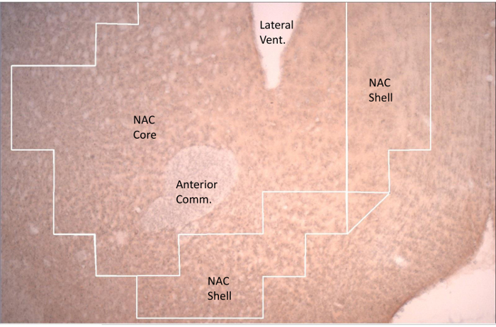
图1. 伏隔核(NAC)代表性切片,显示通过网格划定的兴趣区域,用于c-fos计数。 伏隔核壳部位于伏隔核核心的内侧和腹侧。伏隔核核心环绕前连合(Anterior Comm.)。侧脑室(Lateral Vent.)的腹侧部分清晰可见。 请点击此处查看该图的放大版本。
- 在所有测试条件下,每只动物每个共同位点至少分析三个具有代表性的切片。
- 使用软件和光学显微镜,通过勾画轮廓来分析每个感兴趣区域(ROI)的整个区域(图1)。
- 对于特定位点,打开应用程序,点击采集下拉菜单,选择“实时图像”(Live Image)。将感兴趣区域调至清晰,点击屏幕以设定参考点。然后以网格为参考,勾画选定的脑区。完成勾画后,开始计数细胞(步骤 5.3.1.1 - 5.3.1.3)。
- 双击软件图标。进入菜单栏,点击“Acquisition”(采集),然后选择“Live Image”(实时图像)。将感兴趣区域调至清晰,点击屏幕以设定参考点。
- 进入网格工具栏,点击“Display Grid”(显示网格)和“Use grid labels”(使用网格标签)。按照预设的路径勾画感兴趣区域的轮廓。
- 对每个感兴趣区域内的所有细胞进行计数,在左侧边栏中选择“+”以记录c-fos阳性细胞数量。逐一点击每个细胞以登记计数。当观察到明确的深红色圆形信号时,判定该细胞为c-fos阳性(图1)。
- 对每个位点重复上述过程。
- 将计数结果记录在实验记录本和计算机中,以供后续分析。进入菜单栏,点击“File”(文件),选择“Save Data File”(保存数据文件),以保存勾画结果和计数数据。
- 对于特定位点,打开应用程序,点击采集下拉菜单,选择“实时图像”(Live Image)。将感兴趣区域调至清晰,点击屏幕以设定参考点。然后以网格为参考,勾画选定的脑区。完成勾画后,开始计数细胞(步骤 5.3.1.1 - 5.3.1.3)。
- 确保两名不知情评分者对每个感兴趣区域各切片的细胞计数结果之间的评分者间信度(采用计数相关性评估)始终高于0.8。
6. 统计学
- 使用重复测量单因素方差分析(ANOVA)比较第1、2、3和第4天的糖精摄入量,评估前四天的基础糖精摄入水平69。
- 采用随机区组双因素方差分析(ANOVA)比较六组动物在第4天的糖精摄入量与第5天测试摄入量69。
- 使用Tukey检验(p < 0.05)确定各组间的显著性差异效应69。
- 确定评分者间信度,然后采用共同观察者的计数结果。
- 对每个脑区三个代表性切片的c-fos计数取平均值69。
- 对摄入六种不同溶液(3.5%玉米油、8%葡萄糖、8%果糖、0.2%调味糖精、黄原胶对照和水)后边缘下前额叶皮层(perilimbic mPFC)的c-fos激活情况进行单因素方差分析(ANOVA)69。
- 对边缘下mPFC、伏隔核核心(NAC core)、伏隔核壳部(NAC shell)、基底外侧杏仁核(basolateral AMY)、中央-皮质-髓质杏仁核(central-cortico-medial AMY)、腹侧被盖区(VTA)和背侧纹状体进行六组平行分析。使用Tukey检验(p < 0.05)揭示各显著性效应69。
- 将玉米油摄入量分别与水摄入量及其悬浮剂黄原胶的摄入量进行比较。将果糖和葡萄糖摄入量分别与水摄入量及非营养性甜味剂糖精的摄入量进行比较。
- 使用Bonferroni相关性分析(p < 0.05)确定各脑区溶液摄入量与c-fos激活之间是否存在显著相关性。
- 系统性地比较3.5%玉米油组每只动物边缘下和边缘前额叶皮层的c-fos计数。
- 对3.5%玉米油组,系统性地重复进行六脑区(VTA、背侧纹状体、边缘下mPFC、边缘下mPFC、NAC核心、NAC壳部、基底外侧杏仁核、中央-皮质-髓质杏仁核)中每对脑区的平行分析。
- 对其他五种实验摄入条件(8%葡萄糖、8%果糖、0.2%调味糖精、黄原胶对照和水),系统性地重复上述六个脑区的平行分析。
- 利用同一溶液条件下动物在所有脑区均被评估的特点,通过Bonferroni相关性分析(p < 0.05)确定不同溶液间以及每种溶液内部c-fos激活之间的显著相关关系。
访问受限。请登录或开始试用以查看此内容。
结果
下文所述所有代表性结果此前均已发表69,此处重新展示旨在提供“概念验证”,以说明该技术的有效性。
溶液摄入量
在实验开始的前四天中,所有动物的基础糖精摄入量存在显著差异(F(3,108) = 57.27,p < 0.001),摄入量呈显著(p < 0.05,Tukey HSD 检验)且逐步上升趋势(第1天:1.3 (± 0.2) ml;第2天:3.9 (± 0.4) ml;第3天:5.9 (± 0.6) ml;第4天:7.1 (± 0.6) ml)。与第4天的糖精摄入量相比,第5天的果糖和葡萄糖摄入量显著增加(p < 0.05,Tukey HSD 检验),而玉米油和糖精摄入量则无显著变化(p < 0.05,Tukey HSD 检验);其中果糖(9.6 (± 0.4) ml)和葡萄糖(9.4 (± 0.6) ml)的摄入量显著高于糖精摄入量。此外,玉米油摄入量(7.4...
访问受限。请登录或开始试用以查看此内容。
讨论
本研究旨在利用细胞c-fos技术,确定大鼠初次摄入脂肪和糖后,多巴胺奖赏相关神经元的来源(腹侧被盖区,VTA)及其前脑投射靶区(伏隔核,NAC;杏仁核,AMY;内侧前额叶皮层,mPFC)是否同时被激活。本研究详细描述了先前发表的一项研究的实验方案69。我们假设,VTA及其向 prelimbic 和 infralimbic 区域的mPFC、NAC的核心与壳部、AMY的基底外侧部及中央-皮质-髓质区,以及背侧纹状体的主要投射区域将构成一个分布式的脑网络24–27,并在初次摄入葡萄糖(8%)、果糖(8%)或玉米油(3.5%)溶液后,相对于糖精(0.2%)、水及其他对照溶液,表现出协调且同步的即刻早期基因荧光激活(FLI)。结果表明,玉米油、葡萄糖和果糖的摄入均显著且差异性地激活了VTA、prelimbic和infralimbic mPFC、NAC的核心与壳部、AMY的基底外侧部和中央-皮质-髓质区以及背侧纹状体,而糖精摄入则未产生此类激活。除c-fos技术外,本研究还采用了行为学方法评估糖、脂肪和人工甜味剂的摄入情况。
一个关键步骤是及时采集摄...
访问受限。请登录或开始试用以查看此内容。
披露
作者无竞争性财务利益。
致谢
感谢戴安娜·伊卡萨-库拉基、克里斯塔尔·桑普森和特奥洛吉亚·卡拉吉奥里斯为本项目付出的辛勤努力。
访问受限。请登录或开始试用以查看此内容。
材料
| 姓名 | 公司 | 目录编号 | 评论 |
|---|---|---|---|
| 仪器设备 | |||
| Sprague-Dawley 大鼠 | Charles River Laboratories | CD-1 | |
| 金属丝网笼 | Lab Products, Seaford, DE | 30-Cage rack | |
| 大鼠饲料 | PMI Nutrition International | 5001 | |
| 张紧金属弹簧 | Lab Products, Seaford, DE | n/a | |
| 大鼠电子秤 | Fisher Scientific Company | n/a | |
| Nalgene 离心管 | Lab Products, Seaford, DE | 10-0501 | |
| 橡胶塞 | Lab Products, Seaford, DE | n/a | |
| 金属饮水管 | Lab Products, Seaford, DE | n/a | |
| 糖精 | Sigma Chemical Co | 82385-42-0 | |
| Kool-Aid,樱桃味 | Kool-Aid | Commercial | |
| Kool-Aid,葡萄味 | Kool-Aid | Commercial | |
| 果糖 | Sigma Chemical Co | F0127 | |
| 葡萄糖 | Sigma Chemical Co | G8270 | |
| 玉米油 | Mazzola | Commercial | |
| 黄原胶 | Sigma Chemical Co | 11138-66-2 | |
| 滑动式切片机 | Microm International | n/a | |
| Neurolucida 成像系统 | MBF Bioscience | Software application | |
| 明胶包被载玻片 | Fisher Scientific Company | 12-550-343 | |
| 盖玻片 | Fisher Scientific Company | 12-545-M | |
| 金色尼龙刷 | Loew-Cornell | 2037 | |
| 天然毛松鼠毛刷 | Loew-Cornell | 2022 | |
| 24 孔板 | Fisher Scientific | 3527 | |
| 6 孔板 | Fisher Scientific | 3506 | |
| 1 L Pyrex 瓶 | Fisher Scientific | 1395-1L | |
| 组织嵌入盒(组织筛) | Fisher Scientific | 7200214 | |
| Eagle 移液器 | World Precision Instruments | E10(1–10μl) | |
| Eagle 移液器 | World Precision Instruments | E100(20–100μl) | |
| Eagle 移液器 | World Precision Instruments | E200(50–200μl) | |
| Eagle 移液器 | World Precision Instruments | E1000(100–1000μl) | |
| Eagle 移液器 | World Precision Instruments | E5000(1000–5000μl ) | |
| 通用吸头 0.1–10 μl | World Precision Instruments | 500192 | |
| 通用吸头 5–200 μl | World Precision Instruments | 500194 | |
| 通用吸头 500–5,000 μl | World Precision Instruments | 500198 | |
| Blade Vibroslice 100 刀片 | World Precision Instruments | BLADE | |
| DPX 封片剂 | Electron Microscopy | 13510 | |
| 15 ml 离心管 | Biologix Research Co. | 10-0501 | |
| 载玻片盒 | Biologix Research Co. | 41-6100 | |
| 旋转摇床 | Madell Corporation | ZD-9556 | |
| 称量舟 | Fisher Scientific | 02-202-100 | |
| 5 ml 一次性移液管 | Fisher Scientific | 13-711-5AM | |
| Stereo Investigator 软件 | Micro Bright Field | Software application | |
| 名称 | 公司 | 货号 | 备注 |
| 试剂 | |||
| 颗粒状多聚甲醛 | Fisher Scientific | 19210 | |
| NaCl | Fisher Scientific | S271-1 | |
| 磷酸二氢钠 | Fisher Scientific | S468-500 | |
| 磷酸氢二钠 | Fisher Scientific | BP332-500 | |
| 过氧化氢 | Fisher Scientific | H324-500 | |
| SafeClear II | Fisher Scientific | 23-044-192 | |
| 甲醇 | Fisher Scientific | A412-1 | |
| 正常山羊血清 | Vector | S-1000 | |
| 生物素化抗兔 IgG(H+L) | Vector | BA-1000 | |
| ABC 试剂盒(过氧化物酶标准型) | Vector | PK-4000 | |
| 抗 c-Fos(Ab-5)兔单克隆抗体 | EMD chem/Cal Biochem | PC38 | |
| 吐温 X-100 | SigmaAldrich | X-100 | |
| 3,3' 二氨基联苯胺四盐酸盐 | SigmaAldrich | D5905 | |
| 氢氧化钠 | SigmaAldrich | 5881 | |
| 一抗 TH 抗体 | EMD Millipore | AB152 | |
| Euthosol | Virbac AH |
参考文献
- Koob, G. F. Neural mechanisms of drug reinforcement. Ann. N.Y. Acad. Sci. 654, 171-191 (1992).
- Wise, R. A. Role of brain dopamine in food reward. Philos. Trans. R. Soc. Lond. B. Biol. Sci. 361, 1149-1158 (2006).
- Salamone, J. D., Correa, M. The mysterious motivational functions of mesolimbic dopamine. Neuron. 76, 470-485 (2012).
- Horvitz, J. C., Choi, W. Y., Morvan, C., Eyny, Y., Balsam, P. D. A "good parent" function of dopamine: transient modulation of learning and performance during early stages of training. Ann. N.Y. Acad. Sci. 1104, 270-288 (2007).
- Wickens, J. R., Horvitz, J. C., Costa, R. M., Killcross, S. Dopaminergic mechanisms in actions and habits. J. Neurosci. 27, 8181-8183 (2007).
- Bjorklund, A., Dunnett, S. B. Dopamine neuron systems in the brain: an update. Trends Neurosci. 30, 194-202 (2007).
- Swanson, L. W. The projections of the ventral tegmental area and adjacent regions: a combined fluorescent retrograde tracer and immunofluorescence study in the rat. Brain Res. Bull. 9, 321-353 (1982).
- Cacciapaglia, F., Wrightman, R. M., Careli, R. M. Rapid dopamine signaling differentially modulates distinct microcircuits within the nucleus accumbens during sucrose-directed behavior. J. Neurosci. 31, 13860-13869 (2011).
- Martinez-Hernandez, J., Lanuza, E., Martinez-Garcia, F. Selective dopaminergic lesions of the ventral tegmental area impair preference for sucrose but not for male sex pheromones in female mice. Eur. J. Neurosci. 24, 885-893 (2006).
- Bassareo, V., Di Chiara, G. Differential influence of associative and nonassociative learning mechanisms on the responsiveness of prefrontal and accumbal dopamine transmission to food stimuli in rats fed ad libitum. J. Neurosci. 17, 851-861 (1997).
- Bassareo, V., Di Chiara, G. Differential responsiveness of dopamine transmission to food-stimuli in nucleus accumbens shell/core compartments. Neurosci. 89, 637-641 (1999).
- Cheng, J., Feenstra, M. G. Individual differences in dopamine efflux in nucleus accumbens shell and core during instrumental conditioning. Learn. Mem. 13, 168-177 (2006).
- Genn, R. F., Ahn, S., Phillips, A. G. Attenuated dopamine efflux in the rat nucleus accumbens during successive negative contrast. Behav. Neurosci. 118, 869-873 (2004).
- Hajnal, A., Norgren, R. Accumbens dopamine mechanisms in sucrose intake. Brain Res. 904, 76-84 (2001).
- Hajnal, A., Smith, G. P., Norgren, R. Oral sucrose stimulation increases accumbens dopamine in the rat. Am. J. Physiol. 286, R31-R37 (2003).
- Bassareo, V., Di Chiara, G. Modulation of feeding-induced activation of mesolimbic dopamine transmission by appetitive stimuli and its relation to motivational state. Eur. J. Neurosci. 11, 4389-4397 (1999).
- Hajnal, A., Lenard, L. Feeding-related dopamine in the amygdala of freely moving rats. Neuroreport. 8, 2817-2820 (1997).
- Bassareo, V., De Luca, M. A., Di Chiara, G. Differential expression of motivational stimulus properties by dopamine in nucleus accumbens shell versus core and prefrontal cortex. J. Neurosci. 22, 4709-4719 (2002).
- Feenstra, M., Botterblom, M. Rapid sampling of extracellular dopamine in the rat prefrontal cortex during food consumption, handling, and exposure to novelty. Brain Res. 742, 17-24 (1996).
- Hernandez, L., Hoebel, B. G. Feeding can enhance dopamine turnover in the prefrontal cortex. Brain Res. Bull. 25, 975-979 (1990).
- Liang, N. C., Hajnal, A., Norgren, R. Sham feeding corn oil increases accumbens dopamine in the rat. Am. J. Physiol. Regul. Integr. Comp. Physiol. 291, R1236-R1239 (2006).
- Dunnett, S. B., Iversen, S. D. Regulatory impairments following selective kainic acid lesions of the neostriatum. Behav. Brain Res. 1, 497-506 (1980).
- Salamone, J. D., Zigmond, M. J., Stricker, E. M. Characterization of the impaired feeding behavior in rats given haloperidol or dopamine-depleting brain lesions. Neurosci. 39, 17-24 (1990).
- Kelley, A. E. Ventral striatal control of appetitive motivation: role in ingestive behavior and reward-related learning. Neurosci. Biobehav. Rev. 27, 765-776 (2004).
- Kelley, A. E. Memory and addiction: shared neural circuitry and molecular mechanisms. Neuron. 44, 161-179 (2004).
- Kelley, A. E., Baldo, B. A., Pratt, W. E. A proposed hypothalamic-thalamic-striatal axis for the integration of energy balance, arousal and food reward. J. Comp. Neurol. 493, 72-85 (2005).
- Kelley, A. E., Baldo, B. A., Pratt, W. E., Will, M. J. Corticostriatal-hypothalamic circuitry and food motivation: integration of energy, action and reward. Physiol. Behav. 86, 773-795 (2005).
- Berendse, H. W., Galis-de-Graaf, Y., Groenewegen, H. J. Topographical organization and relationship with ventral striatal compartments of prefrontal corticostriatal projections in the rat. J. Comp. Neurol. 316, 314-347 (1992).
- Brog, J. S., Salyapongse, A., Deutch, A. Y., Zahm, D. S. The patterns of afferent innervation of the core and shell in the "accumbens" part of rat ventral striatum: immunohistochemical detection of retrogradely transported fluoro-gold. J. Comp. Neurol. 338, 255-278 (1993).
- McDonald, A. J. Organization of amygdaloid projections to the prefrontal cortex and associated stritum in the rat. Neurosci. 44, 1-14 (1991).
- McGeorge, A. J., Faull, R. L. The organization of the projection from the cerebral cortex to the striatum in the rat. Neurosci. 29, 503-537 (1989).
- Sesack, S. R., Deutch, A. Y., Roth, R. H., Bunney, B. S. Topographical organization of the efferent projections of the medial prefrontal cortex in the rat: an anterograde tract-tracing study with Phaseolus vulgaris leucoagglutinin. J. Comp. Neurol. 290, 213-242 (1989).
- Wright, C. I., Beijer, A. V., Groenewegen, H. J. Basal amygdaloid complex afferents to the rat nucleus accumbens are compartmentally organized. J. Neurosci. 16, 1877-1893 (1996).
- Wright, C. I., Groenewegen, H. J. Patterns of convergence and segregation in the medial nucleus accumbens of the rat: relationships of prefrontal cortical, midline thalamic and basal amygdaloid afferents. J. Comp. Neurol. 361, 383-403 (1995).
- Geary, N., Smith, G. P. Pimozide decreases the positive reinforcing effect of sham fed sucrose in the rat. Pharmacol. Biochem. Behav. 22, 787-790 (1985).
- Muscat, R., Willner, P. Effects of selective dopamine receptor antagonists on sucrose consumption and preference. Psychopharmacol. 99, 98-102 (1989).
- Schneider, L. H., Gibbs, J., Smith, G. P. D-2 selective receptor antagonists suppress sucrose sham feeding in the rat. Brain Res. Bull. 17, 605-611 (1986).
- Baker, R. W., Osman, J., Bodnar, R. J. Differential actions of dopamine receptor antagonism in rats upon food intake elicited by mercaptoacetate or exposure to a palatable high-fat diet. Pharmacol. Biochem. Behav. 69, 201-208 (2001).
- Rao, R. E., Wojnicki, F. H., Coupland, J., Ghosh, S., Corwin, R. L. Baclofen, raclopride and naltrexone differentially reduce solid fat emulsion intake under limited access conditions. Pharmacol. Biochem. Behav. 89, 581-590 (2008).
- Weatherford, S. C., Smith, G. P., Melville, L. D. D-1 and D-2 receptor antagonists decrease corn oil sham feeding in rats. Physiol. Behav. 44, 569-572 (1988).
- Azzara, A. V., Bodnar, R. J., Delamater, A. R., Sclafani, A. D1 but not D2 dopamine receptor antagonism blocks the acquisition of a flavor preference conditioned by intragastric carbohydrate infusions. Pharmacol. Biochem. Behav. 68, 709-720 (2001).
- Baker, R. M., Shah, M. J., Sclafani, A., Bodnar, R. J. Dopamine D1 and D2 antagonists reduce the acquisition and expression of flavor-preferences conditioned by fructose in rats. Pharmacol. Biochem. Behav. 75, 55-65 (2003).
- Dela Cruz, J. A., Coke, T., Icaza-Cukali, D., Khalifa, N., Bodnar, R. J. Roles of NMDA and dopamine D1 and D2 receptors in the acquisition and expression of flavor preferences conditioned by oral glucose in rats. Neurobiol. Learn. Mem. 114, 223-230 (2014).
- Dela Cruz, J. A., et al. Roles of dopamine D1 and D2 receptors in the acquisition and expression of fat-conditioned flavor preferences in rats. Neurobiol. Learn. Mem. 97, 332-337 (2012).
- Yu, W. Z., Silva, R. M., Sclafani, A., Delamater, A. R., Bodnar, R. J. Pharmacology of flavor preference conditioning in sham-feeding rats: effects of dopamine receptor antagonists. Pharmacol. Biochem. Behav. 65, 635-647 (2000).
- Yu, W. Z., Silva, R. M., Sclafani, A., Delamater, A. R., Bodnar, R. J. Role of D(1) and D(2) dopamine receptors in the acquisition and expression of flavor-preference conditioning in sham-feeding rats. Pharmacol. Biochem. Behav. 67 (1), 537-544 (2000).
- Touzani, K., Bodnar, R. J., Sclafani, A. Activation of dopamine D1-like receptors in nucleus accumbens is critical for the acquisition, but not the expression, of nutrient-conditioned flavor preferences in rats. Eur. J. Neurosci. 27, 1525-1533 (2008).
- Touzani, K., Bodnar, R. J., Sclafani, A. Dopamine D1-like receptor antagonism in amygdala impairs the acquisition of glucose-conditioned flavor preference in rats. Eur. J. Neurosci. 30, 289-298 (2009).
- Touzani, K., Bodnar, R. J., Sclafani, A. Acquisition of glucose-conditioned flavor preference requires the activation of dopamine D1-like receptors within the medial prefrontal cortex in rats. Neurobiol. Learn. Mem. 94, 214-219 (2010).
- Malkusz, D. C., et al. Dopamine signaling in the medial prefrontal cortex and amygdala is required for the acquisition of fructose-conditioned flavor preferences in rats. Behav. Brain Res. 233, 500-507 (2012).
- Bernal, S. Y., et al. Role of dopamine D1 and D2 receptors in the nucleus accumbens shell on the acquisition and expression of fructose-conditioned flavor-flavor preferences in rats. Behav. Brain Res. 190, 59-66 (2008).
- Bernal, S. Y., et al. Role of amygdala dopamine D1 and D2 receptors in the acquisition and expression of fructose-conditioned flavor preferences in rats. Behav. Brain Res. 205, 183-190 (2009).
- Dragunow, M., Faull, R. The use of c-fos as a metabolic marker in neuronal pathway tracing. J. Neurosci. Methods. 29, 261-265 (1989).
- VanElzakker, M., Fevurly, R. D., Breindel, T., Spencer, R. L. Environmental novelty is associated with a selective increase in Fos expression in the output elements of the hippocampal formation and the perirhinal cortex. Learn. Mem. 15, 899-908 (2008).
- Norgren, R., Hajnal, A., Mungarndee, S. S. Gustatory reward and the nucleus accumbens. Physiol. Behav. 89, 531-535 (2006).
- Park, T. H., Carr, K. D. Neuroanatomical patterns of fos-like immunoreactivity induced by a palatable meal and meal-paired environment in saline- and naltrexone-treated rats. Brain Res. 805, 169-180 (1998).
- Zhao, X. L., Yan, J. Q., Chen, K., Yang, X. J., Li, J. R., Zhang, Y. Glutaminergic neurons expressing c-Fos in the brainstem and amygdala participate in signal transmission and integration of sweet taste. Nan.Fang Yi.Ke.Da.Xue.Xue.Bao. 31, 1138-1142 (2011).
- Mungarndee, S. S., Lundy, R. F., Norgren, R. Expression of Fos during sham sucrose intake in rats with central gustatory lesions. Am. J. Physiol. Regul. Integr. Comp. Physiol. 295, R751-R763 (2008).
- Otsubo, H., Kondoh, T., Shibata, M., Torii, K., Ueta, Y. Induction of Fos expression in the rat forebrain after intragastric administration of monosodium L-glutamate, glucose and NaCl. Neurosci. 196, 97-103 (2011).
- Yamamoto, T., Sako, N., Sakai, N., Iwafune, A. Gustatory and visceral inputs to the amygdala of the rat: conditioned taste aversion and induction of c-fos-like immunoreactivity. Neurosci. Lett. 226, 127-130 (1997).
- Mitra, A., Lenglos, C., Martin, J., Mbende, N., Gagne, A., Timofeeva, E. Sucrose modifies c-fos mRNA expression in the brain of rats maintained on feeding schedules. Neurosci. 192, 459-474 (2011).
- Pecoraro, N., Dallman, M. F. c-Fos after incentive shifts: expectancy, incredulity, and recovery. Behav. Neurosci. 119, 366-387 (2005).
- Hamlin, A. S., Blatchford, K. E., McNally, G. P. Renewal of an extinguished instrumental response: Neural correlates and the role of D1 dopamine receptors. Neurosci. 143, 25-38 (2006).
- Kerfoot, E. C., Agarwal, I., Lee, H. J., Holland, P. C. Control of appetitive and aversive taste-reactivity responses by an auditory conditioned stimulus in a devaluation task: A FOS and behavioral analysis. Learn. Mem. 14, 581-589 (2007).
- Zhang, M., Kelley, A. E. Enhanced intake of high-fat food following striatal mu-opioid stimulation: microinjection mapping and fos expression. Neurosci. 99, 267-277 (2000).
- Teegarden, S. L., Scott, A. N., Bale, T. L. Early life exposure to a high fat diet promotes long-term changes in dietary preferences and central reward signaling. Neurosci. 162, 924-932 (2009).
- Del Rio, D., et al. Involvement of the dorsomedial prefrontal cortex in high-fat food conditioning in adolescent mice. Behav. Brain Res. 283, 227-232 (2015).
- Knapska, E., Radwanska, K., Werka, T., Kaczmarek, L. Functional internal complexity of amygdala: focus on gene activity mapping after behavioral training and drugs of abuse. Physiol. Rev. 87, 1113-1173 (2007).
- Dela Cruz, J. A. D., et al. c-Fos induction in mesotelencephalic dopamine pathway projection targets and dorsal striatum following oral intake of sugars and fats in rats. Brain Res. Bull. 111, 9-19 (2015).
- Paxinos, G., Watson, C. The rat brain in stereotaxic coordinates. , Elsevier. (2006).
- Ranaldi, R., et al. The effects of VTA NMDA receptor antagonism on reward-related learning and associated c-fos expression in forebrain. Behav. Brain Res. 216, 424-432 (2011).
访问受限。请登录或开始试用以查看此内容。
重印与许可
