量化细胞分裂与扩展对理解整株植物生长至关重要。本文介绍了一种计算决定玉米叶片生长速率的细胞参数的实验方案,并强调了利用这些数据通过指导发育阶段特异性的取样策略来研究分子水平上的生长调控机制。
需要JoVE订阅才能观看此内容。 请登录或开始免费试用
方法文章
细胞分裂与扩张的运动学分析:量化生长的细胞基础及发育区域取样 Zea mays 叶片
* These authors contributed equally
本文内容
摘要
摘要
生长分析常用于植物科学研究中,以探究不同基因型以及环境条件对植物的影响。其中,细胞层面的分析至关重要,因为生长过程由细胞分裂和细胞伸长所驱动。运动学分析是一种用于量化这两个过程的方法。此外,该技术易于在非专业实验室中应用。本文介绍了一种在单子叶玉米(Zea mays)叶片中进行运动学分析的实验方案。本方案涵盖两个方面:(1)细胞分裂和扩展参数的量化;(2)发育区段位置的确定。该方法可为取样设计提供依据,或有助于对叶片生长区中具有高空间分辨率的生化与分子测量数据进行解释。玉米叶片的生长区应在稳态生长期间采集。单个叶片可用于通过DAPI染色测定分生组织长度,并利用微分干涉差(DIC)显微镜获取细胞长度分布图谱。该实验方案适用于在稳态生长期间采集的已展开单子叶叶片,其生长区应至少延伸数厘米。为了更深入理解植物生长调控机制,必须将生长数据与分子研究结果相结合。因此,运动学分析的一个重要优势在于,能够将分子水平的变化与明确的细胞发育阶段相关联。此外,它还支持针对特定发育阶段进行更有针对性的取样,这在预算或时间有限的情况下尤为有用。
引言
生长分析依赖于一系列植物科学家常用的工具,用于描述由基因型决定的生长差异和/或对环境因素的表型响应。这些工具包括对整株植物或某一器官的大小和重量进行测量,以及计算生长速率,以探究生长的内在机制。器官的生长在细胞水平上由细胞分裂和细胞扩张共同决定。因此,在生长分析中纳入对这两个过程的定量分析,是理解整个器官生长差异的关键1。因此,建立一种相对简便、适用于非专业实验室的细胞生长参数测定方法至关重要。
运动学分析已被确立为一种方法,可为器官生长模型的构建提供强大的框架2该技术已针对线性系统(如 拟南芥 根和单子叶植物叶片,也适用于双子叶植物叶片等非线性系统3如今,该方法正越来越多地被用于研究遗传、激素、发育和环境因素如何影响不同器官中的细胞分裂与扩展表1)。此外,它还提供了一个将细胞过程与其潜在的生化、分子及生理调控机制相联系的框架(表2),尽管对于需要较大量植物材料的技术而言,器官大小和空间结构可能会带来一定限制例如, 代谢物测定,蛋白质组学 等等。).
单子叶植物叶片(例如玉米(Zea mays)叶片)呈线性结构,细胞从叶片基部向顶端移动,依次经过分生区和伸长区,最终到达成熟区。这使其成为研究生长空间模式的理想定量模型系统4。此外,玉米叶片具有较大的生长区域(分生区和伸长区可达数厘米5),并为其他组织层次的研究提供了可能。因此,可通过运动学分析结合多种分子技术、生理学测量和细胞生物学方法,对调控细胞分裂与扩展的(潜在)调控机制进行研究(Table 2)。
本文提供了一种对单子叶植物叶片进行运动学分析的实验方案。首先,我们阐述了如何沿叶片纵轴方向,针对不同位置的细胞分裂和细胞伸长进行准确分析,并介绍了运动学参数的计算方法。其次,我们还展示了如何以此为基础进行取样设计。本文讨论了两种取样策略:高分辨率取样和靶向取样,分别有助于提高数据解读的准确性以及节省时间和成本。
表1. 用于量化不同器官中细胞分裂与扩展的运动学分析方法概述。
| 器官 | 参考文献 |
| 单子叶植物叶片 | 16, 20, 21, 22 |
| 根尖 | 2, 23, 24, 25, 26, 27, 28, 29 |
| 双子叶植物叶片 | 21, 30, 31 |
| 茎尖分生组织 | 32 |
表1. 用于量化不同器官中细胞分裂与扩展的运动学分析方法概述。
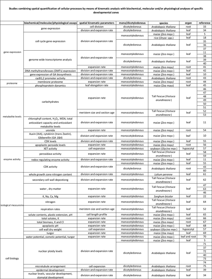
表2. 通过运动学分析量化细胞过程与其在分子水平调控之间的关联。 各项研究的参考文献,这些研究将不同物种和器官中细胞过程的量化结果与生物化学和分子检测结果相联系。木葡聚糖内转糖基酶(XET)、丙二醛(MDA)、周期蛋白依赖性激酶(CDK)。请点击此处查看此表格的放大版本。
访问受限。请登录或开始试用以查看此内容。
方案
注意:以下运动学分析方案仅适用于处于稳态生长阶段的叶片。这意味着在数天内,叶片的伸长速率以及细胞长度和扩展的空间模式保持稳定6。
1. 植物生长及叶片伸长速率(LER)的测量
- 选择处于稳态生长且处于目标发育阶段的叶片。
注意:稳态生长与重复生长之间存在差异,后者意味着同一轴上连续叶片具有相似的空间生长模式。在幼苗生长的早期阶段,由于生长区尺寸不断增加,连续叶片的生长速度通常会逐渐加快7。尽管少数较高叶位可能表现出相似的生长模式8,但这是一种短暂的阶段,可能受到所研究处理的影响。因此,即使叶片在不同时间发育,也必须严格比较相同叶位的品系和处理。即使伸长速率恒定,不同发育阶段的生长速率分布也可能不同。因此,应在相同的发育阶段分析叶片8,通常以出苗后的天数来定义。 - 为对单子叶植物叶片生长进行完整的运动学分析,在生长室的受控条件下,每种处理和基因型至少培养15株植物。
- 当目标叶片出现(从周围叶片的叶鞘中伸出)时,开始每天使用刻度尺测量叶片长度,直至叶片完全展开(图1i)。叶片长度指从土壤表面到叶尖的长度。测量时需小心,避免折断或损伤叶片,因为这可能改变其生长特性。
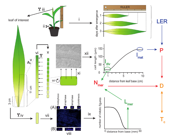
图 1:玉米叶片运动学分析的示意图概览。 感兴趣的叶片需连续三天用尺子测量,以计算叶片伸长速率(LER)。随后收获叶片,并取一个三厘米长的片段用于测定分生组织大小。该测量通过DAPI染色后,从叶片基部测量至最远端可见有丝分裂图像的位置来完成。(A) 增殖型有丝分裂图像示例;(B) 成形型有丝分裂图像示例。在叶片中脉另一侧,从叶片基部起的前十一厘米用于切割成十个一厘米长的片段,以进行细胞长度测量。这些测量数据用于构建细胞长度分布曲线,进而确定成熟细胞长度(lmat)以及离开分生组织时的细胞长度(ldiv)。利用LER和lmat可计算细胞生成速率(P),而ldiv与Lmer则用于计算分生组织中的细胞数量(Nmer)。进一步地,P和Nmer可用于计算平均细胞分裂速率(D),该速率是细胞周期持续时间(Tc)的倒数。相同颜色的箭头表示用于计算箭头指向参数的输入参数。比例尺 = 40 µm。罗马数字用于指代本实验方案中描述的特定实验步骤。 请点击此处查看该图的放大版本。
2. 收获
- 在感兴趣的发育阶段(例如, 在羽化后的第三天,从该批次中选取至少五株具有代表性的植株进行运动学分析。按照步骤1.3所述方法继续测量其余植株,以确定最终的叶片长度。
- 剪去植物的地上部分。为保持分生组织完整,应尽可能靠近根部切割。图1ii).
- 从外层叶片开始,逐个轻轻展开并移除所有叶片,直至目标叶片。如有必要,可从基部再切除几毫米以分离叶片。同时移除目标叶片内部包裹的顶端及小型叶片图1iii).
- 从主脉一侧基部开始,截取一段3 cm的组织,放入装有3:1(v:v)无水乙醇:冰醋酸溶液的1.5 ml离心管中(注意:佩戴手套),并于 4 °C 24 小时至几个月图1iv该片段后续将用于确定分生组织的长度。
- 从血管的另一端,从基部截取一段11 cm的节段(图1 v)并放入装有无水乙醇的 15 ml 离心管中 4 °C 至少 6 小时以去除色素(图1 vi).
注意:后续仅使用前10 cm来确定细胞长度分布(见讨论部分)。 - 更换无水乙醇,进行下一轮清洗 4 °C 至少 24 小时(图1vi).
- 最后,用纯乳酸替代乙醇进行清洗和储存(注意:需戴手套) 4 °C 24 小时或直至进一步使用(图1vi).
3. 分生组织长度测量
- 配制洗涤缓冲液,其中含有 50 mM 氯化钠(NaCl)、5 mM 乙二胺四乙酸(EDTA;注意:需佩戴手套)和 10 mM 三(羟甲基)氨基甲烷-盐酸(TRIS-HCl;pH 7)。
- 取第 2.4 节中的 3 cm 节段,将其浸入缓冲液中 20 分钟(图 1vii)。
- 在等待期间,用洗涤缓冲液配制浓度为 1 µg/ml 的 4',6-二脒基-2-苯基吲哚(DAPI)染色液,并将其置于冰上避光保存。
- 将分生组织节段放入 DAPI 染色液中染色 2–5 分钟,以染核。操作过程需在冰上并避光进行(图 1vii)。
- 将节段快速置于显微镜载玻片上,加盖盖玻片,观察荧光信号。表皮细胞应显示荧光,而下层细胞层则不应显示。
- 若染色不足,可将节段重新放回 DAPI 染色液中继续染色数分钟。
- 为终止染色,将节段置于显微镜载玻片上的一滴洗涤缓冲液中,并加盖盖玻片。
- 使用配备紫外荧光装置的显微镜,以 20X 放大倍数观察,可同时看到约 1,000 个表皮细胞。在整个节段范围内移动视野,寻找增殖性有丝分裂相(中期、后期、末期和胞质分裂),但应避免发育中气孔的成形性细胞分裂(图 1viii)9。确定最远端有丝分裂相的位置。
- 通过测量叶片基部与最远端表皮有丝分裂相之间的距离,确定分生组织的长度。使用图像分析软件(例如,ImageJ)测量图像视野的全长。
- 统计覆盖整个分生组织长度(从叶片基部到最远端有丝分裂相)的视野数量,并将该数值乘以单个视野的长度,得到完整的分生组织长度(图 1ix)。
4. 细胞长度分布图
- 取出保存在乳酸中的叶段(步骤 2.5),小心地放置在实验台上。用解剖刀将叶段切成 10 段,每段长 1 cm(图 1x)。
- 将连续的叶段置于载玻片上的一小滴乳酸中。确保所有叶段均保持同一面(上表面或下表面)朝上。原则上,朝上面的选择无特定要求。
- 使用配备微分干涉差(DIC)光学系统的显微镜,从叶片基部开始分析叶段。利用图像分析软件,测量气孔列附近纵向排列的表皮细胞列中至少 20 个重复细胞的长度,以确保始终选择同一类型的细胞。
- 在每一段上等间距的位置进行测量(每段测量 4 个位置即可),并记录每个测量值在叶片上的相应位置(图 1xi)。
- 采用 R 脚本中实现的局部多项式平滑法,确定沿叶片轴每毫米位置的平均细胞长度(图 1xii;补充文件 1)。
注意:R 脚本会输出一系列不同程度平滑的数据。所需平滑程度具有一定主观性,理想情况下应仅消除局部噪声,而不影响整体曲线形态。务必在同一实验的所有样品中使用相同的平滑程度。 - 计算不同植株间每个位置的细胞长度平均值,并计算标准误,以绘制沿叶片轴的细胞长度分布图谱。
5. 运动学参数的计算(参见补充文件2)
- 计算 LER 通过测量两个连续时间点之间叶片长度的变化(例如, 24 小时,如步骤 1.3 所述)并除以时间间隔。
- 计算生长区的长度(Lgz对应于细胞在平滑的细胞长度曲线上达到其成熟长度95%的位置,该位置位于基部远端。
- 在平滑后的细胞长度分布曲线上,对每个位置取该位置之后所有细胞长度平均值的95%图2).
- 将平滑处理后的细胞长度(步骤 4.4)与各位置计算得到的 95% 细胞长度进行比较。从叶片基部开始,生长区结束于实际细胞长度等于后续细胞长度 95% 的位置。图2;参见补充数据2。
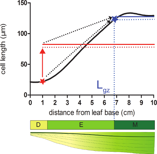
图 2:确定生长区的终点。分生区:在红星所示位置,实际细胞大小小于此后所有细胞平均大小的95%(红色虚线)(红色实线)。生长区的终点(Lgz;蓝星所示)位于此后所有细胞平均大小的95%(蓝色虚线)(蓝色实线)等于实际细胞大小的位置。分裂区(D)、伸长区(E)和成熟区(M)。虚线箭头表示从叶片基部向顶端移动时,局部细胞大小与远端区域平均大小的95%逐渐趋近。请点击此处查看该图的放大版本。
- 将生长区长度(Lgz)与分生组织大小(Lmer;在步骤3中确定)相减,计算伸长区长度(Lel)。
- 将成熟区的平均细胞长度作为成熟细胞长度(lmat)。
- 将细胞伸长速率(LER)除以成熟细胞长度(lmat),得到细胞生产速率(P)。
- 通过 Ngz 与 Nmer 相减计算伸长区中的细胞数量(Nel)。分生区中的细胞数量(Nmer)等于位于对应分生区区间的细胞累积总数;生长区中的细胞数量(Ngz)等于位于对应生长区区间的细胞累积总数。
- 将细胞生产速率(P)除以分生区细胞数量(Nmer),计算平均细胞分裂速率(D)。细胞周期持续时间(Tc)等于 ln(2)/D。
- 将伸长区细胞数量(Nel)除以细胞生产速率(P),计算细胞在伸长区的时间(Tel)。细胞在分裂区的时间等于 log2(Nmer)×Tc。离开分生区时的细胞长度(ldiv)等于分生区末端处平滑化细胞长度曲线中的细胞长度。
- 使用以下公式计算平均细胞扩展速率(Rel):[ln(lmat)−ln(ldiv)]/Tel。
访问受限。请登录或开始试用以查看此内容。
结果
本研究比较了充分供水植物(对照组,土壤含水量(SWC)为54%)与干旱胁迫条件下植物(干旱组,SWC为34%)的叶片生长情况。所有植物均在人工气候室中于受控条件下培养(16小时光照/8小时黑暗,昼夜温度分别为25 °C/18 °C,光合有效辐射为300–400 µEm-2sec-1)。通过停止浇水直至达到目标SWC,并维持该水分水平以建立干旱条件。在前期研究中确定,第五片叶是首个在此胁迫条件下萌发并发育的叶片,因此被选为目标叶片。干旱胁迫植株的最终叶片长度(LL)比充分供水植株低40%,这主要归因于叶片伸长速率(LER)的降低(表3)。为了理解叶片生长响应的细胞学基础,我们进行了运动学分析。总体而言,叶片伸长速率取决于分生组织中的细胞生成量以及伸长区决定的成熟细胞大小(图3)。因此,叶片缩短必然由其中一个或两个参数的变化所致。我们的结果表明,尽管成熟细胞长度(lmat)未受影响,但细胞生...
访问受限。请登录或开始试用以查看此内容。
讨论
对玉米叶片进行完整的运动学分析,能够确定叶片生长的细胞学基础,并有助于设计高效的取样策略。尽管该实验方案相对简单,但在以下关键步骤中仍需谨慎操作:(1)在分离较幼嫩的包埋叶片时(步骤2.3),必须避免损伤分生组织,因为分生组织长度的测定(步骤3)需要完整的分生组织存在,操作前可能需要一定的练习。(2)分生组织长度的测定依赖于对有丝分裂图像的判读,因此建议在同一实验中由同一人完成这些测量,以减少因操作者不同而引入的变异。(3)尽管实际细胞长度测量仅使用10厘米的叶片区段,但在采集用于细胞长度测量的样品时(步骤2.5),应包含11厘米的长度,因为样品边缘并不总能完全浸入乙醇和/或乳酸中;因此,额外增加的1厘米可确保生长区前10厘米部分被充分透明化。(4)最后,建议检查所有运动学参数的计算结果,以确认其正确性。例如,在表3中,干旱胁迫植株的细胞产生速率(P)降低了71%,换句话说,P干旱为P对照的0.29倍。由于细胞产生速率取决于平均细胞分裂速率(D;下降63%)和分生组织中的细胞数量(...
访问受限。请登录或开始试用以查看此内容。
披露
作者声明不存在任何竞争性经济利益。
致谢
本工作获得了安特卫普大学向 V.A. 提供的博士生奖学金;弗拉芒科学基金会(FWO,11ZI916N)向 K.S. 提供的博士生奖学金;弗拉芒科学基金会(FWO,G0D0514N)的项目资助;安特卫普大学研究委员会资助的协同研究活动(GOA)研究项目“"A Systems Biology Approach of Leaf Morphogenesis"”;以及比利时联邦科学政策办公室(BELSPO)资助的大学间吸引力计划(IUAP VII/29,MARS)项目“"Maize and Arabidopsis Root and Shoot Growth"”,受资助者为 G.T.S.B。Han Asard、Bulelani L. Sizani 和 Hamada AbdElgawad 均对本视频的制作做出了贡献。
访问受限。请登录或开始试用以查看此内容。
材料
| 姓名 | 公司 | 目录编号 | 评论 |
|---|---|---|---|
| 花盆 | 任何 | 任何 | 我们使用以下规格的花盆,但可根据处理/研究需要而有所不同:底部直径:11 cm,开口直径:15 cm,高度:12 cm。每盆种植一株玉米植株。 |
| 种植基质 | 任何 | 任何 | 我们使用盆栽培养基(Jiffy,荷兰),但也可根据处理或研究需要选用其他基质。 |
| 尺子 | 任何 | 任何 | 需要一把至少1.5米长的延长尺来测量植物的最终叶片长度。 |
| 种子 | 任何 | NA | 种子可从育种者处订购。 |
| 手术刀 | 任何 | 任何 | 手术刀在叶片采收过程中用于将目标叶片与其周围的叶片分离,并在采收后立即用于切割合适的样品,以进行细胞长度和分生组织长度的测量。 |
| 15 mL 离心管 | 任何 | 任何 | 15 mL Falcon离心管用于在使用无水乙醇和乳酸进行样本透明化处理期间,储存用于细胞长度测量的样本。 |
| Eppendorf管 | 任何 | 任何 | Eppendorf管用于储存以乙醇:醋酸3:1(v:v)溶液固定的分生组织长度测量样品。 |
| 手套 | 任何 | 任何 | 乳胶手套,用于防护腐蚀性试剂。 |
| 乙酸 | 任何 | 任何 | 警告:对金属有腐蚀性,类别1;皮肤腐蚀,类别1A、1B、1C;严重眼损伤,类别1;易燃液体,类别1、2、3 |
| 无水乙醇 | 任何 | 任何 | 警告:皮肤接触(刺激物)、眼睛接触(刺激物)、吸入时有危险。皮肤接触(渗透剂)、吞食时有轻度危险。 |
| 乳酸 >98% | 任何 | 任何 | 警告:对金属具有腐蚀性,类别1;皮肤腐蚀,类别1A、1B、1C;严重眼损伤,类别1 |
| 氯化钠(NaCl) | 任何 | 任何 | |
| 乙二胺四乙酸(EDTA) | 任何 | 任何 | 警告:急性毒性(经口、经皮、吸入),类别4;皮肤刺激,类别2;眼刺激,类别2;皮肤致敏,类别1;特异性靶器官毒性 – 单次暴露,3类 |
| 三(羟甲基)氨基甲烷盐酸盐(Tris-HCl) | 任何 | 任何 | 该物质可能具有刺激性,应避免接触眼睛和皮肤。吸入粉尘可能对呼吸道产生刺激。 |
| 4′,6-二脒基-2′-苯基吲哚二盐酸盐(DAPI) | 任何 | 任何 | 细胞可渗透的荧光DNA小沟结合探针。引起皮肤刺激。可能引起过敏性皮肤反应。可能引起呼吸道刺激。 |
| 冰 | 任何 | NA | DAPI 溶液必须置于冰上保存。 |
| 荧光显微镜 | AxioScope A1、Zeiss 的 Axiocam ICm1 或其他同类设备 | 任何荧光显微镜均可用于测定分生组织长度。 | |
| 显微镜载玻片 | 任何 | 任何 | |
| 盖玻片 | 任何 | 任何 | |
| 镊子 | 任何 | 任何 | 镊子在收获后用于展开卷曲的玉米叶片,以便切割出合适的样品,用于细胞长度和分生组织长度的测量。 |
| 图像分析软件 | 蔡司 Axiovision(版本 4.8) | NA | 软件可从以下网址下载:http://www.zeiss.com/microscopy/en_de/downloads/axiovision.html。也可使用其他软件,如 ImageJ(https://imagej.nih.gov/ij/)。 |
| 配备微分干涉差(DIC)的显微镜 | AxioScope A1、Zeiss 的 Axiocam ICm1 或其他同类设备 | 任何配备微分干涉差显微镜(DIC)的显微镜均可用于测量细胞长度。 | |
| R 统计分析软件 | R 统计计算基金会 | NA | 开源;可从 https://www.r-project.org/ 下载 |
| R 脚本 | NA | NA | 我们使用 Kern Smooth 软件包中的核平滑函数 locpoly(Wand MP, Jones MC)。 核平滑:查普曼 & Hall/CRC(1995))。该脚本可根据要求向通讯作者索取,适用于 Mac 和 Windows 系统。 |
参考文献
- Fiorani, F., Beemster, G. T. S. Quantitative analyses of cell division in plants. Plant Mol. Biol. 60, 963-979 (2006).
- Silk, W. K., Erickson, R. O. Kinematics of Plant-Growth. J. Theor. Biol. 76, 481-501 (1979).
- Rymen, B., Coppens, F., Dhondt, S., Fiorani, F., Beemster, G. T. S. Kinematic Analysis of Cell Division and Expansion. Plant Developmental Biology. Hennig, L., Köhler, C. , Chapter 14 (2010).
- Avramova, V., Sprangers, K., Beemster, G. T. S. The Maize Leaf: Another Perspective on Growth Regulation. Trends Plant Sci. 20, 787-797 (2015).
- Rymen, B., et al. Cold nights impair leaf growth and cell cycle progression in maize through transcriptional changes of cell cycle genes. Plant Physiol. 143, 1429-1438 (2007).
- Muller, B., Reymond, M., Tardieu, F. The elongation rate at the base of a maize leaf shows an invariant pattern during both the steady-state elongation and the establishment of the elongation zone. J. Exp. Bot. 52, 1259-1268 (2001).
- Beemster, G. T. S., Masle, J., Williamson, R. E., Farquhar, G. D. Effects of soil resistance to root penetration on leaf expansion in wheat (Triticum aestivum L): Kinematic analysis of leaf elongation. J. Exp. Bot. 47, 1663-1678 (1996).
- Bernstein, N., Silk, W. K., Lauchli, A. Growth and Development of Sorghum Leaves under Conditions of Nacl Stress - Spatial and Temporal Aspects of Leaf Growth-Inhibition. Planta. 191, 433-439 (1993).
- Sylvester, A. W., Smith, L. G. Cell Biology of Maize Leaf Development. Handbook of maize: It's Biology. Bennetzen, J. L., Hake, S. C. , Springer. NY. (2009).
- Nelissen, H., et al. A Local Maximum in Gibberellin Levels Regulates Maize Leaf Growth by Spatial Control of Cell Division. Curr. Biol. 22, 1183-1187 (2012).
- Avramova, V., et al. Drought Induces Distinct Growth Response, Protection, and Recovery Mechanisms in the Maize Leaf Growth Zone. Plant Physiol. 169, 1382-1396 (2015).
- Picaud, J. C., et al. Total malondialdehyde (MDA) concentrations as a marker of lipid peroxidation in all-in-one parenteral nutrition admixtures (APA) used in newborn infants. Pediatr. Res. 53, 406(2003).
- Basu, P., Pal, A., Lynch, J. P., Brown, K. M. A novel image-analysis technique for kinematic study of growth and curvature. Plant Physiol. 145, 305-316 (2007).
- Vander Weele, C. M., et al. A new algorithm for computational image analysis of deformable motion at high spatial and temporal resolution applied to root growth. Roughly uniform elongation in the meristem and also, after an abrupt acceleration, in the elongation zone. Plant Physiol. 132, 1138-1148 (2003).
- Nelissen, H., Rymen, B., Coppens, F., Dhondt, S., Fiorani, F., Beemster, G. T. S. Plant Organogenesis. DeSmet, I. , Chapter 17 (2013).
- Ben-Haj-Salah, H., Tardieu, F. Temperature Affects Expansion Rate of Maize Leaves without Change in Spatial-Distribution of Cell Length - Analysis of the Coordination between Cell-Division and Cell Expansion. Plant Physiol. 109, 861-870 (1995).
- Fiorani, F., Beemster, G. T. S., Bultynck, L., Lambers, H. Can meristematic activity determine variation in leaf size and elongation rate among four Poa species? A kinematic study. Plant Physiol. 124, 845-855 (2000).
- Pettko-Szandtner, A., et al. Core cell cycle regulatory genes in rice and their expression profiles across the growth zone of the leaf. J. Plant Res. 128, 953-974 (2015).
- Poorter, H., Remkes, C. Leaf-Area Ratio and Net Assimilation Rate of 24 Wild-Species Differing in Relative Growth-Rate. Oecologia. 83, 553-559 (1990).
- Macadam, J. W., Volenec, J. J., Nelson, C. J. Effects of Nitrogen on Mesophyll Cell-Division and Epidermal-Cell Elongation in Tall Fescue Leaf Blades. Plant Physiol. 89, 549-556 (1989).
- Tardieu, F., Granier, C. Quantitative analysis of cell division in leaves: methods, developmental patterns and effects of environmental conditions. Plant Mol. Biol. 43, 555-567 (2000).
- Bernstein, N., Silk, W. K., Lauchli, A. Growth and Development of Sorghum Leaves under Conditions of Nacl Stress - Possible Role of Some Mineral Elements in Growth-Inhibition. Planta. 196, 699-705 (1995).
- Erickson, R. O., Sax, K. B. Rates of Cell-Division and Cell Elongation in the Growth of the Primary Root of Zea-Mays. P. Am. Philos. Soc. 100, 499-514 (1956).
- Beemster, G. T. S., Baskin, T. I. Analysis of cell division and elongation underlying the developmental acceleration of root growth in Arabidopsis thaliana. Plant Physiol. 116, 1515-1526 (1998).
- Goodwin, R. H., Stepka, W. Growth and differentiation in the root tip of Phleum pratense. Am. J. Bot. 32, 36-46 (1945).
- Hejnowicz, Z. Growth and Cell Division in the Apical Meristem of Wheat Roots. Physiologia Plantarum. 12, 124-138 (1959).
- Gandar, P. W. Growth in Root Apices .1. The Kinematic Description of Growth. Bot. Gaz. 144, 1-10 (1983).
- Baskin, T. I., Cork, A., Williamson, R. E., Gorst, J. R. Stunted-Plant-1, a Gene Required for Expansion in Rapidly Elongating but Not in Dividing Cells and Mediating Root-Growth Responses to Applied Cytokinin. Plant Physiol. 107, 233-243 (1995).
- Sacks, M. M., Silk, W. K., Burman, P. Effect of water stress on cortical cell division rates within the apical meristem of primary roots of maize. Plant Physiol. 114, 519-527 (1997).
- Granier, C., Tardieu, F. Spatial and temporal analyses of expansion and cell cycle in sunflower leaves - A common pattern of development for all zones of a leaf and different leaves of a plant. Plant Physiol. 116, 991-1001 (1998).
- De Veylder, L., et al. Functional analysis of cyclin-dependent kinase inhibitors of Arabidopsis. Plant Cell. 13, 1653-1667 (2001).
- Kwiatkowska, D. Surface growth at the reproductive shoot apex of Arabidopsis thaliana pin-formed 1 and wild type. J. Exp. Bot. 55, 1021-1032 (2004).
- Kutschmar, A., et al. PSK-alpha promotes root growth in Arabidopsis. New Phytol. 181, 820-831 (2009).
- Vanneste, S., et al. Plant CYCA2s are G2/M regulators that are transcriptionally repressed during differentiation. Embo J. 30, 3430-3441 (2011).
- Eloy, N. B., et al. Functional Analysis of the anaphase-Promoting Complex Subunit 10. Plant J. 68, 553-563 (2011).
- Eloy, N. B., et al. SAMBA, a plant-specific anaphase-promoting complex/cyclosome regulator is involved in early development and A-type cyclin stabilization. P. Natl. Acad. Sci. USA. 109, 13853-13858 (2012).
- Dhondt, S., et al. SHORT-ROOT and SCARECROW Regulate Leaf Growth in Arabidopsis by Stimulating S-Phase Progression of the Cell Cycle. Plant Physiol. 154, 1183-1195 (2010).
- Baute, J., et al. Correlation analysis of the transcriptome of growing leaves with mature leaf parameters in a maize RIL population. Genome Biol. 16, (2015).
- Andriankaja, M., et al. Exit from Proliferation during Leaf Development in Arabidopsis thaliana: A Not-So-Gradual Process. Dev. Cell. 22, 64-78 (2012).
- Beemster, G. T. S., et al. Genome-wide analysis of gene expression profiles associated with cell cycle transitions in growing organs of Arabidopsis. Plant Physiol. 138, 734-743 (2005).
- Spollen, W. G., et al. Spatial distribution of transcript changes in the maize primary root elongation zone at low water potential. Bmc Plant Biol. 8, (2008).
- Candaele, J., et al. Differential Methylation during Maize Leaf Growth Targets Developmentally Regulated Genes. Plant Physiol. 164, 1350-1364 (2014).
- West, G., Inze, D., Beemster, G. T. S. Cell cycle modulation in the response of the primary root of Arabidopsis to salt stress. Plant Physiol. 135, 1050-1058 (2004).
- Zhang, Z., Voothuluru, P., Yamaguchi, M., Sharp, R. E., Peck, S. C. Developmental distribution of the plasma membrane-enriched proteome in the maize primary root growth zone. Front. Plant Sci. 4, (2013).
- Bonhomme, L., Valot, B., Tardieu, F., Zivy, M. Phosphoproteome Dynamics Upon Changes in Plant Water Status Reveal Early Events Associated With Rapid Growth Adjustment in Maize Leaves. Mol. Cell Proteomics. 11, 957-972 (2012).
- Schnyder, H., Nelson, C. J. Growth-Rates and Assimilate Partitioning in the Elongation Zone of Tall Fescue Leaf Blades at High and Low Irradiance. Plant Physiol. 90, 1201-1206 (1989).
- Schnyder, H., Nelson, C. J., Spollen, W. G. Diurnal Growth of Tall Fescue Leaf Blades .2. Dry-Matter Partitioning and Carbohydrate-Metabolism in the Elongation Zone and Adjacent Expanded Tissue. Plant Physiol. 86, 1077-1083 (1988).
- Schnyder, H., Nelson, C. J. Growth-Rates and Carbohydrate Fluxes within the Elongation Zone of Tall Fescue Leaf Blades. Plant Physiol. 85, 548-553 (1987).
- Vassey, T. L., Shnyder, H. S., Spollen, W. G., Nelson, C. J. Cellular Characterisation and Fructan Profiles in Expanding Tall Fescue. Curr. T. Pl. B. 4, 227-229 (1985).
- Allard, G., Nelson, C. J. Photosynthate Partitioning in Basal Zones of Tall Fescue Leaf Blades. Plant Physiol. 95, 663-668 (1991).
- Spollen, W. G., Nelson, C. J. Response of Fructan to Water-Deficit in Growing Leaves of Tall Fescue. Plant Physiol. 106, 329-336 (1994).
- Volenec, J. J., Nelson, C. J. Carbohydrate-Metabolism in Leaf Meristems of Tall Fescue .1. Relationship to Genetically Altered Leaf Elongation Rates. Plant Physiol. 74, 590-594 (1984).
- Volenec, J. J., Nelson, C. J. Carbohydrate-Metabolism in Leaf Meristems of Tall Fescue .2. Relationship to Leaf Elongation Rates Modified by Nitrogen-Fertilization. Plant Physiol. 74, 595-600 (1984).
- Silk, W. K., Walker, R. C., Labavitch, J. Uronide Deposition Rates in the Primary Root of Zea-Mays. Plant Physiol. 74, 721-726 (1984).
- Granier, C., Inze, D., Tardieu, F. Spatial distribution of cell division rate can be deduced from that of p34(cdc2) kinase activity in maize leaves grown at contrasting temperatures and soil water conditions. Plant Physiol. 124, 1393-1402 (2000).
- Voothuluru, P., Sharp, R. E. Apoplastic hydrogen peroxide in the growth zone of the maize primary root under water stress.1. Increased levels are specific to the apical region of growth maintenance. J. Exp. Bot. 64, 1223-1233 (2012).
- Wu, Y. J., Jeong, B. R., Fry, S. C., Boyer, J. S. Change in XET activities, cell wall extensibility and hypocotyl elongation of soybean seedlings at low water potential. Planta. 220, 593-601 (2005).
- Macadam, J. W., Nelson, C. J., Sharp, R. E. Peroxidase-Activity in the Leaf Elongation Zone of Tall Fescue .1. Spatial-Distribution of Ionically Bound Peroxidase-Activity in Genotypes Differing in Length of the Elongation Zone. Plant Physiol. 99, 872-878 (1992).
- Macadam, J. W., Sharp, R. E., Nelson, C. J. Peroxidase-Activity in the Leaf Elongation Zone of Tall Fescue .2. Spatial-Distribution of Apoplastic Peroxidase-Activity in Genotypes Differing in Length of the Elongation Zone. Plant Physiol. 99, 879-885 (1992).
- Beemster, G. T. S., De Vusser, K., De Tavernier, E., De Bock, K., Inze, D. Variation in growth rate between Arabidopsis ecotypes is correlated with cell division and A-type cyclin-dependent kinase activity. Plant Physiol. 129, 854-864 (2002).
- Kavanova, M., Lattanzi, F. A., Schnyder, H. Nitrogen deficiency inhibits leaf blade growth in Lolium perenne by increasing cell cycle duration and decreasing mitotic and post-mitotic growth rates. Plant Cell Environ. 31, 727-737 (2008).
- Macadam, J. W., Nelson, C. J. Secondary cell wall deposition causes radial growth of fibre cells in the maturation zone of elongating tall fescue leaf blades. Ann. Bot-London. 89, 89-96 (2002).
- Schnyder, H., Nelson, C. J. Diurnal Growth of Tall Fescue Leaf Blades .1. Spatial-Distribution of Growth, Deposition of Water, and Assimilate Import in the Elongation Zone. Plant Physiol. 86, 1070-1076 (1988).
- Gastal, F., Nelson, C. J. Nitrogen Use within the Growing Leaf Blade of Tall Fescue. Plant Physiol. 105, 191-197 (1994).
- Vanvolkenburgh, E., Boyer, J. S. Inhibitory Effects of Water Deficit on Maize Leaf Elongation. Plant Physiol. 77, 190-194 (1985).
- Silk, W. K., Hsiao, T. C., Diedenhofen, U., Matson, C. Spatial Distributions of Potassium, Solutes, and Their Deposition Rates in the Growth Zone of the Primary Corn Root. Plant Physiol. 82, 853-858 (1986).
- Meiri, A., Silk, W. K., Lauchli, A. Growth and Deposition of Inorganic Nutrient Elements in Developing Leaves of Zea-Mays L. Plant Physiol. 99, 972-978 (1992).
- Neves-Piestun, B. G., Bernstein, N. Salinity-induced inhibition of leaf elongation in maize is not mediated by changes in cell wall acidification capacity. Plant Physiol. 125, 1419-1428 (2001).
- Bouchabke, O., Tardieu, F., Simonneau, T. Leaf growth and turgor in growing cells of maize (Zea mays L.) respond to evaporative demand under moderate irrigation but not in water-saturated soil. Plant Cell Environ. 29, 1138-1148 (2006).
- Westgate, M. E., Boyer, J. S. Transpiration-Induced and Growth-Induced Water Potentials in Maize. Plant Physiol. 74, 882-889 (1984).
- Horiguchi, G., Gonzalez, N., Beemster, G. T. S., Inze, D., Tsukaya, H. Impact of segmental chromosomal duplications on leaf size in the grandifolia-D mutants of Arabidopsis thaliana. Plant J. 60, 122-133 (2009).
- Fleury, D., et al. The Arabidopsis thaliana homolog of yeast BRE1 has a function in cell cycle regulation during early leaf and root growth. Plant Cell. 19, 417-432 (2007).
- Vlieghe, K., et al. The DP-E2F-like gene DEL1 controls the endocycle in Arabidopsis thaliana. Curr. Biol. 15, 59-63 (2005).
- Boudolf, V., et al. The plant-specific cyclin-dependent kinase CDKB1;1 and transcription factor E2Fa-DPa control the balance of mitotically dividing and endoreduplicating cells in Arabidopsis. Plant Cell. 16, 2683-2692 (2004).
- Baskin, T. I., Beemster, G. T. S., Judy-March, J. E., Marga, F. Disorganization of cortical microtubules stimulates tangential expansion and reduces the uniformity of cellulose microfibril alignment among cells in the root of Arabidopsis. Plant Physiol. 135, 2279-2290 (2004).
访问受限。请登录或开始试用以查看此内容。
重印与许可
