本方案概述了采用图像引导的激光降解技术,在聚乙二醇二丙烯酸酯(PEGDA)水凝胶中构建源自血管、具有仿生特性的微流体网络的方法。此类仿生微流体系统可应用于组织工程、组织构建 体外 疾病模型,以及先进的“芯片上”设备的制备。
本文内容
摘要
摘要
本详细方案概述了采用图像引导的激光降解法,在聚乙二醇二丙烯酸酯(PEGDA)水凝胶中构建血管衍生的微流控网络的制备方法。本文描述了虚拟掩模的创建,以实现图像引导下的激光控制;微模塑 PEGDA 水凝胶的光聚合过程,适用于微流控网络的构建及压力驱动流体流动;商用激光扫描共聚焦显微镜与飞秒脉冲钛宝石(Ti:S)激光器的组装与使用,以诱导水凝胶降解;以及利用荧光物质和共聚焦显微镜对所制备的微流控网络进行成像。本方案的大部分内容聚焦于显微镜软件及显微镜宏程序的正确设置与实施,这些步骤在利用商用显微镜进行微流控结构制造时至关重要,且包含诸多技术细节。该技术的图像引导组件支持三维图像堆栈或用户自定义三维模型的导入,从而实现灵活的微流控设计,并可制备几乎任意构型的复杂微流控系统。该方法在组织工程领域具有重要应用前景,有望推动类器官和类人芯片装置中高级仿生微组织结构的构建。通过模拟复杂结构、迂曲度、尺寸及密度的 体内 血管系统,这些结构中可复制重要的生物转运过程,从而实现更精确的模拟 体外 药物药代动力学与疾病建模
引言
血管系统由淋巴管和心血管系统组成,形成高度密集的网络,对营养物质和氧气的输送以及代谢废物的清除至关重要。因此,血管化组织中的细胞与血管的距离从不超过50–100 µm。1. 可重复性能力 体内 血管结构 体外 准确建模的关键 体内 利用工程化构建体研究转运过程。随着近年来推动器官芯片设备的开发2 用于高通量药物筛选3,4 和疾病建模5,6,用于构建可重现微流控网络的方法 体内类组织运输在合成或天然水凝胶中正受到广泛关注。为了增强这些装置中所用微组织的仿生性,我们开发了一种基于图像引导的激光水凝胶降解方法,该方法利用天然血管系统的三维(3D)图像序列作为模板,在聚乙二醇二丙烯酸酯(PEGDA)水凝胶中构建出源自血管的微流体网络。7本方案概述了使用市售的配备飞秒脉冲激光器的激光扫描共聚焦显微镜,通过图像引导的激光降解方法,在聚乙二醇二丙烯酸酯(PEGDA)水凝胶中制备血管来源的仿生微流控网络。
目前使水凝胶液化的方法包括诱导血管生成8-10 或促血管生成的10-12 内皮细胞自组装与微加工技术用于构建预设的内皮化通道13-16尽管自组装网络能够重现微血管的密度和复杂结构,但其通透性通常高于 体内 网络11,17,18这在药物筛选应用的运输建模中可能存在问题。自组装网络包含生理学上相关的、毛细血管尺寸的管道,但由于难以生成更大尺寸的小动脉级管道,其与整体流体流动的整合可能较为困难。由于无法直接控制这些网络的组装过程,最终结构可能在不同样本间存在差异,导致难以重复制备出具有相同流体流动和传输特性的网络。
为了构建具有可重复几何形状和明确定义结构的三维水凝胶包埋微流体网络,已开发出多种微加工技术,包括模块化组装13、牺牲材料的三维打印16、直接书写组装14以及全向打印15。利用这些方法,可在多个结构中重复制造微流体结构,从而实现对流体流动和传输特性的精确控制。然而,这些方法的主要局限在于无法构建具有毛细血管尺寸特征(4至10 µm19)的微流体网络。大多数微加工技术通常仅能实现直径在150至650 µm范围内的结构特征13,16。部分现有技术能够生成具有宽直径范围通道的分级网络,例如直接书写组装可实现10至300 µm的通道14,全向打印可实现18至600 µm的通道15,但它们在生成密集网络或在同一结构内近距离构建多个微流体网络方面仍存在局限7。
为了克服其中一些局限性,我们开发了一种基于激光的图像引导水凝胶降解技术,可重复地构建出仿生的、具有层级结构的微流控网络,以重现目标结构的架构 体内 微血管结构。为此,根据水凝胶内预设的三维位置图像,使用波长为790 nm、脉冲宽度为140飞秒(fs)、重复频率为80 MHz的脉冲激光在目标区域进行光栅扫描, 体内 血管系统。我们推测,该降解过程通过激光诱导的水光学击穿、随之产生的等离子体形成、水的快速热弹性膨胀,以及水膨胀过程中水凝胶的局部降解而实现20该机制与基于激光的蛋白质水凝胶降解机制略有不同21-24与PEGDA不同,蛋白质通常具有较大的双光子截面,因此会通过双光子吸收诱导的化学断裂而发生降解。23通过图像衍生的虚拟掩模控制激光快门,以生成图像引导的微流控网络,该虚拟掩模由多个感兴趣区域拼接而成9 定义微流控结构。利用该方法,我们已成功制备出三维血管来源的仿生微流控网络,可重现密集且迂曲的血管结构。 在体 血管系统,以便通过调节降解过程中传递的能量来局部控制水凝胶的孔隙率。我们还成功构建了两个相互交织且紧密相邻(15 µm)但从未直接连通的独立微流控网络。7我们还证明了通过在激光降解后对微通道进行功能化修饰,引入整合素配体肽序列精氨酸-甘氨酸-天冬氨酸-丝氨酸(RGDS),可实现降解微通道的内皮化,促进内皮细胞黏附和管腔形成。7.
通过本方案,可利用商业显微镜(在多数高校校园内均可获得)结合基于图像引导的激光降解技术,在聚乙二醇二丙烯酸酯(PEGDA)水凝胶中构建复杂的微流控网络。由于降解过程由数字化的虚拟掩模引导,该制造技术适用于创造性地设计微流控网络,因而可广泛应用于多种场景。我们预计,本文所述方法在设计仿生类器官和类人芯片装置方面将具有显著优势,能够模拟药物转运过程中关键的生物传输过程。2该制备技术也可能对生成 体外 疾病模型,包括癌症转移5,6 以及血脑屏障模型25由于基于激光的水凝胶降解技术此前已被用于为神经突起的生长导向创建路径21-23该技术的图像引导延伸在高级组织工程策略中可能具有重要应用价值,可用于将细胞定位在用户自定义的三维空间结构中。
方案
1. 生成虚拟掩模
注意:本方案选用小鼠脑微血管系统的三维图像堆栈作为微流控模型,该数据选自包含完整小鼠脑微血管系统图像的更大规模数据集。微血管图像通过刀锋扫描显微镜(KESM)26,2 获得;类似的微血管数据可通过KESM小鼠脑图谱公开获取28。
- 打开图像处理软件29 并打开和裁剪天然血管的3D TIF图像堆栈,使将在水凝胶内生成的所需微流控网络适配于450 µ米 × 450 µm × 1.5 mm(x × y × z)窗口。操作时,在目标区域周围绘制一个正方形,然后单击 "图像" → "裁剪". 通过点击移除 z 方向上的切片 "图像" → "复制" 并输入期望的切片范围。
注意:这可确保目标特征位于视场范围内(x 乘以 y)(531.2 × 531.2 µ使用激光扫描共聚焦显微镜,配备20倍(数值孔径1.0)水浸物镜、2,884 × 2,884像素的帧尺寸和0.8倍缩放时的视场范围(m),以及该20倍(数值孔径1.0)水浸物镜的工作距离(z)。若其他系统参数未知,可通过使用目标物镜和设定参数采集图像来确定其视场范围。 - 旋转图像堆栈,使特征主要与光栅扫描方向或x方向对齐,方法是单击 "图像" → "转化" → "旋转"这减少了制造所需的时间。
- 将裁剪后的图像堆栈进行缩放,使像素尺寸匹配或超过共聚焦显微镜降解时所用的设置(0.184 µ当使用20倍(NA 1.0)水浸物镜、帧尺寸为2,884 × 2,884像素、缩放倍数为0.8时,为每像素1 μm(m/pixel)。为此,请单击 "图像" → "调节" → "尺寸" 并输入 "2446" 用于 "宽度" (即, 450 µ米,或图像尺寸除以 0.184 µ微米/像素
- 通过输入对图像堆栈进行阈值化/二值化 "Ctrl + Shift + T"取消选择 "暗背景",然后单击 "应用" → "ok". 通过点击完成该过程 "图像" → "查找表" → "反转LUT".
- 使用编程软件中的自定义算法(源代码可在参考文献的手稿补充材料中获取)9将二值化(白色)特征拟合为与光栅扫描方向(x方向)平行的单像素高四边形镶嵌,以生成引导激光位置和快门的感兴趣区域(ROIs)9,7,30,31,32.
注意:该自定义算法9 将 TIF 图像堆栈中的每个单独平面转换为 RLS 文件。 - 将RLS文件的扩展名更改为.OVL,以用于降解过程。
2. 配置激光扫描共聚焦显微镜
注意:尽管可使用配备脉冲激光器的其他激光扫描显微镜,但此处的设置和方案描述的是激光扫描显微镜(LSM)与其配套软件的联合使用方法。
- 开启激光扫描共聚焦显微镜后,打开显微镜软件,点击“启动系统”。在“维护”选项卡中选择物镜“W Plan-Apochromat 20x/1.0 DIC VIS-IR M27 75mm”。
- 设置“水凝胶可视化”配置
- 在“采集”选项卡中,确保在“激光”窗口中已选择并开启激光线(514 nm 氩离子激光)。注意:该通道用于通过曙红Y荧光成像水凝胶,以实现定位目的7。
- 在“成像设置”窗口中,将“模式”设为“通道模式”,将“切换轨道间隔”设为“帧”,并选择“Track1”。
- 在“光路”窗口中,仅选择Ch1荧光探测器,检测范围为516至735 nm;可见光路使用主分束器(MBS)458/514滤光片;不可见光路插入滤光板。取消选择明场光电倍增管探测器“T-PMT”。
- 在“采集模式”窗口中,确认已选择正确的物镜,并将“扫描模式”设为“帧”,X和Y方向的“帧尺寸”设为“2884”,“线步进”设为“1”,“平均次数”设为“1”,“平均模式”设为“线”,“平均方法”设为“均值”,“位深度”设为“16位”,“方向”设为“<->”,以实现双向扫描。
- 在“采集模式”窗口中,根据需要设置“速度”,并将“Corr X”和“Y”设为“0.05”,或根据所用显微镜的具体情况进行调整。取消选择“HDR”。
- 在“采集模式”窗口中,确保“扫描区域”显示“图像尺寸”为“531.2 µm x 531.2 µm”,“像素尺寸”为“0.18”,“缩放”设为“0.8”;将其他所有“扫描区域”参数设为零。
- 在“通道”窗口中,选择“Track1”作为Ch1荧光探测器。选择激光线“514”,功率百分比设为“10.0”,“针孔”设为“1AU”,初始“增益(主)”设为“800”,“数字偏移”设为“0”,“数字增益”设为“1”。
注意:根据所用显微镜的具体情况,必要时调整这些参数。 - 将此配置保存为“水凝胶可视化”。
- 设置“通道形成”配置
- 在“采集”选项卡中,确保在“激光”窗口中已选择并开启激光线(790 nm 140 fs脉冲Ti:S激光)。将“GDD校正”设为“4000”,以在790 nm处获得最大输出功率。
- 将“成像设置”窗口设置为与步骤2.2.2中所述一致。
- 在“光路”窗口中,确保未选择任何荧光探测器,可见光路使用MBS 458/514/561/633滤光片,不可见光路使用MBS 760+滤光片。取消选择明场光电倍增管探测器“T-PMT”。
- 在“采集模式”窗口中,确认列出正确的物镜。将“速度”设为“3”,其余设置与步骤2.2.4中所述一致。
- 在“通道”窗口中,选择“Track1”作为T-PMT探测器。选择激光线“790”,功率百分比设为“100.0”,初始“增益(主)”设为“800”,“数字偏移”设为“0”,“数字增益”设为“1”。
- 在“载物台”窗口中,点击“设为零”以将载物台归零。点击“标记”以记录位置。此步骤对于在第3步中将虚拟掩模上传至显微镜宏至关重要。
- 在“时间序列”窗口中,将“循环次数”设为“1”,“时间间隔”设为“0”。
- 在“漂白”窗口中,勾选“在#次扫描后开始漂白”并将其设为“0 of 1”。选择“不同扫描速度”,数值设为“3”(对应像素驻留时间为8.96 µs/像素)。选择“GaAsP安全漂白”。
- 在“漂白”窗口的激光线部分,选择790激光线并将功率百分比设为“100.0”。漂白窗口中的其他选项保持未勾选状态。保存“漂白”窗口中的漂白设置。
- 将此配置保存为“通道形成”。
注意:“Z-Stack”、“区域”、“焦点”和“载物台”窗口在本方案中同样重要,但在初始配置期间无需设置或调整。
3. 创建配方并将虚拟掩模上传至显微镜软件与宏
- 在激光扫描共聚焦显微镜持续运行且显微镜软件仍处于开启状态时,仅选择 "区域" 窗口(旁边) "开始实验" 按钮)在 "通道形成" 构型
- 为确保掩膜能正确加载至显微镜宏中,请设置百分比功率 "通道" 窗口到 "0.2" 以及 "速度" 在 "采集模式" 窗口到 "8",然后单击 "快照" 位于屏幕左上方。
- 检查图像尺寸是否仍然 "531.2 µm × 531.2 µm",像素尺寸为 "0.18",在 "信息" 图像的标签。如果这些值不正确,请检查 "帧尺寸" 和 "缩放" 再次输入数值。
- 关键:检查 X 和 Y 位置是否在 "阶段" 窗口归零并标记位置后,再打开显微镜宏。参见步骤 2.3.6。
- 要打开显微镜宏,请输入 "Alt+F8",单击 "加载",选择正确的版本。详见特定材料/设备列表。将打开一个包含大量子宏的列表 "宏控制" 窗口。
- 在 "宏控制" 窗口,单击 "运行" 打开显微镜宏,然后关闭 "宏控制" 窗口
注意:此方案中使用的显微镜宏的其他版本可能与基于激光的水凝胶降解不兼容。 - 在 "保存" 显微镜宏的选项卡中,提供一个任意的 "基础文件名",选择 "单文件输出",并选择了一个 "临时文件" 文件夹。设置 "打开临时文件夹" 至指定位置 "1",选择 "单个临时图像文件夹",并为该配方命名 "储存/应用配方" 与 "回忆位置列表" 选择。单击 "储存" 用于初步创建配方。
- 在 "采集" 显微镜宏的选项卡中,确保 "扫描配置" 与步骤 2.3 中为显微镜软件配置集指定的名称相匹配。设置 "区块内时间点数量" 到 "1",具有 "间隔" 的 "0"确保 "标记 Z - Z 堆栈顶部" 以绿色突出显示。所有其他默认设置保持不变即可。
- 在 "时间安排" 显微镜宏的选项卡中,确保 "等待延迟" 呈绿色高亮,设置 "实验重复次数" 以及 "组重复" 至 "1" 和 "仅在第一个位置执行前等待间隔时间" 至 "0"所有其他默认设置均无需更改,保持原样即可。
注意: "组重复" (而不是 "实验重复次数")框用于设置激光对某一区域扫描的次数即, 重复实验配方的次数。 - 在 "Z轴列表" 在显微镜宏的选项卡中,指定z方向的降解平面。设置z间距, "dZ",以 "1 µm",与 "位置数量" 设置为与将要使用的虚拟掩模数量相匹配。
- 单击 "创建Z轴堆栈" 按钮以制作 "Z轴列表" 出现在下拉菜单中 "位置列表" 窗口底部。检查位置数量和z轴间距是否正确,并确认X和Y位置已归零。
注意:如果未满足条件,请勿保存该方案;关闭显微镜宏,重新执行步骤 2.3.6,然后再次打开显微镜宏并重新设置方案。
注意:无需将 100 个掩膜按 1 间隔排列 µ例如,拥有50个掩膜,每个使用两次且间隔1 m,可能是有利的 µm,以获得等效的微流体结构,因为将单个掩模逐个加载到显微镜宏中可能耗时较长。 - 在 "漂白" 显微镜宏的选项卡中,必须加载所有掩模并将其与特定编号位置匹配 "地点列表"。首先,选择 "漂白" 盒中,并确保 "配置" 与漂白设置的名称相匹配 "漂白" 显微镜主软件界面中的窗口(参见步骤 2.3.8)。设置 "漂白后等待延迟" 至 "0",取消选择 "点",并留下 "感兴趣区域" empty.
- 请勿更改以下内容中的任何部分 "位置", "拼接图像", "网格", "自动对焦", "区块",以及 "选项" 从默认设置中移除标签页
- 要将掩模加载到显微镜宏中,请仅选择 "区域" 显微镜软件中的窗口并打开 "漂白" 显微镜宏的选项卡中。在 "区域" 窗口,单击 "加载" 并选择位于待降解 z-stack 底部的 .OVL 文件作为掩模。
注意:序列中的第一个位置 "Z 列表" 是 z 轴堆栈的底部,显微镜宏将从该位置逐步运行至 z 轴堆栈或水凝胶的顶部。 - 一旦ROI列表出现在 "区域" 窗口中,点击箭头按钮以查看视野内掩模的感兴趣区域(ROI)(即步骤 3.2 中拍摄的图像)。确认 ROI 位于视野范围内。
- 为在显微镜宏中保存已加载的掩膜,请为该掩膜指定一个名称和编号 "将当前区域添加到感兴趣区域列表" 框上的 "漂白" 标签,然后单击 "将当前区域添加到感兴趣区域列表" 按钮。名称和编号将出现在 "感兴趣区域" 下拉列表中包含任何其他掩模(包括来自先前创建的其他配方的掩模)。删除该掩模 "区域" 显微镜软件中的窗口
- 重复步骤 3.14 和 3.16,将所有掩模上传并保存至显微镜宏中。
- 将上传的掩模分配给每个Z位置时,在下拉菜单中高亮选择位置1 "位置列表". 从ROI中选择适当的区域 "感兴趣区域" 下拉列表。确保 "漂白" 选中顶部的框 "漂白" 显微镜宏中的选项卡
- 对下拉菜单中的所有位置重复步骤 3.18 "位置列表",然后单击 "储存" 在 "保存" 按 Tab 键以最终形式保存方案。
4. 光聚合 PEGDA 水凝胶
- 制备含三乙醇胺(TEOA)的无菌 HEPES 缓冲盐水(HBS)
- 在 1 L 烧杯中加入 500 mL 去离子 H2O,在通风橱中使用磁力搅拌子搅拌,依次加入 2.922 g 氯化钠、1.196 g HEPES 和 7.5 mL TEOA。
- 用巴斯德移液管逐滴加入 1 N 氢氧化钠溶液,调节 pH 至 8.3。
- 使用 0.2-µm 真空过滤杯将溶液无菌过滤至一个 500 mL 的已灭菌玻璃培养瓶中。分装后于 4 °C 保存。
- 将一圈带背衬的双面胶粘附到一个特制的 60 mm 培养皿底部(该培养皿底部已切出一个 20 mm 的孔)。保持背衬不撕除,将培养皿与双面胶之间的气泡压出。
- 准备材料。从 -80 °C 冰箱中取出合成的33 3.4 kDa PEGDA,使其升至室温。在进行水凝胶光聚合前至少 15 分钟开启白光光源。
- 在通风橱中,将 1 mL 含 TEOA 的 HBS 加入一个用铝箔包裹的 2 mL 棕色离心管中(用于防止光引发剂曙红 Y 暴露于光下)。加入 10 µL 1 mM 曙红 Y 二钠盐水溶液(DI H2O 配制)。
- 在通风橱中,用巴斯德移液管吸取少量 1-乙烯基-2-吡咯烷酮(NVP)至玻璃烧杯中。从此液体中移取 3.5 µL NVP 加入含 HBS、TEOA 和曙红 Y 的棕色离心管中。
- 在另一个用铝箔包裹的 2 mL 棕色离心管中(用于防止预聚物溶液意外光活化),加入 10 mg 3.4 kDa PEGDA。加入 0.2 mL 含 HBS、TEOA、曙红 Y 和 NVP 的溶液,配制成 5% w/v 的 PEGDA 预聚物溶液。涡旋振荡至 PEGDA 完全溶解。使用功率计和探测器探头将白光光源强度调节至 95 mW。
- 制备聚二甲基硅氧烷(PDMS)模具
- 在较大表面(如玻璃或聚苯乙烯细胞培养皿)上制备 PDMS 模具7,以便于操作,并组装模具使成型的水凝胶可容纳于直径 20 mm 的圆形区域内。
- 为制备厚度为 500 µm 的矩形水凝胶,将两个 500 µm 厚的 PDMS 间隔片置于 PDMS 表面上,以形成两个确定的水凝胶边缘。
- 加入一个 300 µm 厚的 PDMS 间隔片,使水凝胶表面形成一个 300 µm 深的凹槽,便于后续引入流体。
- 将 60 µL 的 5% w/v PEGDA 预聚物溶液移取至 PDMS 模具上。移液时注意避免引入气泡。
- 将一片经[3-(甲基丙烯酰氧基)丙基]三甲氧基硅烷(TMPSA)功能化处理7、直径 40 mm、#1.5 玻璃盖玻片,居中放置于溶液上方。确保盖玻片落在 500 µm 厚的 PDMS 间隔片上。水凝胶将与甲基丙烯酰化盖玻片键合,防止其在溶液中漂浮。
- 将 PDMS、PEGDA 预聚物溶液和盖玻片的组装体置于白光光源下照射 3 分钟。
- 在模具与盖玻片之间通入去离子 H2O,使水凝胶与 PDMS 分离。用去离子 H2O 冲洗水凝胶。用实验用纸巾擦干盖玻片边缘。注意不要触碰水凝胶或从中吸走水分。
- 撕去双面胶背衬后,将盖玻片粘附至已准备好的培养皿上,并轻轻压出胶层与玻璃之间的气泡。将水凝胶完全浸没于培养皿中的去离子 H2O 中。
注:若将 PDMS 模具用去离子 H2O 冲洗、氮气吹干并防尘保存,则可重复使用以制备更多水凝胶。
5. 通过激光降解法构建血管衍生的微流控网络
- 打开激光扫描共聚焦显微镜及显微镜软件后,选择物镜 "W Plan-Apochromat 20x/1.0 DIC VIS-IR M27 75mm" 在 "维持" 标签。在 "采集" 标签,确保必要的激光线(514 nm 氩激光和 790 nm 140 fs 脉冲 Ti:S 激光)已开启并完成预热。
- 设置水凝胶以形成入口通道
- 将水凝胶和培养皿安装到载物台插件上,并将载物台插件放置在显微镜载物台上。向培养皿中加入至少5 mL的去离子水2O. 在将水浸物镜浸入去离子水之前,先用肉眼将水凝胶和孔的特征居中2O.
注意:切勿用物镜挤压水凝胶——两者绝不可接触! - 要找到水凝胶,切换到 "水凝胶可视化" 步骤 2.2 中的配置。单击 "活体" 打开激光线,并将物镜靠近水凝胶,直至观察到水凝胶顶部的曙红Y信号(由Ch1荧光检测器检测)。
注意:为便于找到水凝胶,可提高激光功率百分比或增大针孔。然而,一旦物镜聚焦到水凝胶上,应立即降低激光功率百分比以保护荧光探测器,并将针孔减小至1 AU,以避免探测器过饱和,从而准确定位水凝胶表面。 - 在 "阶段" 窗口,标记水凝胶顶部和底部以及孔底部的位置。
注:此处的 z 值将有助于确定 hydrogel 的厚度和孔的深度。 - 使用操纵杆在 X 和 Y 方向上移动,找到孔的边缘。将水凝胶置于屏幕右侧,孔置于左侧,或 反之亦然。 让水凝胶填充不超过视野的三分之二。
- 将焦平面下移150 µm进入水凝胶,通过设置 "步长" 至 "150" 在 "聚焦" 窗口并点击其旁边的下拉箭头 "Z轴位置" 方框。如有必要,重复步骤 5.2.4。
注意:入口的 x、y 和 z 位置完全取决于虚拟掩模的设计。 - 关键步骤:将 X 和 Y 位置归零 "阶段" 窗口,删除所有其他标记位置,仅保留此位置。
注意:如果未执行此步骤,显微镜宏将无法正确恢复先前保存的方案中的位置,导致水凝胶无法在预期位置发生降解。
- 将水凝胶和培养皿安装到载物台插件上,并将载物台插件放置在显微镜载物台上。向培养皿中加入至少5 mL的去离子水2O. 在将水浸物镜浸入去离子水之前,先用肉眼将水凝胶和孔的特征居中2O.
- 形成进样通道
- "快照" 一幅图像,并使用矩形按钮绘制一个矩形区域,作为血管网络的入口 "区域" 窗口。
注意:确保部分区域延伸至孔中,以保证入口能够连接孔与血管网络。 - 紧邻在 ROI 生成的 "区域" 窗口,选择 "成像" 并取消选择 "漂白" 和 "分析".
- 保存 "水凝胶可视化" 配置,取消选择 "区域",并切换到 "通道形成" 构型,于步骤2.3中设置。在 "通道形成" 配置,选择 "区域" 再次。
注意:在不同模式之间切换时 "水凝胶可视化" 以及 "通道形成" 配置时,务必取消选择屏幕左上角的区域。如果未执行此操作,区域的缩放比例在不同配置之间可能会意外发生变化。 - 在 "通道形成" 配置,确保仅 "区域" 以及 "Z-堆栈" 选定。在 "Z-轴堆栈" 窗口,单击 "中心" 顶部的按钮,然后 "中心" 上方按钮 "偏移量" 将z-stack的中心设置为当前Z位置。根据入口所需大小,设置层数 "切片" 与一个 "间隔" 的 "1"Z-stack 的大小将显示在下方 "范围".
- 检查 "速度" 在 "采集模式" 窗口被设置为 "3" 且该 "功率百分比" 在 "通道" 窗口被设置为 "100"保存配置并单击 "开始实验" 按钮。
注意:在多个平面上降解相同简单形状(如此处用于生成入口通道的操作)时,无需使用显微镜宏。仅当在每个单独平面上降解不同区域时才需要使用该宏,其作用是将虚拟掩模存储于一个方案中。 - 为了在降解过程中观察水凝胶,可选择提高 "增益(主控)" 在 "通道" 如果视野中的区域为黑色,则减小窗口 "增益(主控)" 如果该区域为白色。
注意:此处气泡的形成是激光诱导水凝胶降解的迹象。 - 若需对入口进行多次降解而非仅一次,可调整 "循环次数" 在 "时间序列" 窗口
- 在 "水凝胶可视化" 配置,单击 "活体" 以确保入口已形成。
- "快照" 一幅图像,并使用矩形按钮绘制一个矩形区域,作为血管网络的入口 "区域" 窗口。
- 设置水凝胶以生成微流控网络
- 在 "区域" 窗口中,加载 z 轴虚拟掩模堆栈中的任意 .OVL 文件,然后点击箭头按钮以查看视野中的感兴趣区域。
- 点击 "活体" 用于活体扫描的 "水凝胶可视化" 配置并使用操纵杆或 "阶段" 调整水凝胶在 X 和 Y 方向的位置,使入口与血管网络中的正确位置相连。
注意:确保虚拟掩模的感兴趣区域(ROI)与先前形成的入口略微重叠。 - 将焦平面下移50 µμm 从先前的中心 Z 位置,或 200 µm 从水凝胶表面,使用 "步长" 在 "聚焦" 窗口并点击其旁边的下拉箭头 "Z轴位置" 盒子。这将是显微镜宏中的第一个位置。
注意:在 z 方向上的实际移动距离取决于网络设计。确保目标网络在 z 方向上不超过水凝胶的顶面或底面。 - 关键步骤:将 X 和 Y 位置归零 "阶段" 窗口,删除所有其他标记位置,仅标记此位置。
注意:这将是显微镜宏中的起始点。该程序将从该点逐步回退至水凝胶表面。 - "快照" 一幅图像在 "水凝胶可视化" 配置、删除和取消选择 "区域",并保存配置。
- 生成微流控网络
- 切换到 "通道形成" 构型,并仅选择 "时间序列" 以及 "漂白" 窗口。验证设置是否正确 "时间序列" 以及 "漂白" 窗口设置与步骤 2.3.7 和 2.3.8 中的初始设置保持一致。
- 设置 "速度" 在 "采集模式" 窗口到 "8" 以及激光线的功率百分比 "0.2" 在 "通道" 保存配置。
- 按照步骤 3.5 打开显微镜宏。在 "保存" 在宏的选项卡中,选择步骤3中预先制作的配方,然后单击 "应用".
注意:不要点击 "储存" 否则配方将被覆盖! - 检查所有选项卡中的设置,确认虚拟掩膜(.OVL 文件)仍正确关联到下拉列表中的 Z 位置。同时检查第一个 Z 位置是否与标记位置一致 "阶段" 显微镜软件的窗口
注意:与直觉相反,下拉列表中的第二个位置在物理上将位于第一个位置的上方,因为显微镜宏程序将从水凝胶的底部(距离物镜最远)向顶部(距离物镜最近)依次进行操作。 - 保存 "通道形成" 再次配置,然后单击 "更新" 在显微镜宏中。点击 "否" 至 "覆盖当前位置列表?",然后单击 "开始" 在显微镜宏中启动该实验方案。
6. 微流控网络的可视化
- 若要观察降解后的水凝胶及形成的微流控网络,请先关闭显微镜宏程序。切换至“水凝胶可视化”配置,以观察水凝胶的曙红Y信号;降解形成的网络区域将呈现为暗色。
注意:在显微镜宏程序运行期间无法观察水凝胶的降解过程。 - 若需专门观察微流控网络,可使用“水凝胶可视化”配置,并调低激光功率,以便成像引入的荧光素(FITC)标记葡聚糖;其信号强度高于水凝胶自身曙红Y的荧光信号。
- 在引入荧光标记葡聚糖前,使用实验室纸巾通过毛细作用从水凝胶的孔中吸除水分。随后加入10 µL浓度为5 mg/mL、分子量为2,000 kDa的FITC标记葡聚糖水溶液(DI H2O),并预先通过0.2 µm针筒滤器过滤。
注意:应使用分子量等于或大于2,000 kDa的葡聚糖,以防止其扩散进入凝胶内部。若成像时间超过30分钟,建议使用带有60%或更高湿度的孵育载物台,以防止水凝胶干燥及FITC标记葡聚糖溶液中的水分蒸发。 - 将水凝胶与培养皿从载物台上取下,并在培养皿上做标记,以便在后续实验中快速定位所形成的血管样网络结构。
结果
为了演示基于图像引导的激光降解水凝胶技术以构建三维血管来源的微流控网络,我们采用了一组通过刀锋扫描显微镜(KESM)获取的小鼠脑血管三维图像序列26,27。从该较大的微血管数据集中选取一个三维区域,并如上所述将其转换为一系列用于图像引导微流控网络构建的虚拟掩模。制造完成后,将所得微流控网络用含有FITC标记的2,000 kDa葡聚糖溶液灌注,并通过共聚焦显微镜成像。定义网络结构的体内血管图像序列(图1A和C)与所制备的微流控网络(图1B和D)被用于生成Z轴投影图像(图1A和B)以及三维重建图像(图1C和D)。图像对比表明,基于图像引导的激光水凝胶降解技术能够制造出可重现体内血管尺寸、迂曲度及复杂结构特征的三维仿生微流控网络。该技术适用于构建包含多种直径范围的网络;在图1中,所生成的通道直径范围为3.3至28.8 µm。请注意,图1B左上方以及图1D左侧角落中较大的矩形结构是用于向网络引入流体的进样通道。
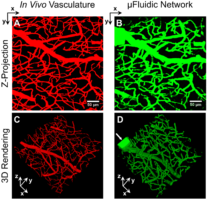
图1:血管来源的微流控网络 一系列源自小鼠脑血管图像堆栈的虚拟掩膜(A 和 C),用于构建嵌入 PEGDA 水凝胶中的三维仿生微流控网络(B 和 D) 通过图像引导的激光降解实现。微流控网络灌注了2,000 kDa FITC标记的葡聚糖,并通过共聚焦显微镜成像。Z投影(A 和 B)和三维重建图像(C 和 D两个网络的密度、组织结构和整体构型展示了生成微流控网络的能力,可重现目标结构的密度、组织和整体架构 体内 血管系统。箭头(D) 标记为引入葡聚糖所创建的矩形入口。比例尺代表 50 µm. 请点击此处以查看此图的放大版本。
我们采用了两种方法——压力头和注射泵,以在这些嵌入式微流控网络中启动流体流动。例如,一个30乘以 30 µm 矩形通道被制备用于连接微模塑PEGDA水凝胶中的两个孔洞(图 2A),通过压力差驱动流体流动7左侧小孔中产生的压力头驱动液体流经微通道并进入右侧小孔。在 图2A,通过延时显微镜观察到四甲基罗丹明(TRITC)标记的70 kDa葡聚糖初始前沿在通道中移动。随后,在 图2B,10-µm 直径荧光聚苯乙烯微球从左侧孔经通道流向右侧孔。通过控制入口微模压孔中液体的体积来调控流动持续时间,通过调节入口与出口孔之间的流体静压梯度来控制流速。这种由压力差驱动的微流体流动在微模压水凝胶中提供了一种无需外部装置或注射泵的简单流动诱导方法。
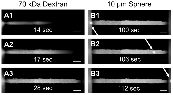
图 2:微模压 PEGDA 水凝胶中的压差驱动流动。通过微模压技术制备 PEGDA 水凝胶,形成包含两个储液井并通过嵌入式微通道相连的结构。在左侧储液井中施加压差,驱动 TRITC 标记的 70 kDa 葡聚糖(A1-A3)和 10 µm 聚苯乙烯微球(B1-B3)通过横截面为 30 × 30 µm 的矩形通道从一个储液井流向另一个储液井。比例尺为 50 µm。请点击此处查看该图的放大版本。
或者,为了在微流控网络中产生稳定的液体流动,可通过在具有进样口和出样口的二级微流控装置内对水凝胶进行光聚合,以实现由注射泵驱动的流动。在图3A中,展示了一种市售的预制微流控装置,其中一条1 mm宽的水凝胶带在装置宽度方向上通过光聚合形成7。采用基于激光的降解方法,按照本文所述用于独立水凝胶的相同方案,在封装于装置内的水凝胶中构建微流控通道和网络。在图3B中可观察到由注射泵产生的流动,其中在装置内的水凝胶中构建了一条直径为20 µm的圆柱形通道。在此直径为20 µm的通道中,无流体流动时,单个直径为2 µm的荧光聚苯乙烯微球可被清晰分辨(图3B1);而当施加5 µL/s的流速时,微球穿过视野,表现为拖尾现象(图3B2)。该方法同样被用于诱导流体通过一个二维的、源自血管结构的网络,以监测TRITC标记的65 kDa葡聚糖在该网络中的流动及其向周围水凝胶的扩散情况(图3C)。
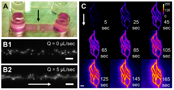
图 3:在微流控装置中光聚合的 PEGDA 水凝胶内由注射泵驱动的流体流动。 一种市售的预制微流控装置(A),其微通道尺寸为 17 × 3.8 × 0.53 mm(x, y, z),内部包含一个光聚合形成的 1 × 3.8 × 0.53 mm(x, y, z)PEGDA 水凝胶,黑色箭头所示。在水凝胶中降解出一条直径为 20 µm 的圆柱形通道,并将直径为 2 µm 的聚苯乙烯微球泵入装置中(B)。在无流体流动时,微球清晰可分辨(B1);但在流速为 5 µL/s 时,微球呈现为条纹状(B2)。在另一块封装的水凝胶中,构建了一个二维血管衍生网络,并将 TRITC 标记的 65 kDa 右旋糖酐泵入该网络(C)。采用延时显微技术监测荧光标记的右旋糖酐填充通道并扩散至周围水凝胶的过程。图中(B)和(C)中的白色箭头指示流体流动方向。(C)中的标定条表示荧光右旋糖酐的强度。比例尺在(B1)和(B2)中代表 20 µm,在(C)中代表 50 µm。请点击此处查看该图的放大版本。
讨论
关键在于覆盖显微镜软件的标准功能,以实现利用虚拟掩模进行图像引导的激光降解,因此必须仔细设置和操作显微镜宏。显微镜软件与显微镜宏之间的交互方式有时可能违反直觉,为此我们投入了大量精力来确定两个程序中的最佳设置,从而开发出本方法。建议使用者对激光扫描共聚焦显微镜的基本操作具有基本了解,尤其是在尝试激光降解之前,应熟练使用"Stage"和"Focus"窗口,在x、y和z维度上找到水凝胶并将其准确定位。此外,入口通道的制备对本实验方案至关重要;悬浮的荧光物质必须能够进入微流控系统,才能成功成像并表征网络结构。需要注意的是,本方案适用于配备特定飞秒脉冲激光器、运行特定版本显微镜软件和显微镜宏的特定激光扫描共聚焦显微镜(详见“特定材料/设备清单”中的说明)。我们发现,较新版本的显微镜宏缺乏图像引导激光控制所必需的控制功能和操作特性。尽管可使用其他显微镜平台和脉冲激光源,但本方案专门针对该系统设计,若更换平台、激光源或软件,则需相应调整。然而,本方案中的基本原理仍然普遍适用。
对该方案的一种可能改进是改变水凝胶的组成。本研究中采用的是5 wt%、3.4 kDa的PEGDA水凝胶,但基于激光的降解技术(除用于生成微流控网络外)已被应用于降解多种合成与天然水凝胶,包括聚乙二醇化纤维蛋白原21,22、丝蛋白水凝胶23以及胶原蛋白24。调整激光功率、扫描速度、Z层间间距以及重复次数,将有助于确定在其他水凝胶体系中构建微流控网络的最佳参数。
该技术目前的一个局限性在于,在合理的时间内可降解的水凝胶总体积有限。为了在水凝胶内创建开放的空腔或微流控结构,当使用37.7 nJ/µm2的激光能量密度时,激光驻留时间必须达到或超过8.96 µs/像素(或激光扫描速度为0.021 µm/µs)。在这些参数下,降解0.014 mm3体积内的血管结构需要1.4小时(如图1所示)。若采用4.48 µs/像素或更低的激光驻留时间,所提供的能量不足以完全降解本研究所使用的水凝胶配方。通过采用不同的水凝胶组成,有望克服这一限制。例如,使用含有光敏组分的光可裂解凝胶34-36,或具有较大双光子吸收截面的水凝胶21-24,都是可行的选择,这些材料可在较低能量下实现更快的降解。在尺寸限制方面,已有研究在水凝胶中加工出深达1.5 mm的结构7。可实现的加工深度取决于物镜的工作距离,使用专为体内成像优化的超长工作距离物镜可进一步增加深度。使用20X(NA1.0)水浸物镜所制备的最小通道7,其宽度为3.3 µm,深度为8.9 µm,与图1中生成的最小通道尺寸相当。尽管其他实验室曾使用较低数值孔径(NA)的物镜21,23,24,但我们预计,采用更高NA的物镜可在牺牲工作距离的前提下,制造出特征尺寸更小的微流控网络。然而,最终该技术的分辨率取决于聚焦激光束的点扩散函数、激光扫描特性、激光输出能量以及被降解材料对激光的吸收特性。
此外,必须根据水凝胶的特性(交联密度、大分子/单体分子量、质量百分比以及水凝胶类型:蛋白质基与合成型)优化激光参数(扫描速度、能量密度、虚拟掩模间距及重复次数),因为这些因素会固有地改变水凝胶与激光之间的相互作用,从而影响最终加工分辨率。由于激光能量输出还受所用物镜的影响,在更换不同物镜时还需进行额外的参数优化。就相关成本而言,需要配备带有脉冲激光器的显微镜,尽管可使用多种不同物镜,但应优先选择高数值孔径(NA)且工作距离较长的物镜(用于 体内 成像可能成本较高。
除了本文详细描述的方案外,使用该技术制备的微流控网络还可以在通道成型后通过功能化修饰细胞黏附肽,以促进内皮细胞的黏附和管腔形成7。此外,在通道降解前,若在体相材料中引入细胞可降解的肽序列9,则可研究细胞向水凝胶内部及穿过水凝胶的迁移过程。为了实现水凝胶内微流控网络的持续流体化,可将水凝胶在较大的流体装置或外壳中进行光聚合,如图2和图3所示7。
通过血管发生或血管生成的自组装形成血管网络8-12 提供了一种简单的方法,可在相对较大的水凝胶体积内诱导血管化。尽管该方法能够形成可灌注的流体网络,但难以直接控制网络的尺寸、迂曲度、密度以及整体结构。由于这一局限性,不同实验之间的血管内流动轮廓和剪切速率可能存在差异。另一种方法是采用微加工技术13-16 可直接控制网络结构,但通常受限于无法生成毛细血管尺寸的微小通道,或难以制造出模拟密集网络的结构 体内 血管结构。本文所述基于激光的水凝胶降解技术虽为新近重新应用于微流控生成,但其同时克服了现有微加工方法在结构控制和尺寸限制方面的不足,能够构建出仿生的三维微流控网络,真实再现血管系统的密度、迂曲度、尺寸范围及整体架构 体内 血管系统。此外,可在单个水凝胶中生成多个流体网络,从而实现对网络间物质运输的研究7. 对于致力于更精确模拟的组织工程应用 体内 转运过程 体外,此处所述的高分辨率水凝胶包埋微流控网络非常适用。我们预计该方案将有助于开发出更精确模拟 体内 用于药物筛选装置的运输 体外 疾病模型
披露
作者无任何利益冲突需要披露。
致谢
作者感谢特拉华生物技术研究所生物成像中心的 Jeff Caplan 博士和 Michael Moore 先生在共聚焦显微镜使用方面提供的支持。显微镜设备的使用得到了美国国立卫生研究院(NIH)共享仪器资助(S10 RR0272773 和 S10 OD016361)以及特拉华州联邦研发资助计划(16A00471)的支持。本研究获得了美国国立卫生研究院普通医学科学国家研究所(NIH NIGMS)机构发展奖(IDeA)(P20GM103446)、美国癌症协会(14-251-07-IRG)、特拉华大学研究基金会(14A00778)、德克萨斯州癌症预防与研究所(CPRIT)(RR140013)以及美国国立卫生研究院国家医学图书馆(4 R00 LM011390-02)资助项目的支持。
材料
| 姓名 | 公司 | 目录编号 | 评论 |
|---|---|---|---|
| MATLAB | Mathworks | R2015a | 在实验方案中称为 "编程软件";有关算法的详细信息,请参见文献来源9 |
| FIJI(Fiji is Just Image J) | NIH | 版本 1.51a | 在实验方案中称为 "图像处理软件" |
| LSM 780 共聚焦显微镜 | Zeiss | 在实验方案中称为 "激光扫描共聚焦显微镜";用于基于激光的水凝胶降解 | |
| Zen 2010B SP1 | Zeiss | 发布版本 6.0 | 在实验方案中称为 "显微镜软件";用于 Zeiss LSM-780 |
| Multitime, 2010 | Zeiss | v16.0 | 在实验方案中称为 "显微镜宏";用于 Zeiss LSM-780(需与 Zen 配合使用) |
| W Plan-Apochromat 20X/1.0 DIC D=0.17 M27 75 mm 物镜 | Zeiss | 421452-9880-000 | 用于 Zeiss LSM-780 |
| Chameleon Vision II 锁模 Ti:S 激光器 | Coherent | 在实验方案中称为 "高功率脉冲激光器" | |
| 氯化钠 | Sigma-Aldrich | S5886-1KG | |
| HEPES | Sigma-Aldrich | H3375-250G | |
| 三乙醇胺(TEOA) | Sigma-Aldrich | 90279-100ML | 易燃;对皮肤和眼睛有刺激性;应在通风橱中操作 |
| 氢氧化钠 | Sigma-Aldrich | S5881-500G | |
| 150 mL 真空过滤杯,带 0.2 µm PES 膜 | VWR | 10040-460 | |
| PEGDA | 实验室自制 | 合成方法请参见文献来源33;需在氩气环境下储存 | |
| 伊红Y二钠盐 | Sigma-Aldrich | E6003-25G | |
| 1-乙烯基-2-吡咯烷酮(NVP) | Sigma-Aldrich | V3409-5G | 需在氩气环境下储存;具有致癌性;应在通风橱中操作 |
| 3M 双面胶带,9500PC,6.0 mil | Thomas Goldkamp | 37728 | |
| Flexmark 90 PFW 衬垫 | FLEXcon | FLX000620 | 用于双面胶带的操作背衬 |
| SC 型号绘图仪(胶粘剂切割器) | USCutter | SC631E | 用于将胶粘剂切割成环形,以连接盖玻片与培养皿 |
| 带 20 mm 孔的 60 mm 培养皿 | MatTek Corporation | P60-20-F-NON | |
| 高强度照明器(白光光源) | Fiber-Lite | 4715MS-12WB10 | |
| 功率计 | Newport | 1916-R | 使用白光光源时检测 524 nm 处的功率 |
| 超薄型探头检测器 | Newport | 918D-ST | 与功率计配合使用 |
| Sylgard 184 硅橡胶弹性体试剂盒 | Dow Corning | 3097358-1004 | 用于制作 PDMS 模具;制备方法请参见文献来源7 |
| TMPSA功能化#1.5盖玻片,40 mm圆形 | 实验室自制 | 制备方法请参见文献来源7 | |
| 葡聚糖,荧光素标记,分子量2,000,000,阴离子型,可与赖氨酸交联 | Life Technologies | D7137 | 可使用其他标记葡聚糖;2,000 kDa 的分子不易扩散进入5% 3.4 kDa PEGDA水凝胶 |
| 1 mL 注射器,鲁尔锁扣式 | BD | 309628 | |
| Acrodisc 注射器滤器,0.2 µm Supor 膜,低蛋白结合 | Pall | PN 4602 | |
| sticky-Slide VI0.4 | Ibidi | 80601 | 可用于承载水凝胶的微流控装置 |
参考文献
- Baish, J. W., Stylianopoulos, T., et al. Scaling rules for diffusive drug delivery in tumor and normal tissues. Proc. Natl. Acad. Sci. USA. 108 (5), 1799-1803 (2011).
- Bhatia, S. N., Ingber, D. E. Microfluidic organs-on-chips. Nature Biotechnol. 32 (8), 760-772 (2014).
- Esch, E. W., Bahinski, A., Huh, D. Organs-on-chips at the frontiers of drug discovery. Nat. Rev. Drug Discov. 14 (4), 248-260 (2015).
- Ghaemmaghami, A. M., Hancock, M. J., Harrington, H., Kaji, H., Khademhosseini, A. Biomimetic tissues on a chip for drug discovery. Drug Discov. Today. 17 (3-4), 173-181 (2012).
- Jeon, J. S., Bersini, S., et al. Human 3D vascularized organotypic microfluidic assays to study breast cancer cell extravasation. Proc. Natl. Acad. Sci. USA. 112 (1), 214-219 (2015).
- Pisano, M., Triacca, V., Barbee, K. A., Swartz, M. A. An in vitro model of the tumor-lymphatic microenvironment with simultaneous transendothelial and luminal flows reveals mechanisms of flow enhanced invasion. Integr. Biol. 7 (5), 525-533 (2015).
- Heintz, K. A., Bregenzer, M. E., Mantle, J. L., Lee, K. H., West, J. L., Slater, J. H. Fabrication of 3D Biomimetic Microfluidic Networks in Hydrogels. Adv. Healthc. Mater. , (2016).
- Cuchiara, M. P., Gould, D. J., McHale, M. K., Dickinson, M. E., West, J. L. Integration of Self-Assembled Microvascular Networks with Microfabricated PEG-Based Hydrogels. Adv. Funct. Mater. 22 (21), 4511-4518 (2012).
- Culver, J. C., Hoffmann, J. C., Poché, R. A., Slater, J. H., West, J. L., Dickinson, M. E. Three-Dimensional Biomimetic Patterning in Hydrogels to Guide Cellular Organization. Adv. Mater. 24 (17), 2344-2348 (2012).
- Kim, S., Lee, H., Chung, M., Jeon, N. L. Engineering of functional, perfusable 3D microvascular networks on a chip. Lab Chip. 13 (8), 1489(2013).
- Saik, J. E., Gould, D. J., Keswani, A. H., Dickinson, M. E., West, J. L. Biomimetic Hydrogels with Immobilized EphrinA1 for Therapeutic Angiogenesis. Biomacromolecules. 12 (7), 2715-2722 (2011).
- Zisch, A. H., Lutolf, M. P., et al. Cell-demanded release of VEGF from synthetic, biointeractive cell-ingrowth matrices for vascularized tissue growth. FASEB J. , (2003).
- He, J., Zhu, L., Liu, Y., Li, D., Jin, Z. Sequential assembly of 3D perfusable microfluidic hydrogels. J. Mater. Sci. Mater. Med. 25 (11), 2491-2500 (2014).
- Therriault, D., White, S. R., Lewis, J. A. Chaotic mixing in three-dimensional microvascular networks fabricated by direct-write assembly. Nat. Mater. 2 (4), 265-271 (2003).
- Wu, W., DeConinck, A., Lewis, J. A. Omnidirectional Printing of 3D Microvascular Networks. Adv. Mater. 23 (24), H178-H183 (2011).
- Kolesky, D. B., Truby, R. L., Gladman, A. S., Busbee, T. A., Homan, K. A., Lewis, J. A. 3D Bioprinting of Vascularized, Heterogeneous Cell-Laden Tissue Constructs. Adv. Mater. 26 (19), 3124-3130 (2014).
- Zervantonakis, I. K., Hughes-Alford, S. K., Charest, J. L., Condeelis, J. S., Gertler, F. B., Kamm, R. D. Three-dimensional microfluidic model for tumor cell intravasation and endothelial barrier function. Proc. Natl. Acad. Sci. USA. 109 (34), 13515-13520 (2012).
- Roudsari, L. C., West, J. L. Studying the influence of angiogenesis in in vitro cancer model systems. Adv. Drug Deliv. Rev. , (2015).
- Dawson, T. H. Scaling laws for capillary vessels of mammals at rest and in exercise. Proc. R. Soc. Long. [Biol.]. 270 (1516), 755-763 (2003).
- Vogel, A., Noack, J., Hüttman, G., Paltauf, G. Mechanisms of femtosecond laser nanosurgery of cells and tissues. Appl. Phys. B. 81 (8), 1015-1047 (2005).
- Sarig-Nadir, O., Livnat, N., Zajdman, R., Shoham, S., Seliktar, D. Laser Photoablation of Guidance Microchannels into Hydrogels Directs Cell Growth in Three Dimensions. Biophys. J. 96 (11), 4743-4752 (2009).
- Three-dimensional guidance of DRG neurite outgrowth using multi-photon photo-ablation. Livnat, N., Sarig-Nadir, O., Seliktar, D., Shoham, S. 2009 4th International IEEE/EMBS Conference on Neural Engineering, , 116-119 (2009).
- Applegate, M. B., Coburn, J., et al. Laser-based three-dimensional multiscale micropatterning of biocompatible hydrogels for customized tissue engineering scaffolds. Proc. Natl. Acad. Sci. USA. 112 (39), 12052-12057 (2015).
- Ilina, O., Bakker, G. J., Vasaturo, A., Hoffman, R. M., Friedl, P. Two-photon laser-generated microtracks in 3D collagen lattices: principles of MMP-dependent and -independent collective cancer cell invasion. PB. 8 (2), (2011).
- Naik, P., Cucullo, L. In Vitro Blood-Brain Barrier Models: Current and Perspective Technologies. J. Pharm. Sci. 101 (4), 1337-1354 (2012).
- Choe, Y., Mayerich, D., et al. Specimen Preparation, Imaging, and Analysis Protocols for Knife-edge Scanning Microscopy. JoVE. (58), (2011).
- Mayerich, D., Kwon, J., Sung, C., Abbott, L., Keyser, J., Choe, Y. Fast macro-scale transmission imaging of microvascular networks using KESM. Biomed. Opt. Express. 2 (10), 2888-2896 (2011).
- Chung, J. R., Sung, C., et al. Multiscale Exploration of Mouse Brain Microstructures Using the Knife-Edge Scanning Microscope Brain Atlas. Front. Neuroinform. 5, (2011).
- Schindelin, J., Arganda-Carreras, I., et al. Fiji: an open-source platform for biological-image analysis. Nat. Methods. 9 (7), 676-682 (2012).
- Shukla, A., Slater, J. H., Culver, J. C., Dickinson, M. E., West, J. L. Biomimetic Surface Patterning Promotes Mesenchymal Stem Cell Differentiation. ACS Appl. Mater. Interfaces. , (2015).
- Slater, J. H., Culver, J. C., et al. Recapitulation and Modulation of the Cellular Architecture of a User-Chosen Cell of Interest Using Cell-Derived, Biomimetic Patterning. ACS Nano. 9 (6), 6128-6138 (2015).
- Slater, J. H., West, J. L. Fabrication of Multifaceted, Micropatterned Surfaces and Image-Guided Patterning Using Laser Scanning Lithography. Methods Cell Biol. 119, 193-217 (2014).
- DeLong, S. A., Gobin, A. S., West, J. L. Covalent immobilization of RGDS on hydrogel surfaces to direct cell alignment and migration. J. Control. Release. 109 (1-3), 139-148 (2005).
- DeForest, C. A., Anseth, K. S. Cytocompatible click-based hydrogels with dynamically-tunable properties through orthogonal photoconjugation and photocleavage reactions. Nat. Chem. 3 (12), 925-931 (2011).
- Kloxin, A. M., Kasko, A. M., Salinas, C. N., Anseth, K. S. Photodegradable Hydrogels for Dynamic Tuning of Physical and Chemical Properties. Science. 324 (5923), 59-63 (2009).
- Tibbitt, M. W., Kloxin, A. M., Dyamenahalli, K. U., Anseth, K. S. Controlled two-photon photodegradation of PEG hydrogels to study and manipulate subcellular interactions on soft materials. Soft Matter. 6 (20), 5100-5108 (2010).
重印与许可
