对人多能干细胞来源的心肌细胞(hPSC-CMs)进行电生理表征,对于心脏疾病建模及评估药物反应至关重要。本方案提供了将hPSC-CMs解离并接种于多电极阵列上的必要信息,用于测量其场电位,并介绍了一种分析QT间期和RR间期的方法。
本文内容
摘要
摘要
现在可以高效地从人胚胎干细胞和人诱导性多能干细胞(hPSC)分化获得心肌细胞。人多能干细胞来源的心肌细胞(hPSC-CMs) increasingly 被认为在人类心血管疾病建模中具有重要价值,尤其是心律失常综合征的研究。此外,它们在以下方面也显示出相关性: 体外 用于预测药物反应的系统,使其在药物筛选与发现、安全性药理学,甚至最终在个体化医疗中具有潜在应用价值。通过从患者或易感个体获取人诱导多能干细胞(hiPSCs)来衍生人多能干细胞来源心肌细胞(hPSC-CMs),将有助于实现上述应用。然而,对于所有应用而言,精确测量和分析hPSC-CMs的电生理特性对于识别由心脏离子通道突变和/或作用于离子通道并可能导致猝死的药物所引起的变化至关重要。与人工膜片钳技术相比,多电极阵列(MEA)设备具有可实现中至高通量记录的优势。本方案描述了如何将hPSC-CMs的二维培养物解离为小聚集体和单细胞,并将其接种至MEA上,以记录其自发性场电位活动。本文还介绍了分析记录数据以提取特定参数(如QT间期和RR间期)的方法。携带导致心律失常突变的hPSC-CMs,或在添加特定药物后,这些参数会发生预期变化,从而可检测出具有心脏毒性的药物。
引言
人类多能干细胞(hPSCs)具有自我更新的能力,并可通过分化产生人体几乎所有类型的细胞1,2。已有详细方案描述了如何将hPSCs定向分化为多种心肌细胞谱系(如心室样、心房样及起搏细胞样心肌细胞)3,4,5,6,7。心肌细胞是电活性细胞,深入了解其电生理活动对于认识心脏 发育与疾病具有重要意义8。患者特异性的诱导多能干细胞来源的心肌细胞(hiPSC-CMs)已成功用于建模并研究多种心脏心律失常的细胞、分子和电生理特征,包括长QT综合征(LQTS)9,10,11,12,13、Brugada综合征14以及儿茶酚胺敏感性多形性室性心动过速15,16。此外,已有多种药物被施加于病变的hiPSC-CMs中,以模拟治疗干预并挽救细胞的病理表型10,15,20,21,22。近年来,为满足药物发现早期阶段对人类模型系统的需求,研究人员开发了基于野生型hiPSC-CMs的筛选平台23,24,25,因为啮齿类动物心肌细胞在离子通道表达和生物物理特性方面与人类存在显著差异26。
为此,适合中高通量应用的技术正在被开发和实施。这些技术包括膜电位的光学记录、Ca2+ 瞬变与应变、阻抗测量(作为细胞收缩力的间接指标)以及细胞外场电位(FP)测量(综述见参考文献)24)。多电极阵列(MEA)设备可用于记录由心肌细胞单层或小细胞簇产生并形成的电波形信号(即场电位,FP)。场电位的波形轮廓与心脏动作电位相关,并在一定程度上与心电图(ECG)记录相对应。27;它们通常表现出初始的快速上升支,对应于 Na+ 内流和膜去极化(R/Q峰),一个缓慢的去极化波/平台期,可能对应于Ca2+ 内流,以及对应于主要为 K⁺ 外流的复极化阶段+ 外流(T峰)。FP波形的扰动可与动作电位特定时相的变化相关联28.
尽管膜片钳记录动作电位可提供更丰富的信息,尤其是关于上升速度和静息膜电位等参数,但在中、高通量规模的实验中,手动测量并不可行,而自动膜片钳技术直到最近才被应用于人多能干细胞来源的心肌细胞(hPSC-CMs)29。然而,由于多电极阵列(MEA)上的长时间记录能够同时研究化合物的急性与慢性暴露效应,目前已有可能将hPSC-CM平台用于药物筛选、药物发现24,30以及安全性药理学研究31,32。这为未来实现精准医疗或个体化医疗带来了希望33。
本实验方案的目的是提供在MEA芯片上分离和接种hPSC-CMs并测量其动作电位(FP)所需的全部信息。该流程中每一步均已优化,以确保细胞在解离后具有最佳的存活率和恢复能力,细胞在MEA板上的贴附效果最佳,并实现参数的标准化分析与定量。特别地,本文详细解释并举例说明了细胞外动作电位记录、QT间期和RR间期的分析方法以及药物效应的评估。
方案
1. 溶液与试剂的配制
- 使用低胰岛素、牛血清白蛋白、聚乙烯醇、必需脂质(LI-BPEL)培养基34,35,36配制hPSC-CM培养液,具体方法见表1中所述试剂。用0.22 µm孔径滤器过滤培养基,4 °C保存,最长可保存2周。
- 将1 mg人重组纤连蛋白溶于5 mL无菌蒸馏水中,配制成200 µg/mL的储存液,分装后于-80 °C保存。
- 配制1% (w/v) 酶洗涤剂溶液(见材料表),用于清除微阵列芯片上的残留细胞:将1 g酶洗涤剂粉末溶于100 mL温热(约40–45 °C)的去离子水中。待溶液冷却后,4 °C保存,最长可保存一年。
2. MEA芯片的灭菌(图1A)
注意:MEA 有多种不同配置,包括单孔或多孔格式。此处描述的方案使用单腔室 MEA,其包含 60 个记录电极,呈 8 × 8 网格排列(参见材料表)。电极直径为 30 µm,电极间距为 200 µm。此外还设有一个参考电极。
- 用去离子水彻底冲洗芯片。
- 将MEA芯片放入可高压灭菌的玻璃培养皿中,用铝箔包裹培养皿。
- 在实验室高压锅中对芯片进行6分钟灭菌。打开前让培养皿冷却。
注意:或者,可将芯片浸入1 mL 80%(v/v)乙醇中,在室温下进行灭菌 15–30 分钟。 - 将芯片置于细胞培养罩中,并用紫外光照射表面约30分钟。
3. MEA芯片的包被(图1B和1C)
- 将洁净的芯片放入标准的 10 cm Ø 塑料无菌培养皿中。
- 向培养皿中加入 8 mL 无菌去离子水,以形成湿润环境,防止芯片上的少量培养基在放入培养箱时干燥。
- 使用定制的聚四氟乙烯(PTFE)环(图 1B),确保将心肌细胞接种在芯片中央,即电极阵列所在的位置。(PTFE 环此前保存在 80% 乙醇中。)在超净台内,将环从乙醇中取出,放入无盖的无菌培养皿中,并在超净台内使其干燥。
- 使用火焰灭菌的镊子将一个干燥的环放入一个 MEA 芯片内(图 1C)。
注意:也可使用 80% 乙醇清洗镊子,并在组织培养超净台内晾干后再使用。 - 解冻一份人重组纤连蛋白原液(200 µg/mL 溶于去离子水),用含 Ca2+ 和 Mg2+ 的 PBS 稀释,配制成 40 µg/mL 的工作液。通过在环内加入 50 µL 的 40 µg/mL 纤连蛋白溶液,对电极进行包被。
注意:使用人重组纤连蛋白包被可确保 hPSC-CM 的最佳贴附效果。然而, 也可使用其他类型的包被蛋白,如牛纤连蛋白或细胞外基质蛋白混合物。 - 盖上 10 cm Ø 塑料培养皿的盖子,小心地将含有 MEA 芯片的培养皿转移至培养箱。在 37 °C 下孵育至少 1 小时,或在 4 °C 过夜。
- 将含有 MEA 芯片的培养皿转移回细胞培养超净台。使用 MEA 芯片前,用 P200 移液器或超净台内的真空系统吸除 50 µL 纤连蛋白溶液,注意不要移动 PTFE 环。此步骤应使用塑料吸头。确保无任何固体物体(例如, 移液器吸头)接触培养皿内部,以免损坏电极。
注意:此操作可延长 MEA 芯片的使用寿命。 - 轻柔地加入 950 µL 的 LI-BPEL 培养基(参见 表 1 和 材料表),确保液体均匀分布且环不会漂浮。将培养皿放回培养箱。
4. hPSC-CM 的解离与铺板(图2)
注意:此处描述的方案使用了通过细胞因子在单层培养中分化的大约18天后的hPSC-CMs34。然而,该方案已被证实适用于任何二维(2D)和三维(3D) hPSC-CM培养体系。当使用更早或更晚时间点分化的细胞时,可能需要调整解离酶(见材料表)的孵育时间。以下体积适用于12孔板中的单个孔(3.8 cm2)。
- 吸出hPSC-CMs培养孔中的培养基。
- 在超净工作台内操作,每孔加入1–2 mL不含Ca2+/Mg2+的PBS洗涤培养物,随后吸除PBS。
- 每孔加入500 µL解离酶溶液,在37 °C孵育5分钟。
- 每孔加入1 mL LI-BPEL以稀释酶液。使用P1000移液器轻轻刮擦,使hPSC-CMs单层细胞脱落。将细胞悬液收集至15 mL离心管中。
- 用1 mL LI-BPEL冲洗孔内残留的细胞及细胞团块,并收集入离心管。
- 再加入2–3 mL LI-BPEL,使终体积达到5–6 mL,使用5 mL移液器轻柔吹打3–5次,以分散细胞团块。
注意:此时无需将细胞完全解离为单细胞,因为这会影响细胞存活率。保留少量细胞簇有助于提高细胞活力。 - 在室温 下以300 × g离心3分钟。
- 弃去上清液,尽量去除多余液体,但避免扰动细胞沉淀。
- 用250 µL LI-BPEL重悬细胞沉淀(使用P1000,吹打动作需极其轻柔)。
- 将细胞悬液分装至MEA芯片,每片约50 µL(一个12孔板中的单孔可制备最多5片MEA),直接将细胞悬液滴加至PTFE环中央、电极阵列上方。
注意:由于存在细胞簇,此时难以准确计数,每片MEA上的细胞总数可能因hPSC-CMs来源不同而有较大差异。接种时应确保解离后的细胞云覆盖电极区域。 - 小心将MEA芯片转移至37 °C培养箱中,过夜静置使细胞贴壁。
5. 去除琼脂环与培养基更换(图3)
- 铺板后第1天,在无菌环境中使用无菌镊子小心移除环状结构(图3A-3B)。
- 用80%(v/v)乙醇冲洗该环,并将其储存于含有新鲜80%(v/v)乙醇的50 mL离心管中。
- 轻柔地从MEA芯片中移除500 µL培养基,并加入500 µL新鲜的LI-BPEL培养基。
- 将MEA芯片转移至37 °C培养箱中。
注意:细胞通常在移除环状结构并更换培养基后的1–7天内开始搏动(图3C)。 - 在移除环状结构后的1–7天内,利用MEA检测hPSC-CMs的电活动。
6. 检查信号质量(图4)
- 打开计算机并启动与MEA系统连接的软件套件:TCX-Control、MC_MEA Select 和 MC_Rack。在TCX-Control中将温度设置为37 °C,以在生理温度下记录测量数据。
- 从培养箱中取出含有MEA芯片的培养皿。打开盖子,取出MEA芯片,并将其放置在纸巾上以吸除残留水分。
- 用纸巾小心擦拭芯片板外部的电极接触点,并使用蘸有100%(v/v)乙醇的棉签清洁这些接触点,以去除可能引起信号噪声的残留水分或碎屑。
- 将MEA芯片板转移至加热(37 °C)的记录头阶段,以检测自发性电活动(例如,硬件:见材料表;软件:MC_Rack)。
- 打开MC_Rack:点击“Edit” → “Add MC_Card”以创建新协议。使用“Edit”下拉菜单向协议中添加不同的Recorder和Display窗口。
注意:建议采样频率至少为10 kHz。我们使用的协议包含一个Longterm Display Tool,显示整个MEA芯片的完整布局,以及一个Spike Sorter,用于实时捕捉药物效应。Spike Cutout参数设置为“Pre Trigger”20 ms、“Post Trigger”800 ms和“Dead Time”2 ms。该协议可保存为“.rck”文件,并在实验开始前重新加载。
- 打开MC_Rack:点击“Edit” → “Add MC_Card”以创建新协议。使用“Edit”下拉菜单向协议中添加不同的Recorder和Display窗口。
- 点击“play”按钮,以播放模式启动协议。
注意:此时可以重新加载步骤6.5中保存的协议。在MC_Rack中,通过点击“File” → “Open”来加载协议(.rck文件扩展名)。 - 如果信号中清晰可见R峰和T峰,则等待10–15分钟以完成适应阶段。良好与低质量信号示例见图4。
注意:如果在任何电极上均无法检测到T峰,则不应继续实验。这通常是由于hPSC-CMs电活动较弱或细胞与电极附着不良所致。
7. 开始实验并记录
- 点击“记录”然后点击“播放”,在基线条件下采集10分钟数据,以确定稳态。对信号最佳的电极进行标注,以便后续能够轻松识别并导出用于分析。
- 为评估药物反应,每隔10分钟加入递增浓度的药物。例如,加入hERG阻断剂E4031,终浓度为1 μM。具体操作为:移除100 μL培养基,加入等体积溶于培养基中的10 μM E4031。
注意:如Cavero及其同事先前所证实31,选择合适的药物溶解体积至关重要,因其可能显著改变药物反应曲线。 - 对所有其他感兴趣的药物浓度重复步骤7.2。
- 在实验方案结束时,点击“停止”以结束记录。
8. 微电极阵列的清洁与重复使用
- 实验记录结束后,使用 P1000 移液器轻柔地移除培养基。切勿触碰培养皿内壁,以免损坏电极。根据当地安全规定处理废液。
- 使用洗瓶以去离子水冲洗 MEA 芯片,并重复清洗 3–4 次。
注意:此时无需确保细胞完全从芯片上脱离。 - 向每个孔中加入 1 mL 1% (v/v) 酶类洗涤剂溶液,于 4 °C 条件下孵育过夜,以促进细胞脱离和清除。
- 第二天,用去离子水彻底冲洗 MEA 芯片,以去除酶类洗涤剂溶液及残留细胞,然后加入 1 mL 去离子水。清洁后的 MEA 芯片可浸泡于去离子水中,于 4 °C 保存。
9. 数据导出
- 打开与MEA系统连接的MC_Data Tool软件。
- 点击“文件” → “打开MCD”。
- 点击“工具” → “将MCD转换为ABF”。
- 选择记录信号最佳的电极用于导出分析。选择要保存导出文件的目录,然后点击“保存”。导出过程可能需要数分钟,具体时间取决于导出的电极数量和记录文件的总大小。
10. 数据分析
- 下载并安装电生理数据采集与分析软件(例如 pClamp)。安装完成后,启动分析软件(例如Clampfit)。
- RR 间期计算(图 5)。
- 放置两个垂直光标以在波形上定义感兴趣区域。选择“事件检测” → “阈值搜索”。水平光标应穿过所有需要量化的事件,如图 5A所示。
- 点击“确定”,然后选择“接受整个类别”。软件将自动搜索波形中所有符合条件的事件。事件上方将标有蓝色标记。
- 通过调整主事件检测窗口中的参数,优化自动选择的灵敏度。
- 当所有事件均被自动识别后,进入结果窗口(窗口 → 结果),复制标题为"Intervent Interval"的列,该列包含频率数据(图 5B)。
- QT 间期计算(图 6–9):
- 在稳态条件下,于单个动作电位(FP)起始前和结束后的波形位置各放置一个光标。选择“事件检测” → “创建模板”(图 6)。
- 确认动作电位(FP)被正确识别后,点击“添加”。模板将移至下方面板。
- 将模板保存为“.atf”文件。由此创建的模板波形将被软件用于在整个记录中进行搜索(图 7)。为每种实验条件单独创建一个模板,因为药物效应可能改变动作电位的形态。如有必要,可对波形进行轻微滤波,确保两个光标之间的数据点不超过 10,000 个。
- 模板以“.atf”扩展名保存后,选择“事件检测” → “模板搜索”,并加载该模板。
- 调整"模板匹配阈值",以准确识别所选区间内的所有动作电位(FP)。
- 当所有事件均被正确识别后,将其保存为一个新的“.abf”文件。
- 使用分析软件打开文件,并自动计算 Q/R 峰和 T 峰的“达峰时间”(图 8、图 9)。若波形噪声较大,可应用滤波处理。在任意电子表格编辑软件中,通过将 T 峰时间减去 Q/R 峰时间,计算 QT 间期。
结果
解离并接种后一天,hPSC-CMs 将形成一层致密的白色薄膜,覆盖在 MEA 腔的中央区域图3A)。移除环状物后(图3B), 该层应保持原位,在光学显微镜下观察可看到MEA电极被(收缩的)hPSC-CM层覆盖图3C由于细胞之间存在物理和电学耦合,分析时仅使用一个电极(金电极)。
或者,在处理三维结构(如类胚体或微组织)时,可将其接种,使其在物理上和电学上相互分离。通过显微镜观察可确认各细胞簇之间无物理连接,而多电极阵列(MEA)上非同步的R波则可证实无电学耦合。在此情况下,可对多个独立电极进行分析。
FP 信号的典型记录结果如图4所示。具体而言,高质量的信号曲线应包含一个清晰的峰,对应 Na+ 内流和细胞膜去极化(R/Q 峰),以及一个清晰的复极化相,对应 K+ 外流(T 峰),同时具有较高的信噪比(图4A,左侧:注意 y 轴刻度,以及图4B)。质量较差的信号曲线(图4A,中间)可能是由于 hPSC-CM 未能良好贴附于 MEA 板,或 hPSC-CM 电活动较弱所致。等待 1–3 天以改善细胞贴附可能有助于提高信号质量;然而,若未观察到信号改善,则建议将该 MEA 排除于实验之外。噪声较大的信号曲线(图4A,右侧)可在滤波后进行分析。
通过目视检查显示峰值检测的屏幕,可判断RR间期分析是否成功(图5A、5B)。在垂直光标所定义的时间区间内,应出现对应于每个峰值的蓝色标记。如果程序未能识别一个或多个峰值,可尝试移动水平光标后重新运行分析,或调整检测设置。类似地,通过目视检查显示FP检测的屏幕,可判断QT间期分析是否成功(图7)。在垂直光标所定义的时间区间内,应出现对应于每次FP检测的蓝色标记。如果程序未能识别一个或多个FP,可尝试重新定义FP模板(图6),或调整检测设置后重新运行分析。
提取的单个荧光蛋白(FP)轨迹或其平均值(图8)可用于在特定设置下获取QT间期数值,如图9所示。分析QT-RR关系是可取的,且在疾病模型和野生型(WT)人多能干细胞(hPSC)系中均具有意义(图10A),也可用于评估是否需要进行QT间期校正及校正的效果(图10B–10D)。携带长QT综合征(LQTS)致病突变的hPSC来源心肌细胞(hPSC-CMs)相较于野生型对照组表现出延长的QT间期(图11A)。使用hERG通道阻断剂处理hPSC-CMs会导致QT间期延长(图11B);相反,使用hERG通道激活剂处理则会导致QT间期缩短(图11C)。最后,使用影响hPSC-CMs搏动频率的药物处理,应表现为RR间期的变化(图11D,RR间期缩短)。
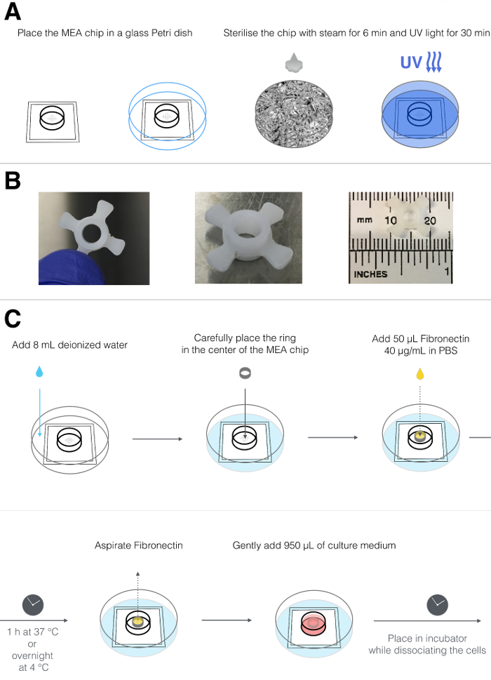
图1:MEA芯片的灭菌与包被。(A)表示灭菌过程的示意图,包括将MEA芯片放入可高压灭菌的玻璃培养皿中,用铝箔包裹,蒸汽处理6分钟,并暴露于紫外线下30分钟。(B)定制PTFE环的顶视图(左图)和侧视图(中图);该环外径为1.2 cm,包含4个凸片,可使其定位在MEA芯片的中心,内径为0.4 cm(右图)。(C)MEA芯片制备的示意图,包括将芯片置于标准塑料培养皿中,在MEA腔室外加入8 mL去离子水,将PTFE环置于腔室中央,并用纤连蛋白包被电极阵列。孵育后,去除纤连蛋白并更换为培养基。请点击此处查看该图的放大版本。
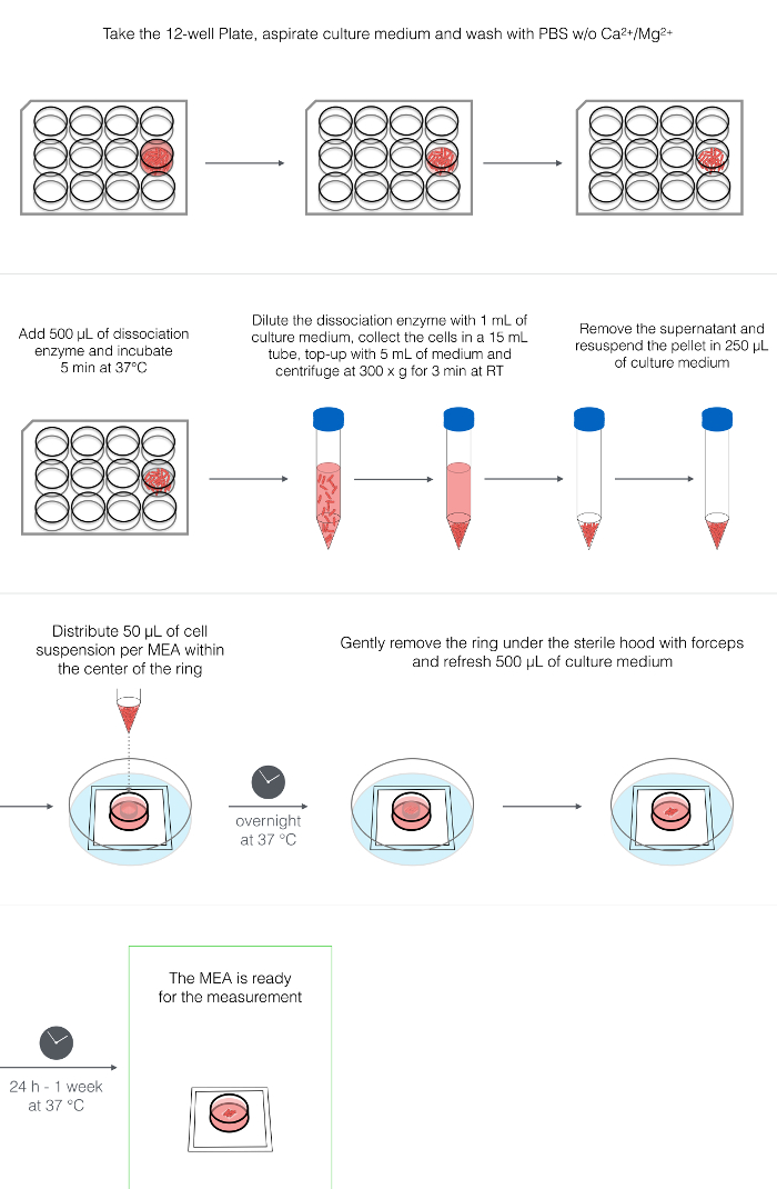
图 2:hPSC-CMs 的解离与接种。 示意图展示了 hPSC-CMs 酶解解离、离心、重悬以及接种至 MEA 腔室中央的过程。请点击此处查看该图的放大版本。
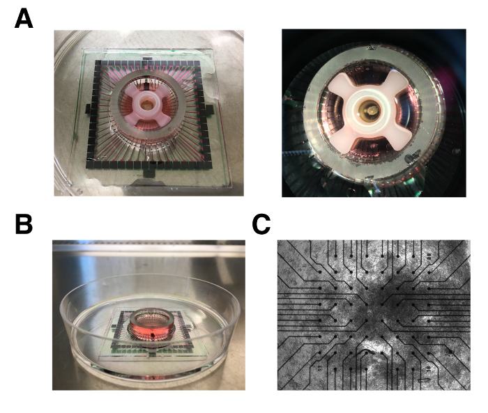
图3:带有hPSC-CM层的MEA芯片。(A)包含环状结构和hPSC-CM层的MEA芯片的顶视图。(B)移除环后MEA芯片的侧视图。(C)接种于微电极阵列上的hPSC-CM层的明场图像;4倍放大。 请点击此处查看该图的放大版本。
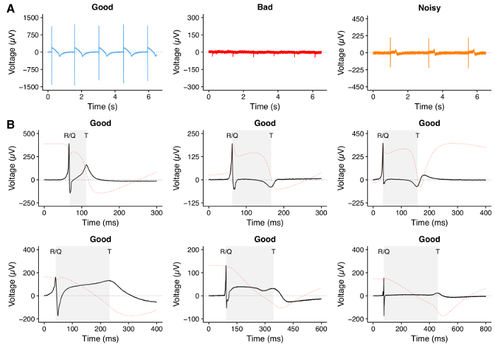
图4:hPSC-CMs的MEA记录。(A)使用MEA记录的代表性轨迹,显示高质量轨迹(左),R/Q波和T波清晰可见且信噪比较高;低质量轨迹(中),R/Q波和T波不清晰;以及噪声较大的轨迹(右),尽管R/Q波和T波清晰可见,但信噪比较低。(B)使用hPSC-CMs进行MEA实验期间可记录到的不同形态的高质量场电位(FP)轨迹的代表性示例。阴影区域表示分析过程中测量的QT间期。由于MEA上的FP类似于动作电位的一阶导数28,我们计算了FP轨迹的积分(以红色虚线表示),作为T波选择接近完整动作电位复极化的理论演示。请点击此处查看该图的放大版本。
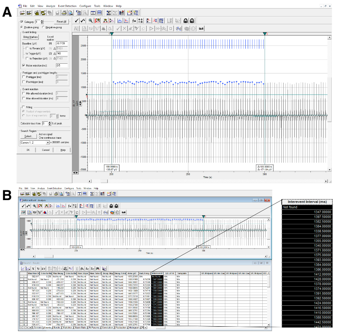
图 5:RR 间期计算。(A)RR 间期分析示例,上图为使用分析软件(见材料表)自动检测波峰,下图为数据提取。垂直游标标示出感兴趣的时程区间,水平游标穿过所有被检测并以蓝色标记识别的事件。(B)放大列显示用于计算 RR 间期的提取数据。请点击此处查看该图的放大版本。
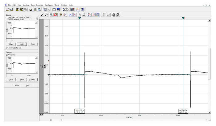
图6:FP模板的创建。 使用垂直光标在单个FP前后进行模板选择的示例。该模板用于FP的自动识别。请点击此处查看此图的放大版本。
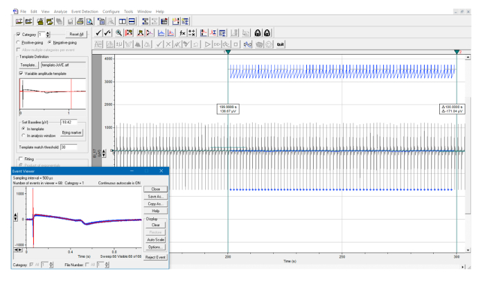
图 7:FP 模板自动识别。 在由两条垂直光标定义的时间区间内进行模板搜索的示例。所有检测到的事件均由蓝色标记标出,并在插图中自动叠加显示。请点击此处查看此图的放大版本。
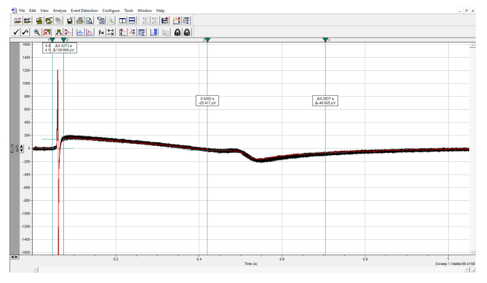
图 8:FP 参数的定量分析。 对保存的事件进行分析,R 峰在前两个光标范围内手动识别,T 峰在后两个光标范围内手动识别。请点击此处查看此图的放大版本。
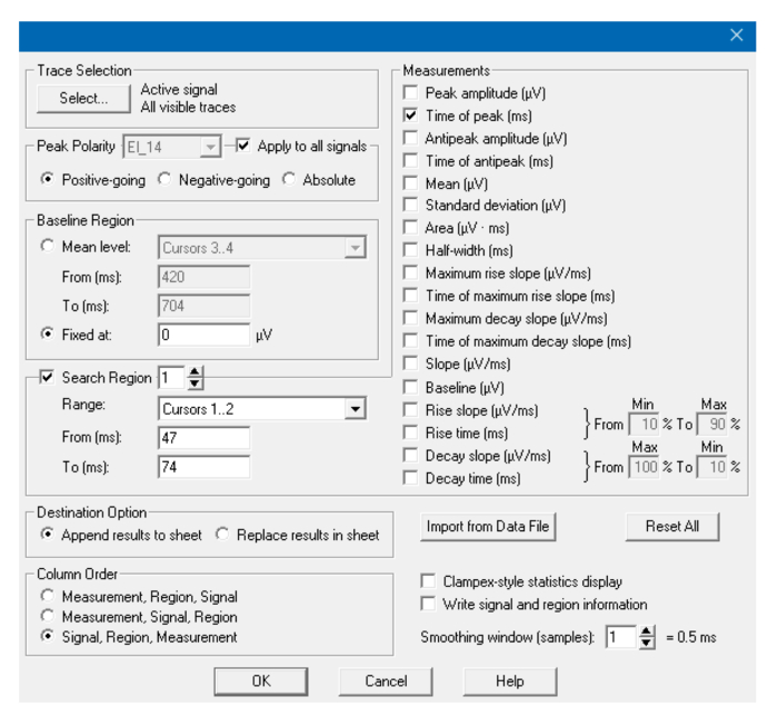
图9:分析窗口。 在“统计”窗口中用于检测R波峰值的参数。如需检测T波峰值,请调整光标位置,必要时调整极性。请点击此处查看此图的放大版本。
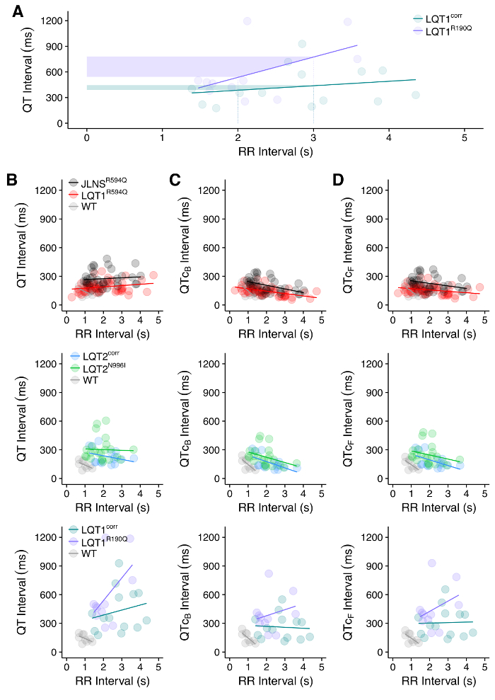
图 10:QT-RR 关系。(A)野生型(LQT1corr)与 1 型长 QT 综合征(LQT1R190Q)人多能干细胞来源心肌细胞(hPSC-CMs)之间 QT 间期与 RR 间期关系差异的示例。阴影区域显示,相同的 RR 间期变化在 LQT1 疾病细胞系中引起更大的 QT 间期变化,因此可能增加心律失常易感性。(B)在来自 7 个不同 hPSC 系的心肌细胞(CMs)中通过多电极阵列(MEA)测量的未校正 QT 间期与 RR 间期之间的关系。QT 间期经 Bazett 公式(C)或 Fridericia 公式(D)校正后的关系以 QT-RR 间期关系斜率的变化形式展示。图改编自参考文献30。请点击此处查看该图的放大版本。
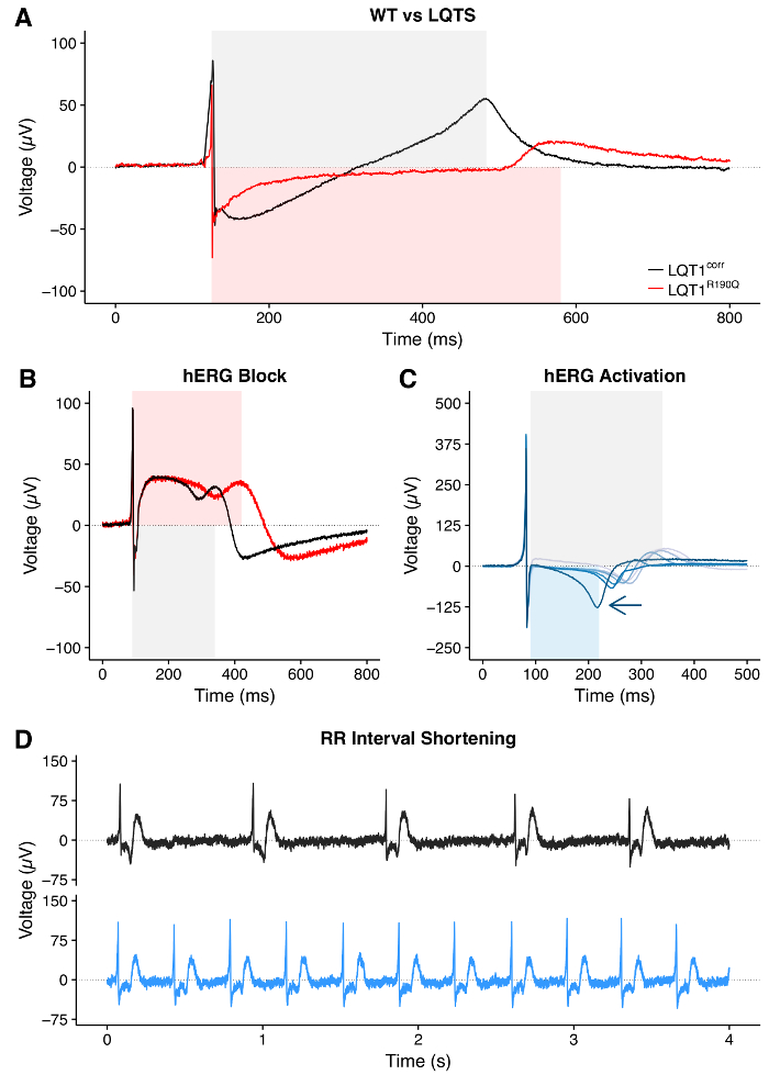
图11:疾病或药物引起的QT间期与RR间期变化。(A)来源于携带长QT综合征(LQTS)突变患者的hPSC-CM与其同基因野生型(WT)对照相比,QT间期延长的示例。(B)hPSC-CM在药物诱导的hERG通道阻断下QT间期延长的示例。(C)使用逐渐增加剂量的hERG激活剂处理后QT间期缩短的示例。箭头指示缩短的方向。(D)药物诱导RR间期缩短的示例。图(C)改编自参考文献30。请点击此处查看该图的放大版本。
| 低胰岛素、牛血清白蛋白、聚乙烯醇、必需脂质(LI-BPEL)培养基 | |||
| 组分 | 每100 mL所需量 | ||
| IMDM | 43 mL | ||
| F12 | 43 mL | ||
| 抗坏血酸-2-磷酸(5 mg/mL,溶于蒸馏水) | 1 mL | ||
| 细胞培养添加剂(L-谷氨酰胺的直接替代物) | 1 mL | ||
| 青霉素/链霉素 | 0.5 mL | ||
| 酚红 | 1 mg | ||
| 无蛋白杂交瘤培养基-II(PFHMII) | 5 mL | ||
| 牛血清白蛋白(10% wt/vol,溶于IMDM) | 2.5 mL | ||
| 聚乙烯醇(5% wt/vol,溶于蒸馏水) | 2.5 mL | ||
| 化学成分明确的脂质浓缩液(CDLC) | 1 mL | ||
| 胰岛素-转铁蛋白-硒-乙醇胺(ITS-X)100倍浓缩液 | 0.1 mL | ||
| α-单硫代甘油(13 µL 溶于 1 mL IMDM) | 0.3 mL | ||
| 将上述试剂混合,用0.22 µm孔径滤膜过滤,4 °C保存,最多可保存2周。 | |||
表1:Li-BPEL培养基成分。
讨论
本方案展示了如何解离和制备人多能干细胞来源的心肌细胞(hPSC-CMs),以使用多电极阵列(MEAs)测量其场电位(FP)。hPSC-CMs通常表现出自发性电活动,该活动可被记录为场电位(FP),并可提供有关搏动频率、QT间期持续时间以及心律失常事件的有意义数据。
二维心肌分化培养物的解离对于在多电极阵列(MEA)上重建搏动细胞层至关重要,是其中的关键步骤。反复吹打造成的机械应力和/或剧烈的解离酶处理可能导致细胞死亡率升高、细胞无法贴附于MEA板以及缺乏自发性电活动。本方案已针对单层培养物进行了优化。然而,对于三维(3D)培养物(例如,类胚体或EBs),采用类似方法并进行少量修改亦可适用,例如先收集EBs,随后用PBS洗涤,并延长解离酶孵育时间。重要的是,无论是二维还是三维分化的培养物,分化细胞的培养时间越长,由于细胞外基质沉积增加,解离细胞所需的孵育时间也可能越长。
本文所述的FP参数定量方案可用于生成心脏活性药物的剂量-反应曲线。正如Cavero 等31最近所述,药物的起始浓度可能显著影响MEA测量的结果。因此,为了提高结果的准确性和可靠性,我们建议采取以下措施:1)对于不可逆的激活剂/阻断剂,应使用含有待测药物的较大体积的培养基。具体而言,从MEA芯片中移除10%-50%的培养基体积,然后加入等体积的、预先将药物溶解至适当浓度的培养基。在此情况下,计算药物最终浓度时,必须考虑移除培养基后浓度的变化。2)对于可逆的激活剂/阻断剂,从100倍浓缩的储备液中加入10 µL的每种药物剂量。
大多数心脏分化方案会产生包含起搏样、心房样和心室样心肌细胞的混合细胞群体,其中以心室样细胞占主导。当用于模拟影响特定心肌细胞亚型的心脏疾病或作用于心肌细胞亚型特异性离子通道的药物时,这可能构成一定局限性。尽管已有若干研究优化了心脏分化过程中的定向诱导条件,以实现更精确的细胞命运控制3,5,37,38,但这些方法的广泛适用性仍有待进一步验证。
此外,在不同实验及不同人多能干细胞系中,可能会观察到分化效率的差异39,40,41,42,43,44基于表面蛋白表达的心肌细胞富集策略35,45 (通过荧光辅助细胞分选或磁珠筛选)46,47,以及代谢筛选44,48 可能代表可应用的有效策略 在接种hPSC-CMs之前,对任何(经基因修饰或未修饰的)hPSC细胞系进行处理,以改善电信号。
尽管人多能干细胞来源的心肌细胞(hPSC-CMs)与人类成人心肌细胞相比具有显著的不成熟性4,49,但它们已被证实可用于重现并识别特定的疾病相关改变(例如,在通道病中)19,20,50,以及药物诱导的反应(例如,心脏离子通道阻滞剂)4,51。此外,不成熟细胞比成人心肌细胞更容易解离,并且在解离和铺板后具有更好的恢复能力44,因此在此方面,hPSC-CMs 的不成熟性可被视为一种优势。然而,为了能够重现例如晚发性心脏疾病,并真实模拟成人心肌细胞对药物的反应,仍需获得机械、代谢和电生理特性更为成熟的 hPSC-CM 状态。促进这些细胞成熟的策略包括延长培养时间52、施加机械应变53、电刺激起搏54、添加小分子化合物55、三维培养56、与其他类型细胞共培养57,甚至联合使用上述多种方法58;迄今为止,这些方法中尚无一种能够诱导出类似成人心肌细胞的表型。
作为不成熟特征的一部分,hPSC-CMs 表现出电生理自动性。本文详细介绍了如何准确量化 QT 间期和 RR 间期。测量自发性电活动的一个局限性在于,当 hPSC-CMs 表现出不同搏动频率时,QT 间期的比较可能变得困难。在此情况下,可采用 Bazett 或 Fridericia 公式对 QT 间期进行频率校正。然而,正如先前报道30,我们强烈建议通过绘制 QT 间期与 RR 间期的关系图,对原始数据和校正后的数据均进行主轴回归分析,以排除校正方法本身可能带来的任何偏差。
本文所述方案结合先前报道的方法59,60,有助于标准化人多能干细胞来源心肌细胞动作电位(hPSC-CM FPs)的实验流程和分析方法,提高数据的可重复性,并实现不同实验室之间结果的更好比较。
披露
C.L.M. 是 Pluriomics bv 的联合创始人兼顾问。部分出版费用由 Multi Channel Systems 承担。
致谢
本工作获得了以下资助:CVON(HUSTCARE):荷兰心血管研究计划(荷兰心脏基金会、荷兰大学医学中心联合会、荷兰健康研究与发展组织以及荷兰皇家艺术与科学学院);欧洲研究理事会(ERCAdG 323182 STEMCARDIOVASC)。 我们感谢E. Giacomelli(LUMC)在hPSC心肌分化方面的帮助。
材料
| 姓名 | 公司 | 目录编号 | 评论 |
|---|---|---|---|
| 生物安全柜/层流罩 | 洁净空气 | ||
| MEA2100 体外 记录系统 | Multi Channel Systems | ||
| CO2 细胞培养箱 | Sanyo | MCO-15A | |
| 离心 | 日立 | himac-CT6EL | |
| Leica 体视显微镜 | Leica Microsystems | MS5 | |
| 手持移液器(P-10(10 μL)、P-200(200 μL),P-1000(1,000 μL)) | 吉尔森国际 | ||
| 滤器吸头(10 μL, 200μL, 1,000μL | 康宁 | 4807 (10 μL),4810(200μL),4809(1000μL) | |
| 一次性瓶顶滤器(0.22 μm) μm 孔径大小) | Millipore | SCGVU02RE | |
| 无菌塑料移液管 | Greiner Bio-One | 606180(5 mL),607180(10 mL),760180(25 mL) | |
| 镊子 | 杜蒙 | ||
| 可高压灭菌的培养皿 | VWR/ Duran 集团 | 391-0860 | |
| MEA芯片 | Multi Channel Systems | MEA200/30iR-Ti-gr | |
| 磷酸盐缓冲盐水(PBS)含钙、含镁 | 赛默飞世尔科技 | 14040-091 | |
| 磷酸盐缓冲液(PBS),无钙,无镁 | 赛默飞世尔科技 | 14190-169 | |
| 人重组纤连蛋白 | Tebu-Bio | J64560 | |
| 定制聚四氟乙烯(PTFE)MEA环 | 莱顿大学医学中心:仪器开发部门 | ||
| 解离酶 - TrypLE Select 1x | 赛默飞世尔科技 | 12563-029 | |
| Tergazyme 酶清洁剂 | Sigma-Aldrich | Z273287-1EA | |
| 蒸馏水 | 赛默飞世尔科技 | 15230-089 | |
| <强>名称 | <强>公司 | <强>目录编号 | <强>评论 |
| LI-BPEL 培养基试剂 | |||
| Iscove改良的DMEM培养基(IMDM) | 赛默飞世尔科技 | 21056-023 | |
| F12营养混合物(Ham) | 赛默飞世尔科技 | 31765-027 | |
| 抗坏血酸-2-磷酸 | Sigma-Aldrich | A8960 | |
| Glutamax | 赛默飞世尔科技 | 35050-038 | |
| 青霉素/链霉素 | 赛默飞世尔科技 | 15070-063 | |
| 酚红 | Sigma-Aldrich | P3532 | |
| 无蛋白杂交瘤培养基-II (PFHMII) | 赛默飞世尔科技 | 12040-077 | |
| 牛血清白蛋白(BSA) | Bovogen Biologicals Australia | BSAS05 | |
| 聚乙烯醇(PVA) | Sigma-Aldrich | P8136 | |
| 化学成分明确的脂质浓缩物(CDLC) | 赛默飞世尔科技 | 11905-031 | |
| 胰岛素-转铁蛋白-硒-乙醇胺(ITS-X)100倍浓缩液 | 赛默飞世尔科技 | 51599-056 | |
| α单硫代甘油 | Sigma-Aldrich | M6145 | |
| <强>名称 | <强>公司 | <强>软件版本 | <强>评论 |
| MC_Rack | Multi Channel Systems | 4.6.2 | 或者,可以使用 Cardio2D 或 MC_Experimenter(MultiChannel Systems)记录数据 |
| TCX 对照 | Multi Channel Systems | 1.3.4 | |
| MEA Select | Multi Channel Systems | 1.3.0 | |
| MC_Data 工具 | Multi Channel Systems | 2.6.15 | 或者,当需要自定义数据导出(HDF5、EDF)时,可使用 Multi Channel Data Manager(MultiChannel Systems)。 等等。) |
| Clampfit | Molecular Devices | 7.0.0 | 在步骤10.1中用于所有数据的分析、绘图和格式化。要使用Clampfit,请从Molecular Devices网站下载并安装电生理数据采集软件pClamp(最新版本10.7.0)。安装完成后,启动Clampfit软件。或者,也可使用Cardio2D(MultiChannel Systems)或MatLab自定义代码进行数据分析。 |
参考文献
- Takahashi, K., et al. Induction of pluripotent stem cells from adult human fibroblasts by defined factors. Cell. 131 (5), 861-872 (2007).
- Thomson, J. A., et al. Embryonic stem cell lines derived from human blastocysts. Science. 282 (5391), 1145-1147 (1998).
- Birket, M. J., et al. Expansion and patterning of cardiovascular progenitors derived from human pluripotent stem cells. Nat Biotechnol. 33 (9), 970-979 (2015).
- Denning, C., et al. Cardiomyocytes from human pluripotent stem cells: From laboratory curiosity to industrial biomedical platform. Biochim Biophys Acta. 1863 (7 Pt B), 1728-1748 (2016).
- Devalla, H. D., et al. Atrial-like cardiomyocytes from human pluripotent stem cells are a robust preclinical model for assessing atrial-selective pharmacology. EMBO Mol Med. 7 (4), 394-410 (2015).
- Lewandowski, J., Kolanowski, T. J., Kurpisz, M. Techniques for the induction of human pluripotent stem cell differentiation towards cardiomyocytes. J Tissue Eng Regen Med. , (2016).
- Mummery, C. L., Zhang, J., Ng, E. S., Elliott, D. A., Elefanty, A. G., Kamp, T. J. Differentiation of human embryonic stem cells and induced pluripotent stem cells to cardiomyocytes: a methods overview. Circ Res. 111 (3), 344-358 (2012).
- Davies, M. P., An, R. H., Doevendans, P., Kubalak, S., Chien, K. R., Kass, R. S. Developmental changes in ionic channel activity in the embryonic murine heart. Circ Res. 78 (1), 15-25 (1996).
- Bellin, M., et al. Isogenic human pluripotent stem cell pairs reveal the role of a KCNH2 mutation in long-QT syndrome. EMBO J. 32 (24), 3161-3175 (2013).
- Moretti, A., et al. Patient-specific induced pluripotent stem-cell models for long-QT syndrome. N Engl J Med. 363 (15), 1397-1409 (2010).
- Ma, D., et al. Modeling type 3 long QT syndrome with cardiomyocytes derived from patient-specific induced pluripotent stem cells. Int J Cardiol. 168 (6), 5277-5286 (2013).
- Yazawa, M., et al. Using induced pluripotent stem cells to investigate cardiac phenotypes in Timothy syndrome. Nature. 471 (7337), 230-U120 (2011).
- Limpitikul, W. B., et al. A Precision Medicine Approach to the Rescue of Function on Malignant Calmodulinopathic Long QT Syndrome. Circ Res. , (2016).
- Liang, P., et al. Patient-Specific and Genome-Edited Induced Pluripotent Stem Cell-Derived Cardiomyocytes Elucidate Single-Cell Phenotype of Brugada Syndrome. J Am Coll Cardiol. 68 (19), 2086-2096 (2016).
- Jung, C. B., et al. Dantrolene rescues arrhythmogenic RYR2 defect in a patient-specific stem cell model of catecholaminergic polymorphic ventricular tachycardia. EMBO Mol Med. 4 (3), 180-191 (2012).
- Novak, A., et al. Cardiomyocytes generated from CPVTD307H patients are arrhythmogenic in response to beta-adrenergic stimulation. J Cell Mol Med. 16 (3), 468-482 (2012).
- Bellin, M., Mummery, C. L. Inherited heart disease - what can we expect from the second decade of human iPS cell research. FEBS Lett. 590 (15), 2482-2493 (2016).
- Sallam, K., Li, Y., Sager, P. T., Houser, S. R., Wu, J. C. Finding the rhythm of sudden cardiac death: new opportunities using induced pluripotent stem cell-derived cardiomyocytes. Circ Res. 116 (12), 1989-2004 (2015).
- Sinnecker, D., Goedel, A., Laugwitz, K. L., Moretti, A. Induced pluripotent stem cell-derived cardiomyocytes: a versatile tool for arrhythmia research. Circ Res. 112 (6), 961-968 (2013).
- Bellin, M., Marchetto, M. C., Gage, F. H., Mummery, C. L. Induced pluripotent stem cells: the new patient? Nat Rev Mol Cell Biol. 13 (11), 713-726 (2012).
- Sinnecker, D., Laugwitz, K. L., Moretti, A. Induced pluripotent stem cell-derived cardiomyocytes for drug development and toxicity testing. Pharmacol Ther. 143 (2), 246-252 (2014).
- Terrenoire, C., et al. Induced pluripotent stem cells used to reveal drug actions in a long QT syndrome family with complex genetics. J Gen Physiol. 141 (1), 61-72 (2013).
- Abi-Gerges, N., et al. Assessment of extracellular field potential and Ca2+ transient signals for early QT/pro-arrhythmia detection using human induced pluripotent stem cell-derived cardiomyocytes. J Pharmacol Toxicol Methods. 83, 1-15 (2016).
- Del Alamo, J. C., et al. High throughput physiological screening of iPSC-derived cardiomyocytes for drug development. Biochim Biophys Acta. 1863 (7 Pt B), 1717-1727 (2016).
- Blinova, K., et al. Comprehensive Translational Assessment of Human Induced Pluripotent Stem Cell Derived Cardiomyocytes for Evaluating Drug-Induced Arrhythmias. Toxicol Sci. , (2016).
- Nerbonne, J. M. Studying cardiac arrhythmias in the mouse--a reasonable model for probing mechanisms? Trends Cardiovasc Med. 14 (3), 83-93 (2004).
- Halbach, M., Egert, U., Hescheler, J., Banach, K. Estimation of action potential changes from field potential recordings in multicellular mouse cardiac myocyte cultures. Cell Physiol Biochem. 13 (5), 271-284 (2003).
- Tertoolen, L. G., Braam, S. R., van Meer, B. J., Passier, R., Mummery, C. L. Interpretation of field potentials measured on a multi electrode array in pharmacological toxicity screening on primary and human pluripotent stem cell-derived cardiomyocytes. Biochem Biophys Res Commun. , (2017).
- Rajamohan, D., et al. Automated Electrophysiological and Pharmacological Evaluation of Human Pluripotent Stem Cell-Derived Cardiomyocytes. Stem Cells Dev. 25 (6), 439-452 (2016).
- Sala, L., et al. A new hERG allosteric modulator rescues genetic and drug-induced long-QT syndrome phenotypes in cardiomyocytes from isogenic pairs of patient induced pluripotent stem cells. EMBO Mol Med. 8 (9), 1065-1081 (2016).
- Cavero, I., Guillon, J. M., Ballet, V., Clements, M., Gerbeau, J. F., Holzgrefe, H. Comprehensive in vitro Proarrhythmia Assay (CiPA): Pending issues for successful validation and implementation. J Pharmacol Toxicol Methods. , (2016).
- Sala, L., Bellin, M., Mummery, C. L. Integrating cardiomyocytes from human pluripotent stem cells in safety pharmacology: has the time come? Br J Pharmacol. , (2016).
- Chen, I. Y., Matsa, E., Wu, J. C. Induced pluripotent stem cells: at the heart of cardiovascular precision medicine. Nat Rev Cardiol. 13 (6), 333-349 (2016).
- Dambrot, C., et al. Strategies for rapidly mapping proviral integration sites and assessing cardiogenic potential of nascent human induced pluripotent stem cell clones. Exp Cell Res. 327 (2), 297-306 (2014).
- Elliott, D. A., et al. NKX2-5(eGFP/w) hESCs for isolation of human cardiac progenitors and cardiomyocytes. Nat Methods. 8 (12), 1037-1040 (2011).
- Ng, E. S., Davis, R., Stanley, E. G., Elefanty, A. G. A protocol describing the use of a recombinant protein-based, animal product-free medium (APEL) for human embryonic stem cell differentiation as spin embryoid bodies. Nat Protoc. 3 (5), 768-776 (2008).
- Karakikes, I., et al. Small molecule-mediated directed differentiation of human embryonic stem cells toward ventricular cardiomyocytes. Stem Cells Transl Med. 3 (1), 18-31 (2014).
- Zhang, Q., et al. Direct differentiation of atrial and ventricular myocytes from human embryonic stem cells by alternating retinoid signals. Cell Res. 21 (4), 579-587 (2011).
- Mummery, C., et al. Differentiation of human embryonic stem cells to cardiomyocytes: role of coculture with visceral endoderm-like cells. Circulation. 107 (21), 2733-2740 (2003).
- Kehat, I., et al. Human embryonic stem cells can differentiate into myocytes with structural and functional properties of cardiomyocytes. J Clin Invest. 108 (3), 407-414 (2001).
- Kattman, S. J., et al. Stage-specific optimization of activin/nodal and BMP signaling promotes cardiac differentiation of mouse and human pluripotent stem cell lines. Cell Stem Cell. 8 (2), 228-240 (2011).
- Zhang, J., et al. Extracellular matrix promotes highly efficient cardiac differentiation of human pluripotent stem cells: the matrix sandwich method. Circ Res. 111 (9), 1125-1136 (2012).
- Lian, X., et al. Directed cardiomyocyte differentiation from human pluripotent stem cells by modulating Wnt/beta-catenin signaling under fully defined conditions. Nat Protoc. 8 (1), 162-175 (2013).
- Burridge, P. W., et al. Chemically defined generation of human cardiomyocytes. Nat Methods. 11 (8), 855-860 (2014).
- Dubois, N. C., et al. SIRPA is a specific cell-surface marker for isolating cardiomyocytes derived from human pluripotent stem cells. Nat Biotechnol. 29 (11), 1011-1018 (2011).
- Fuerstenau-Sharp, M., et al. Generation of highly purified human cardiomyocytes from peripheral blood mononuclear cell-derived induced pluripotent stem cells. PLoS One. 10 (5), e0126596(2015).
- Schwach, V., Passier, R. Generation and purification of human stem cell-derived cardiomyocytes. Differentiation. 91 (4-5), 126-138 (2016).
- Tohyama, S., et al. Distinct metabolic flow enables large-scale purification of mouse and human pluripotent stem cell-derived cardiomyocytes. Cell Stem Cell. 12 (1), 127-137 (2013).
- Veerman, C. C., Kosmidis, G., Mummery, C. L., Casini, S., Verkerk, A. O., Bellin, M. Immaturity of human stem-cell-derived cardiomyocytes in culture: fatal flaw or soluble problem? Stem Cells Dev. 24 (9), 1035-1052 (2015).
- Karakikes, I., Ameen, M., Termglinchan, V., Wu, J. C. Human induced pluripotent stem cell-derived cardiomyocytes: insights into molecular, cellular, and functional phenotypes. Circ Res. 117 (1), 80-88 (2015).
- Avior, Y., Sagi, I., Benvenisty, N. Pluripotent stem cells in disease modelling and drug discovery. Nat Rev Mol Cell Biol. 17 (3), 170-182 (2016).
- Otsuji, T. G., Minami, I., Kurose, Y., Yamauchi, K., Tada, M., Nakatsuji, N. Progressive maturation in contracting cardiomyocytes derived from human embryonic stem cells: Qualitative effects on electrophysiological responses to drugs. Stem Cell Res. 4 (3), 201-213 (2010).
- Mihic, A., et al. The effect of cyclic stretch on maturation and 3D tissue formation of human embryonic stem cell-derived cardiomyocytes. Biomaterials. 35 (9), 2798-2808 (2014).
- Lieu, D. K., et al. Mechanism-based facilitated maturation of human pluripotent stem cell-derived cardiomyocytes. Circ Arrhythm Electrophysiol. 6 (1), 191-201 (2013).
- Yang, X., et al. Tri-iodo-l-thyronine promotes the maturation of human cardiomyocytes-derived from induced pluripotent stem cells. J Mol Cell Cardiol. 72, 296-304 (2014).
- Mannhardt, I., et al. Human Engineered Heart Tissue: Analysis of Contractile Force. Stem Cell Reports. , (2016).
- Kim, C., et al. Non-cardiomyocytes influence the electrophysiological maturation of human embryonic stem cell-derived cardiomyocytes during differentiation. Stem Cells Dev. 19 (6), 783-795 (2010).
- Nunes, S. S., et al. Biowire: a platform for maturation of human pluripotent stem cell-derived cardiomyocytes. Nat Methods. 10 (8), 781-787 (2013).
- Clements, M. Multielectrode Array (MEA) Assay for Profiling Electrophysiological Drug Effects in Human Stem Cell-Derived Cardiomyocytes. Curr Protoc Toxicol. 68 (22), 1-22 (2016).
- Harris, K. A Human Induced Pluripotent Stem Cell-Derived Cardiomyocyte (hiPSC-CM) Multielectrode Array Assay for Preclinical Cardiac Electrophysiology Safety Screening. Curr Protoc Pharmacol. 71, 1-15 (2015).
重印与许可
