生成大规模gRNA文库的方法应具备简单、高效且成本低廉的特点。本文介绍了一种基于靶DNA酶切的gRNA文库制备方案。该方法称为CORALINA(通过可控核酸酶活性实现全面gRNA文库构建),为昂贵的定制寡核苷酸合成提供了替代方案。
本文内容
摘要
摘要
CRISPR/Cas9 系统在基因组和表观基因组编辑中的广泛应用源于其简单性和可适应性。一种效应分子(Cas9 核酸酶或无核酸酶活性的 dCas9 融合蛋白)通过一种称为向导 RNA(gRNA)的小型合成 RNA 被靶向至基因组中的特定位点。CRISPR 系统的双组分特性使其可用于筛选研究,因为含有数千个独立 gRNA 表达盒的质粒文库可在单次实验中对基因组中多个不同位点进行功能探究。
迄今为止,用于文库构建的gRNA序列几乎完全依赖于寡核苷酸合成,这种方法限制了文库中序列的复杂性,且成本相对较高。本文详细介绍了一种名为CORALINA(通过可控核酸酶活性实现全面gRNA文库生成)的方法,这是一种简单且经济高效的gRNA文库构建技术,基于对输入DNA的酶切消化来生成高度复杂的gRNA文库。由于CORALINA文库可由任何来源的DNA制备,因此具备丰富的定制化选择,能够支持多种多样的CRISPR相关筛选实验。
引言
将细菌的 CRISPR/Cas9 系统改造为一种分子靶向工具,引发了分子生物学领域最近的一场革命。以往从未有过如此简便的方法可在基因组特定位点上操控染色质。CRISPR 的常见应用包括靶向基因突变1、基因组编辑2、表观基因组编辑3、转录激活以及基因沉默4。CRISPR 系统的一个显著优势在于,其应用并不局限于已被深入研究的候选位点,因为 gRNA 文库使得无偏倚的筛选成为可能。这有助于在无需任何先验实验知识的情况下发现基因组中的功能位点。然而,目前 gRNA 文库的构建主要依赖于寡核苷酸合成,且可供购买的非人源或非小鼠来源的 gRNA 文库,或靶向开放阅读框以外区域的文库选择十分有限。因此,尽管 CRISPR 筛选已展现出极强的应用潜力5,6,7,8,但其全部潜力尚未被充分挖掘。
为了克服传统gRNA生成方法的局限性,近年来已发展出两种新策略。这两种策略均基于对目标DNA进行可控的酶切消化,而非依赖定制寡核苷酸合成。CORALINA9采用微球菌核酸酶,而目前唯一可替代的方法CRISPR-EATING10则利用限制性内切酶(HpaII、ScrFI、BfaI和MmeI)。重要的是,这两种技术均可应用于任意输入DNA,将其作为gRNA原间隔序列的来源。尽管CRISPR-EATING方法采用策略减少了克隆的gRNA中其靶位点后不含有必需的S.pyogenes PAM(原间隔序列邻近基序)的数量,但它仅能生成特定区域内所有可能功能性gRNA的一小部分。相比之下,CORALINA能够生成来源于序列的所有潜在gRNA,但也包含更高比例的非功能性向导序列。通过可控核酸酶活性进行gRNA文库构建,可简便且经济地为任何物种、任何Cas9蛋白或效应系统生成全面的gRNA文库。此外,CORALINA具有良好的可定制性,通过选择合适的输入材料和载体即可决定文库的类型、大小和内容。本文提供了一套详细的实验方案,可用于从多种DNA来源(图1)——包括细菌人工染色体(BACs)或基因组DNA9——构建全面的gRNA文库。本方案所附的代表性结果是将CORALINA方法应用于BAC DNA后获得的。
方案
1. 使用微球菌核酸酶消化 DNA
- 每次使用新批次的微球菌核酸酶(MNase)时,需先进行优化反应。
注意:应测试所用 MNase 的单位数(可通过系列稀释进行,图 2A)。通常情况下,在下述条件下,5–10 U 的 MNase 可将 1 µg 纯化的基因组 DNA 或 BAC DNA 消化至 5–100 bp 的片段范围。 - 每反应体系中加入 1 µL 10× MNase 缓冲液、1× 牛血清白蛋白(BSA)、1 µg 目标 DNA 以及 1 µL MNase(0.1–50 单位),总体积为 10 µL。
- 在 37 °C 下孵育 15 分钟。
- 立即加入 1 µL 500 mM 乙二醇双(β-氨基乙基醚)四乙酸(EGTA)以终止酶反应。
2. 使用聚丙烯酰胺凝胶电泳(PAGE)分离DNA片段
- 向DNA样本中加入上样缓冲液/上样染料,上样至20% PAGE凝胶中。使用合适的DNA分子量标记物进行片段大小比对(5 bp DNA Ladder)。避免过量上样(1 µ每孔 g DNA)
- 在适当的1x TRIS-硼酸-EDTA(TBE)电泳缓冲液中,以150 V(恒定电压)电泳约1.5小时,或直至迁移至约15 bp位置的下层染料前沿(溴酚蓝,深蓝色)到达凝胶底部。
- 使用超敏核酸染料对凝胶进行染色,并在紫外光下观察。
- 根据凝胶图像,确定将 DNA 消化至 20-30 bp 大小所需的 MNase 最佳浓度。
- 使用优化浓度的MNase通过重复步骤1.2-2.2来消化目标DNA。通常,设置10-12个反应(即 10-12 µg 总起始材料)经聚丙烯酰胺凝胶电泳(PAGE)提取后,可获得足量的消化后 DNA 用于后续步骤。
- 使用无菌手术刀在标记物泳道旁切下凝胶,仅用含有1X超敏核酸染料的新鲜1x TBE电泳缓冲液染色含有DNA标准品的部分。观察DNA标准品,用剃刀切下大小约为18-30 bp的MNase消化DNA片段。
注意:避免将MNase消化的片段暴露于紫外光下。可使用蓝光光源(替代紫外光),或在紫外光下拍摄DNA标准品的图像,按比例打印后用于指导MNase消化的DNA片段的切胶操作。此步骤可避免DNA染色及暴露于紫外光。此步骤中务必使用全新、无菌的一次性解剖刀或剃须刀片,以防止污染。 - 将凝胶切片转移至微量离心管中。
- 按照上述方法用核酸染料对剩余的凝胶进行染色,暴露于紫外光下并记录图像,以留存凝胶切胶步骤的记录。
3. 使用压碎与浸泡法从PAGE凝胶中分离DNA片段
注意:此步骤改编自 Sambrook et al.11
- 配制PAGE胶溶解缓冲液(1.88 mL 4 M醋酸铵,150 μL µ1 M 醋酸镁溶液 30 L µ0.5 M 乙二胺四乙酸(EDTA)(pH 8)的 L 超纯 H2O至总体积15 mL。
- 用无菌的移液器吸头将切下的凝胶片在微型离心管管壁上压碎。
- 加入2倍凝胶体积的PAGE溶解缓冲液,37℃孵育 °在旋转平台上于 C 条件下孵育 16 小时。
- 在微型离心机中以最大速度离心样品1分钟。将上清液转移至新的微型离心管中,注意避免转移任何破碎的凝胶碎片。
- 向凝胶沉淀中加入0.5倍体积的PAGE溶解缓冲液,涡旋混匀,重复离心(步骤3.4)。合并上清液。
- 使用标准的酚-氯仿抽提法提取DNA片段。
警告:苯酚若接触皮肤或吞食具有毒性。必须采取安全防护措施,如佩戴手套、护目镜、实验服,并在通风橱中操作。所有含苯酚的废弃物应依照机构规定进行处理。- 加入等体积的苯酚:氯仿:异戊醇(25:24:1)至样品中,充分涡旋混匀。
- 在室温下,于台式离心机中以最大速度(16,000 × g)离心10分钟。小心将上层水相转移至新的微量离心管中。移液时注意避免带入任何酚相。
- 重复步骤 3.6.1 和 3.6.2 一次。
- 加入少量(0.2 µL)的糖原、0.1 体积的 3 M 乙酸钠(pH 5.2)和 2 体积的 100% 乙醇(EtOH)。
- 涡旋并置于 -20 ℃ 孵育 °C 孵育数小时或过夜。
- 4℃下以最大速度离心30分钟 °使用台式离心机进行离心。
- 小心移除上清液,并用70%乙醇洗涤DNA沉淀。
- 4℃下以最大速度离心30分钟 °使用台式离心机进行离心。
- 去除上清液,将DNA沉淀在空气中干燥。确保所有乙醇均已挥发,但注意不要将沉淀过度干燥。
- 将DNA沉淀溶解于12 µL of H2O.
注意:PAGE胶回收效率很低。每10 µ起始DNA经MNase消化后,预计在凝胶回收后可获得1–20 ng纯化片段。通过上样1/6量的片段来检测其浓度与完整性。th (2 µL)在聚丙烯酰胺凝胶(PAGE)上图2C).
4. 微球菌核酸酶消化后经凝胶纯化的片段的末端修复
- 使用DNA平端化试剂盒配制以下反应体系:10 µL 步骤3.6.10中纯化的DNA,1.5 µL 10X平端化缓冲液,1.5 µL 1 mM dNTP混合物,0.6 µL 平端化酶,1.4 µL H2O。
- 22 °C孵育30分钟,然后在70 °C孵育10分钟以热失活酶。
- 加入85 µL H2O,并采用标准酚/氯仿抽提和乙醇沉淀法(如3.6节所述)进行反应纯化。立即进入接头连接步骤。
5. 接头生成
注意:为了能够立即进行接头连接,接头需与第3节内容平行扩增。以下所用的引物序列必须与所选的gRNA表达载体相匹配。此处提供的引物是为载体pgRNA-pLKO.1设计的。9 从pgRNA-pLKO.1扩增5'接头时,使用引物序列5'-linker-F(TTGGAATCACACGACCTGGA)和5'-linker-R(CGGTGTTTCGTCCTTTCCAC),可得到689 bp的扩增产物。从pgRNA-pLKO.1扩增3'接头时,使用引物3'-linker-F:(GTTTTAGAGCTAGAAATAGCAAGTTAAAATA)和3'-linker-R:(ACTCGGTCATGGTAAGCTCC),可得到848 bp的扩增产物。
- 使用选择的试剂和必要的自定义引物序列,通过 PCR 扩增 gRNA 表达载体中的接头序列。对于 pgRNA-pLKO.1,设置以下 50 µL 的 PCR 反应体系:25 µL PCR 混合液,2.5 µL 正向引物(10 µM),2.5 µL 反向引物(10 µM),0.1 ng gRNA 表达载体(pgRNA-pLKO.1)溶于 H2O 中。
- 将反应置于热循环仪中,按以下条件孵育:98 °C 预变性 30 秒,进行 1 个循环;随后 98 °C 变性 10 秒、59 °C 退火 10 秒、72 °C 延伸 30 秒,共 32 个循环;最后 72 °C 延伸 10 分钟,进行 1 个循环。
- 根据制造商说明书,使用固相可逆固定化磁珠纯化 PCR 产物,用 30 µL H2O 洗脱。
- 为实现接头与末端修复后、经微球菌核酸酶(MNase)消化的 DNA 片段的定向连接,需使用适当的限制性内切酶(此处为 HindIII 和 SacII)消化接头。设置以下反应体系:
- 进行 5' 接头的 HindIII 消化时,使用第 5.3 步纯化的 5' 接头扩增产物 30 µL、缓冲液 5 µL、HindIII(20 U/µL)3 µL 和 H2O 12 µL。
- 进行 3' 接头的 SacII 消化时,使用第 5.3 步纯化的 3' 接头扩增产物 30 µL、缓冲液 5 µL、SacII(20 U/µL)3 µL 和 H2O 12 µL。
- 将消化反应体系在 37 °C 下孵育 3 小时。
- 加入 DNA 上样缓冲液,将限制性内切酶消化产物在 1% 琼脂糖凝胶上电泳。切下 637 bp(5' 接头消化产物)和 295 bp(3' 接头消化产物)处的条带。
- 使用凝胶回收试剂盒从切下的凝胶片段中纯化 DNA。
6. 接头连接与插入片段扩增
- 设置一个14 µ使用等摩尔量的微球菌核酸酶(MNase)消化、末端修复后的片段与接头序列进行L连接反应。
注意:可优化接头与片段的比例。建议包含无片段对照(NFC)反应。- 使用 5 ng 经 MNase 消化并经末端修复和纯化的片段,以及 120 ng 的 5' 接头(HindIII 酶切,纯化),55 ng 3' 接头(SacII 酶切,纯化),1.4 µT4 连接酶缓冲液 L,以及 1.4 µH 中的浓缩 T4 DNA 连接酶 L2O.
- 在16℃下孵育连接反应 °C孵育16小时。做 否 热灭活酶。立即进行切口平移。
注意:经MNase消化并进行末端修复的片段可提供连接子连接所需的5'磷酸基团,因为连接子本身未磷酸化。 - 向连接反应(以及 NFC 反应)中加入以下成分:25 µL 的 Taq 2X 预混液(具有切口平移能力),2.5 µL 引物 Linker-Minus450-F(10 µM,GGGCAAGTTTGTGGAATTGG),2.5 µL 引物 Linker-Plus275-R(10 µM,AAGTGGATCTCTGCTGTCCC)和 6 µL of H2O.
- 包括无模板对照(NTC)。在热循环仪中孵育反应体系,条件如下:1个循环(切刻平移):72 °20分钟95℃,1个循环:95 °C,5 分钟,3-4 个循环:95 °15 秒 58 ℃ °15 秒,72 ℃ °30 秒,1 个循环:72 °5 分钟,温度为 C
- 使用固相可逆固定化磁珠进行反应纯化,样品与磁珠的比例为1:1。用40 μL洗脱液洗脱。 µL of H2O.
- 使用适当的PCR试剂和以下引物进一步扩增目标片段(5'接头 + MNase片段 + 3'接头):
- 使用 12.5 µL 的 PCR 总混合液,1.25 µL of 引物 Linker-Minus450-F(10 µM, GGGCAAGTTTGTGGAATTGG), 1.25 µL 引物 Linker-Plus275-R(10 µM,AAGTGGATCTCTGCTGTCCC),2.5 µ步骤 6.4 和 7.5 中纯化的切口平移产物的 L µL of H2O. 使用以下条件:1 个循环:98 °C,30 秒;10-16 个循环:98 °C,10 秒,63° C,10 秒,72° 15 秒,1 个循环:72 °10 分钟,C
注意:如有必要,可同时设置多个反应,以确保后续步骤有足够的PCR产物。若需在琼脂糖凝胶上观察少量PCR产物,可按照步骤6.5设置额外反应,并将循环数从15次增加至32次。若15个循环后扩增子不可见,该反应可用于质量控制。同时需设置无模板对照(NTC)。
- 使用 12.5 µL 的 PCR 总混合液,1.25 µL of 引物 Linker-Minus450-F(10 µM, GGGCAAGTTTGTGGAATTGG), 1.25 µL 引物 Linker-Plus275-R(10 µM,AAGTGGATCTCTGCTGTCCC),2.5 µ步骤 6.4 和 7.5 中纯化的切口平移产物的 L µL of H2O. 使用以下条件:1 个循环:98 °C,30 秒;10-16 个循环:98 °C,10 秒,63° C,10 秒,72° 15 秒,1 个循环:72 °10 分钟,C
7. 大小筛选
注意:此步骤根据片段大小,将正确连接了5'和3'接头的MNase片段与带有两个5'或两个3'接头的片段分离开来。
- 将步骤6.5中的所有15个循环PCR反应合并,加入DNA上样染料,于0.8%琼脂糖凝胶上电泳。切下869 bp处的明显条带,使用凝胶回收试剂盒纯化DNA。
- 定量 DNA 的量(例如 使用分光光度计)
8. 通过吉布森组装将PCR扩增片段克隆至gRNA表达载体
- 按下述方法制备连接反应主混合液:
注意:以下步骤改编自 Gibson 等12.- 通过将 3 mL 1 M 三(羟甲基)氨基甲烷(Tris)-HCl(pH 7.5)与 300 μL 1 M 氯化镁(MgCl₂)混合,配制 6 mL 等温反应缓冲液 µ1 M MgCl₂ 溶液,60 L µL 100 mM 脱氧鸟苷三磷酸(dGTP),60 µL 100 mM 脱氧腺苷三磷酸(dATP),60 µL 100 mM 脱氧胸苷三磷酸(dTTP),60 µ100 mM 脱氧胞苷三磷酸(dCTP)300 L µ1 M 二硫苏糖醇(DTT)的 L,1.5 g 聚乙二醇(PEG)-8000,300 µ100 mM 烟酰胺腺嘌呤二核苷酸(NAD)的 L 溶液,溶于超纯 H2O.
注意:该缓冲液可分装后于 -20 ℃ 保存 °C. - 通过将320混合,制备1.2 mL的连接反应主混合液 µ5X等温反应缓冲液 L,3 µL,10 U/µL T5 核酸外切酶,20 µ2 U/μLµL DNA聚合酶,160 µL of 40 U/µL DNA连接酶在超纯H2O. 使用 15 µ5 微升连接反应主混合液 µ插入片段的 L
注意:连接反应主混合液可分装后于 -20 ℃ 保存 °C,可在该温度下稳定保存一年以上,并能耐受多次冻融循环。所选的T5外切酶用量适用于长突出末端。
- 通过将 3 mL 1 M 三(羟甲基)氨基甲烷(Tris)-HCl(pH 7.5)与 300 μL 1 M 氯化镁(MgCl₂)混合,配制 6 mL 等温反应缓冲液 µ1 M MgCl₂ 溶液,60 L µL 100 mM 脱氧鸟苷三磷酸(dGTP),60 µL 100 mM 脱氧腺苷三磷酸(dATP),60 µL 100 mM 脱氧胸苷三磷酸(dTTP),60 µ100 mM 脱氧胞苷三磷酸(dCTP)300 L µ1 M 二硫苏糖醇(DTT)的 L,1.5 g 聚乙二醇(PEG)-8000,300 µ100 mM 烟酰胺腺嘌呤二核苷酸(NAD)的 L 溶液,溶于超纯 H2O.
- 载体骨架消化
注意:确保在第 8.3 步中消化足量的载体,以满足所需数量的连接反应。- 每次反应加入以下成分:1.5 µpgRNA-pLKO.1 表达载体的 gRNA(5 μg) µL 缓冲液,1.5 µL of 年龄I (5U/µL)和 38.5 µL of H2O. 37℃孵育消化反应 °2小时,C
- 线性化载体的去磷酸化。
注意:建议执行此步骤,但并非绝对必要。主混合液中的T5核酸外切酶将主要去除 年龄在Taq DNA连接酶发挥作用之前,I型突出端即已存在,因此载体的过度重新连接并不常见。- 加入2.5 µL 虾碱性磷酸酶酶(rSAP,1 U/µL)
- 37℃孵育 °65℃孵育以使酶失活,然后在30分钟内保持在C °5分钟,温度为C
- 进行DNA纯化
注意:建议进行琼脂糖凝胶回收步骤。也可选择柱纯化或磁珠纯化方法。重要的是要通过琼脂糖凝胶电泳等方法确认载体消化完全,同时应平行跑样未消化的载体作为对照。 - 定量测定样品中纯化的经消化的载体的量。
- 以插入片段与载体的2倍摩尔过量比例进行连接反应。每20 µL反应使用100 ng载体(年龄从步骤8.5中消化得到的插入片段为12.2 ng(来自步骤7.1),15 µ步骤 8.1.2 中的 L 组装主混合液2O.
- 在50℃孵育 °C,1 小时。
- 使用柱纯化法纯化反应产物。将 DNA 重悬于 75 µL of H2O(或根据电穿孔规模调整的适当体积)。
注意:转化效率在很大程度上取决于DNA纯度。可增加纯化步骤(例如 酚/氯仿抽提可能提高效率。
9. 电转感受态 TG1 E. coli 细胞的制备
- 在高压灭菌前,用蒸馏水冲洗并注满所有离心瓶和烧瓶,以确保其无去垢剂残留。
注意:此步骤有助于去除可能影响转化效率的杂质。使用前应立即倒掉蒸馏水。或者,建议使用一次性离心瓶。 - 确保用于制备电转感受态细胞的离心瓶、离心管和溶液在使用前均已在冰上预冷。最好在冷室中进行以下操作,以尽量减少温度波动对转化效率的影响。
- 配制2TY培养基。向16 g胰蛋白胨、10 g酵母提取物和5 g NaCl中加入蒸馏水至1 L,混匀后高压灭菌。将培养基于室温保存。
- 制备含适当抗生素的2TY-琼脂包被生物测定皿和10 cm培养皿。将平板储存于4 °C。
注意:抗生素的选择取决于gRNA表达载体的性质,pgRNA-pLKO.1载体使用100 µg/mL氨苄青霉素。 - 从TG1细胞甘油菌种中刮取菌液,接种于10 mL 2TY培养基(不含抗生素)中。
- 在37 °C、225 rpm条件下振荡培养过夜(约16 h)。
- 将过夜培养物10 mL(1/100稀释)接种至1 L 2TY培养基(不含抗生素)中,并均分至两个2 L带挡板的烧瓶中。
- 在37 °C、225 rpm条件下培养,直至OD600 nm达到0.55(约1.5–2 h后)。定期使用分光光度计检测OD600 nm。
- 将菌液在冰上冷却30分钟。
- 将菌液均分至四个500 mL离心瓶(预先在冰上预冷)中。
- 在预冷的离心机中,于4 °C、4,000 x g条件下离心15分钟。
- 倒去上清液,向每个离心瓶中加入1体积(即250 mL)预冷的无菌冰冻蒸馏H2O。通过轻轻摇晃或倒置瓶子(必要时可轻柔吹打)重悬菌体沉淀。
注意:可先加入少量水以更易重悬沉淀。确保最终沉淀完全重悬。 - 在4 °C、4,000 x g条件下离心15分钟。
- 重复洗涤两次(步骤9.12和9.13)。去除上清液。倾倒时需小心,因菌体沉淀在洗涤后会变得越来越松散。
- 将沉淀重悬于50 mL无菌、冰冻的10%甘油溶液中,并转移至预冷的50 mL离心管中。
- 在约4,000 x g、4 °C条件下离心15分钟。小心去除上清液。
- 用2 mL冰冻无菌10%甘油轻柔重悬菌体。
- 若细胞需立即用于电穿孔,则保持在冰上。
注意:细胞可在0.5 mL离心管中以50 µL分装,于干冰-乙醇浴中速冻后储存于-80 °C,但不推荐此方法。
10. TG1 电转感受态 E. coli 细胞的电穿孔
注意:电穿孔是全面文库构建中的一个瓶颈步骤。为保持文库的代表性,建议进行尽可能多的独立电穿孔反应(在可行范围内),并执行下文所述的质量控制步骤(10.6 和 10.8)。
- 将纯化后的反应产物(来自步骤 8.8)分装至无菌且预冷的 PCR 管中,并置于冰上(1 µ每管加入 L。将 1 mm 间隙的电穿孔比色皿置于冰上预冷。
- 加入25 µ将新制备的 TG1 细胞各 1 L 直接加入一份 DNA 样品中,立即转移至电穿孔比色皿。轻弹或轻敲比色皿,确保细胞/DNA 混合物均匀分布于比色皿腔室内(避免残留空气或气泡)。
- 将电穿孔杯放入电穿孔槽室中,并启动相应的电穿孔程序(例如,1次脉冲,1.8 kV(EC1))。
注意:时间常数应介于 5.7 至 6.0 ms 之间。若电穿孔仪发生电弧现象,请轻弹比色皿,确保电击室中无气泡,然后重试。 - 立即加入 975 µ室温的 2TY 培养基 L 加入电转杯中。
- 使用移液管将电穿孔后的细菌转移至一个50 mL离心管中。重复步骤10.2,将用同一文库转化的所有细胞收集至同一个50 mL离心管中。
- 记录总体积。
- 质量控制步骤
- 为定量评估新制备的电转感受态细胞的转化能力,使用已知量的未切割质粒 DNA(例如 10 pg pUC19 对照质粒)进行一次独立的电转化反应。将该反应液转移至 1.5 mL 微离心管中。
注意:转化效率应至少达到 1010 菌落形成单位(cfu)每 µg DNA。新制备的细胞通常表现优于这种情况。
- 为定量评估新制备的电转感受态细胞的转化能力,使用已知量的未切割质粒 DNA(例如 10 pg pUC19 对照质粒)进行一次独立的电转化反应。将该反应液转移至 1.5 mL 微离心管中。
- 将转化后的细菌在37℃下孵育 °225 rpm 振荡条件下 60 分钟
- 质量控制步骤:
- 将经文库转化的细菌以少量均匀涂布于平板例如 10 µ在含有适当抗生素筛选的10 cm琼脂平板上,涂布10-100倍稀释的L样液。
注意:已知总培养体积(来自步骤 10.6),可利用获得的菌落数估算整个文库的总菌落数。理想情况下,文库的表征倍数应维持在 20–30 倍。
- 将经文库转化的细菌以少量均匀涂布于平板例如 10 µ在含有适当抗生素筛选的10 cm琼脂平板上,涂布10-100倍稀释的L样液。
- 将剩余的细菌分散接种到含有适当抗生素筛选的2TY琼脂包被的生物测定培养皿中。
注意:可通过在约4,000 x g条件下离心10分钟(或直至形成可见沉淀且上清液变澄清)来减少培养物体积。此步骤可缩短涂布培养物后平板的干燥时间。使用平板而非液体培养可最大限度减少单个菌落的过度生长。13 - 将适量的pUC19对照电穿孔反应液涂布于另一块含适当抗生素筛选的10 cm琼脂平板上。
- 37℃下孵育琼脂平板过夜(16 h) °C.
- 计数对照组平板(pUC19 和对照文库平板)上的菌落数量,以估算每克菌落形成单位(CFU/)µ用于细菌的gDNA以及文库复杂度。
11. 质粒DNA的提取
- 向过夜培养板中加入 10 mL 的 2-TY 培养基,使用一次性涂布棒刮下平板上的细菌层,并收集至 50 mL 离心管中。重复数次,直至平板表面完全干净。
- 使用质粒大提试剂盒提取 DNA(每块生物测定板需要 2–3 个柱子)。
结果
利用本实验方案,已从人和小鼠基因组DNA中构建出CORALINA gRNA文库9 和 BAC DNA(图1)。为了获得适合克隆至gRNA表达载体中的输入DNA片段,必须确定可控核酸酶消化的最佳条件。图中展示了一个典型的微球菌核酸酶消化优化结果。 图2A. 核酸酶用量不足(0.1、2、3、4、4.5 或 5 单位)时,在目标大小范围(10–100 bp)内未产生明显产物;而使用 5.5–7.5 单位时,产生的片段平均长度仍然过长。当酶用量较大(50 单位)时,10 分钟后会导致输入 DNA 过度降解。因此,选择了一个中间用量(10 单位)。随后将消化反应放大,以获得足量的消化片段用于后续纯化与克隆图2B)。尽管建议根据大小盲选DNA片段,并仅依靠DNA marker进行定位,以尽量减少DNA片段暴露于紫外光下,但凝胶可在后续染色,以对酶切和切割效果进行质量控制。 图2B 展示了一个典型的聚丙烯酰胺凝胶电泳(PAGE)凝胶示例,其中已切取20至30 bp之间的DNA片段。将凝胶纯化的MNase片段上样至20% PAGE凝胶,以检测MNase消化片段的大小选择和纯化是否成功。图2C)。本方案可兼容定制的连接子序列,从而能够将MNase消化的片段克隆至选定的gRNA表达载体中。此处使用gRNA-PLKO9 用作骨架。使用标准PCR从gRNA表达载体中扩增连接子。 图 2D 展示了一个扩增接头序列的代表性示例,其中不含额外的、错误的或无模板的扩增产物。随后,使用限制性内切酶对接头扩增产物进行消化,以确保接头以正确的方向连接到MNase消化的片段上。 图2D 显示5'和3'接头在用限制性内切酶消化前后的琼脂糖凝胶电泳结果 后肢III 和 SacII,表明接头已完全消化为预期的637 bp和295 bp片段。凝胶图像右侧部分显示了消化后接头片段的切除情况。在通过凝胶回收消化后的接头片段后,下一步是将接头连接至末端修复的MNase消化片段。由于接头序列是通过PCR使用未磷酸化的引物扩增获得,因此接头自身不会发生自连。只有经过末端修复的MNase消化DNA片段才能提供连接反应所需的磷酸基团。在完成切口平移后,通过PCR扩增连接产物。为避免过度PCR扩增可能引起的偏差,进而影响文库中gRNA序列的代表性,总扩增循环数限制在20个循环以内。PCR完成后,扩增产物在琼脂糖凝胶上难以观察,因此需另设32个循环的对照PCR以检测产物(但该对照PCR产物不用于文库构建)。该对照PCR的结果如图所示 图2E这有助于优化连接反应,并确保反应中不存在PCR伪影,这些伪影有时会出现在“无片段对照”(NFC)中。 图 2E 显示在使用片段与接头序列等摩尔比(1:1)进行连接反应扩增后,获得的目标扩增子(5'接头 + DNA片段 + 3'接头,长度:869 bp)。
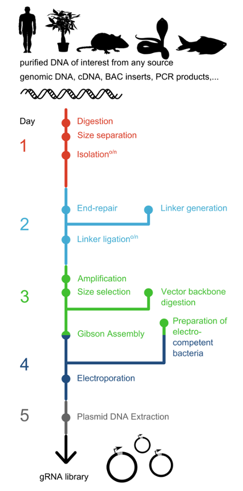
图 1:gRNA 文库制备建议时间线。 CORALINA 提供了一种简单且成本效益高的策略,可从任何生物体的多种不同 DNA 来源中构建全面的 gRNA 文库。本方案可在一周的工作时间内完成。接头生成可与 DNA 末端修复并行进行。制备电转化感受态细菌需要两天时间,包括过夜培养步骤,因此应在组装反应设置之前开始。 请点击此处查看该图的放大版本。
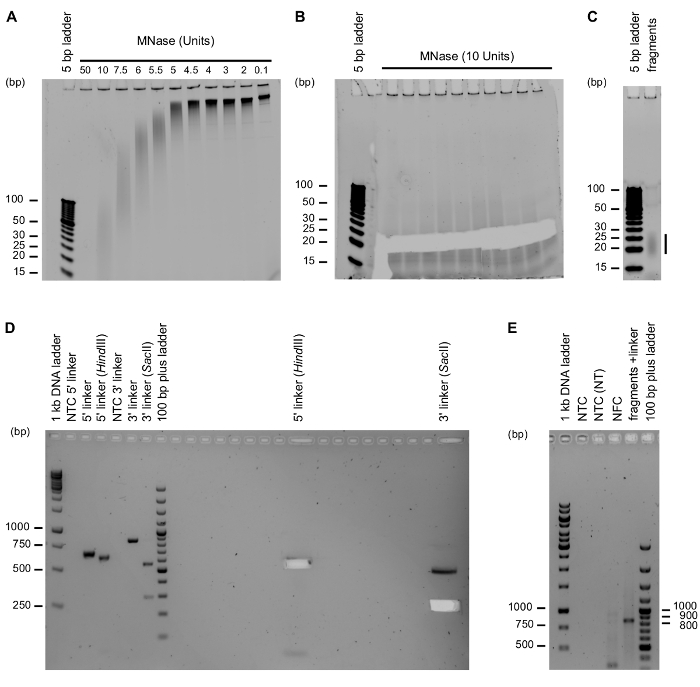
图2:实验方案中的关键步骤。 (A) 对BAC DNA进行可控消化,可产生不同大小的片段。图中展示了MNase消化的优化结果。纯化的BAC DNA用不同量的MNase处理10分钟。10 U的MNase可生成所需长度(20–30 bp)的DNA片段。(B) 通过从聚丙烯酰胺凝胶中切胶,选择20至30 bp大小的片段。纯化的BAC DNA用10 U MNase处理10分钟。图像为切胶后记录。(C) 对凝胶纯化片段的质量控制。凝胶纯化后,取六分之一纯化的MNase片段上样至20% PAGE凝胶,以检测片段大小选择和纯化是否成功。(D) 扩增用于组装的接头序列,并用限制性内切酶消化接头以确保定向克隆。5'和3'接头分别经HindIII和SacII酶切。设置无模板对照(NTC),以排除PCR假象和DNA污染。左侧:分析样品上样;右侧:制备样品上样。图像为凝胶切胶后记录。(E) 可通过增加PCR循环数(32个循环)进行PCR分析,以检测接头与DNA片段的连接是否成功,并设置以H2O作为模板的无模板对照(NTC),或以前一步缺口平移反应的NTC作为输入(NTC NT)进行对照。必须包含无片段对照(NFC),即在连接和缺口平移反应中省略MNase片段的扩增对照。只有将MNase片段与接头DNA结合的样品才能扩增出预期产物(869 bp)。请点击此处查看该图的放大版本。
讨论
CORALINA 可通过受控的核酸酶消化靶 DNA 并对产生的双链片段进行大规模克隆,从而构建大规模的 gRNA 文库。统计推断表明,使用本方案已成功克隆的独立 gRNA 序列数量远超 107 个9。CORALINA 可在多个方面进行定制。模板 DNA 的选择决定了目标区域以及所生成文库的最大复杂度。使用本方案,此前已从人和小鼠的基因组 DNA 成功构建 CORALINA 文库9。本文展示的代表性结果描述了从纯化的 BAC DNA 构建 CORALINA 文库的过程。通过选择不同的 gRNA 表达载体和连接子序列,可进一步实现定制化。我们此前已测试了三种不同长度的连接子组合用于 Gibson 组装,其效率差异较小9。
由于CORALINA gRNA的间隔序列源自整体消化的DNA,其长度通常并非精确的20 bp,而是呈现一定的长度分布,其均值取决于MNase消化的参数以及从PAGE凝胶中切取的片段大小。图2B 和 C 所示的代表性示例中,片段的中位长度介于19至27 bp之间。根据我们的经验,所生成的gRNA间隔序列能够忠实地保留原始片段的长度9。由于过短的片段(短于20 bp)会导致gRNA的脱靶率升高,应予以避免;而较长的片段在后续应用中通常问题较小,已有研究表明,间隔序列长达45 bp的gRNA仍具有功能活性9。
CORALINA 实验方案中最关键的两个步骤是 MNase 消化片段的大小选择和克隆步骤。若产生的片段过短(例如,平均长度低于 18 bp),或引入过多空的 gRNA 表达载体,则会导致文库失效。因此,优化 MNase 消化步骤(图 2A)、监测片段切离情况(图 2B、C)、确认 gRNA 载体骨架被完全消化,并在整个实验流程中设置不含片段的对照组,均至关重要。同时必须特别注意保持 gRNA 文库的代表性。文库构建过程中常见的瓶颈之一是将质粒高效转入细菌以进行扩增。因此,需要大量具有高转化效率的感受态细菌以及大量的独立电穿孔操作,才能获得足够数量的 gRNA 克隆。
在未来几十年内,为了充分挖掘基于CRISPR的筛选方法的全部潜力,必须开发新的gRNA文库构建策略。目前迫切需要成本低廉、操作简便且可定制的方法来生成大规模文库,这是使筛选技术能够应用于更多模式系统以及不同CRISPR工程方法的前提条件。CORALINA为此迈出了第一步。其潜在应用十分广泛,尤其适用于构建基因组的全面文库、来源于非主流模式生物的cDNA文库、高度特异性的文库,以及联合使用多种具有不同PAM要求的CRISPR蛋白的实验设计。
与其他方法不同,CORALINA 能够从输入的 DNA 中生成所有可能的 gRNA。然而,该方法的一个缺点是文库中也包含了缺乏所需 PAM 序列的 gRNA,这一特点与另一种用于生成 gRNA 文库的酶法 CRISPR-EATING 共有(表 1)。选择理想的 gRNA 文库构建方法取决于计划进行的筛选实验的具体要求,尤其是目标区域的性质(基因区、调控区、基因间区)和大小(单个位点、多个区域或全基因组范围)。我们认为,在需要分析大量非编码区或调控区、序列信息不完整或不可靠(如非传统模式系统、物种混合样本(例如 微生物组)或实验获得的输入材料)、联合使用不同的 CRISPR 核酸内切酶,或对较短且明确的位点进行饱和分析(例如 由 BAC 克隆代表的区域)时,使用 CORALINA 具有特殊优势。
披露
作者无任何利益冲突需要披露。
致谢
作者感谢Stephan Beck教授和Magdalena Goetz教授在开发CORALINA方法过程中提供的意见、帮助与支持,感谢Maximilian Wiessbeck和Valentin Baumann提出的有益建议。本工作得到了德国研究基金会(DFG)(STR 1385/1-1)的资助。
材料
| 姓名 | 公司 | 目录编号 | 评论 |
|---|---|---|---|
| 500 mM EGTA | Sigma Aldrich | 03777-10G | 1.4.,Mnase 的失活 |
| Novex Hi-Density TBE 上样缓冲液 | Thermo Fisher Scientific | LC6678 | 2.1. |
| Novex® TBE 凝胶,20%,10 孔 | Thermo Fisher Scientific | EC6315BOX | 2.1.,预制 20% PAGE 凝胶 |
| O'RangeRuler 5 bp DNA 标准品 | Thermo Fisher Scientific | SM1303 | 2.1. |
| Novex® TBE 电泳缓冲液 | Thermo Fisher Scientific | LC6675 | 2.1.,PAGE 凝胶电泳缓冲液 |
| 一次性无菌手术刀 | VWR | 233-5363 | 2.3.,可使用其他等效试剂 |
| SYBR Green I 核酸染料(DMSO 中 1000 倍浓缩液) | Sigma Aldrich | S9430 | 2.3. + 2.5.,Thermo Fisher Scientific 亦有供应(S7563) |
| UltraPure 苯酚:氯仿:异戊醇(25:24:1) | Thermo Fisher Scientific | 15593-031 | 3.6.1. + 4.3.,可使用其他等效试剂 |
| 糖原 | Sigma | 10901393001 | 3.6.4.,可使用其他等效试剂 |
| 3M 乙酸钠,pH 5.2 | Thermo Fisher Scientific | R1181 | 3.6.4.,可使用其他等效试剂 |
| 乙醇 | 3.6.4. + 9.1.8.,分子生物学级 | ||
| 快速平末端修复试剂盒 | New England Biolabs | E1201 | 4.1. |
| 乙酸铵 | Sigma | A1542 | 3.1.,可使用其他等效试剂 |
| 乙酸镁 | Sigma | M5661 | 3.1.,可使用其他等效试剂 |
| 0.5 M EDTA(pH 8.0) | VWR | MOLEM37465520(或 Promega V4231) | 2.2. + 3.1.,可使用其他等效试剂 |
| Agencourt AMPure XP 磁珠 | Beckman Coulter | A63881 | 5.3. + 6.5. |
| 凝胶回收试剂盒 | QIAGEN | 28704 | 5.7. + 7.1. + 8.4.,可使用其他等效试剂 |
| 高浓度 T4 DNA 连接酶 | New England Biolabs | M0202T | 6.1. + 8.1.2. |
| Long Amp Taq 2X 预混液 | New England Biolabs | M0287S | 6.3. |
| 含 HF 缓冲液的 Phusion 高保真 PCR 预混液 | New England Biolabs | M0531S | 5.1. + 6.6.,可使用其他等效试剂 |
| HindIII | New England Biolabs | R0104S | 5.4.1. |
| SacII | New England Biolabs | R0157S | 5.4.2. |
| AgeI | New England Biolabs | R0552S | 8.2.1. |
| Tris 碱 | Sigma | 93362 | 8.1.1. |
| 2M MgCl₂ | Sigma | 93362 | 8.1.1. |
| dGTP、dATP、dCTP、dTTP | New England Biolabs | N0446S | 8.1.1. |
| DTT | Sigma | DTT-RO | 8.1.1. |
| PEG-8000 | Sigma | P5413 | 8.1.1. |
| NAD | Sigma | N6522 | 8.1.1. |
| T5 核酸外切酶 | New England Biolabs | M0363S | 8.1.2. |
| Phusion DNA 聚合酶 | New England Biolabs | M0530S | 8.1.2. |
| Taq DNA 连接酶 | New England Biolabs | M0208L | 8.1.2. |
| rSAP | New England Biolabs | M0371S | 8.3.1. |
| TG1 感受态细胞 | Lucigen | 60502-1 | 9.1. |
| 1 mm 间隙电穿孔比色皿 | VWR | 732-2267 | 10.2. |
| 生物检测皿(聚苯乙烯,245 mm × 245 mm × 25 mm) | Fisher Scientific | DIS-988-010M | 9.4. |
| NaCl | Sigma | S7653 | 9.3. |
| 胰蛋白胨 | BD | 211705 | 9.3. |
| 酵母提取物 | BD | 212750 | 9.3. |
| 琼脂 | Sigma | A1296 | 9.4. |
| 甘油 | Sigma | G5516 | 9.17. |
| MNAse | New England Biolabs | M0247S | 1.1. |
| Nanodrop | Thermo Fisher Scientific | ND-2000 | 全文 |
| MicroPulser | Bio-Rad | 165-2100 | 10.2. |
| 电穿孔比色皿 | Bio-Rad | 732-2267 | 10.2. |
| 250 ml 离心管 | Corning | 430776 | 9.1–9.9. |
参考文献
- Mali, P., Esvelt, K. M., Church, G. M. Cas9 as a versatile tool for engineering biology. Nat Methods. 10 (10), 957-963 (2013).
- Yang, H., et al. One-step generation of mice carrying reporter and conditional alleles by CRISPR/Cas-mediated genome engineering. Cell. 154 (6), 1370-1379 (2013).
- Stricker, S. H., Koferle, A., Beck, S. From profiles to function in epigenomics. Nat Rev Genet. 18 (1), 51-66 (2017).
- Dominguez, A. A., Lim, W. A., Qi, L. S. Beyond editing: repurposing CRISPR-Cas9 for precision genome regulation and interrogation. Nat Rev Mol Cell Biol. 17 (1), 5-15 (2016).
- Shalem, O., et al. Genome-scale CRISPR-Cas9 knockout screening in human cells. Science. 343 (6166), 84-87 (2014).
- Wang, T., Wei, J. J., Sabatini, D. M., Lander, E. S. Genetic screens in human cells using the CRISPR-Cas9 system. Science. 343 (6166), 80-84 (2014).
- Koike-Yusa, H., Li, Y., Tan, E. P., Velasco-Herrera Mdel, C., Yusa, K. Genome-wide recessive genetic screening in mammalian cells with a lentiviral CRISPR-guide RNA library. Nat Biotechnol. 32 (3), 267-273 (2014).
- Hart, T., et al. High-Resolution CRISPR Screens Reveal Fitness Genes and Genotype-Specific Cancer Liabilities. Cell. 163 (6), 1515-1526 (2015).
- Koferle, A., et al. CORALINA: a universal method for the generation of gRNA libraries for CRISPR-based screening. BMC Genomics. 17 (1), 917-940 (2016).
- Lane, A. B., et al. Enzymatically Generated CRISPR Libraries for Genome Labeling and Screening. Dev Cell. 34 (3), 373-378 (2015).
- Sambrook, J., Russell, D. W. Isolation of DNA fragments from polyacrylamide gels by the crush and soak method. CSH Protoc. (1), (2006).
- Gibson, D. G. Enzymatic assembly of overlapping DNA fragments. Methods Enzymol. 498, 349-361 (2011).
- Elsaesser, R., Paysan, J. Liquid gel amplification of complex plasmid libraries. Biotechniques. 37 (2), 200-202 (2004).
重印与许可
