本文介绍了一种利用CRISPR/Cas9技术在人诱导性多能干细胞中对内源性表达的蛋白质进行荧光标记的实验方案。通过荧光激活细胞分选富集可能经过编辑的细胞,并建立单克隆细胞系。
本文内容
摘要
摘要
一种用于生成表达内源性蛋白与同框融合标签的报告基因的人类诱导多能干细胞(hiPSCs)的实验方案 N端或C端荧光标签。原核生物CRISPR/Cas9系统(成簇规律间隔短回文重复序列/CRISPR相关蛋白9)可通过同源定向修复(HDR)将大型外源序列插入基因组位点。为实现目标基因敲入,本方案采用基于核糖核蛋白(RNP)的方法,使用野生型Cas9蛋白 Streptococcus pyogenes Cas9蛋白、合成的双组分向导RNA(gRNA)以及供体质粒通过电穿孔递送至细胞中。通过荧光激活细胞分选(FACS)富集表达荧光标记蛋白的潜在编辑细胞。随后建立单克隆细胞系,并可对其编辑结果进行精确分析。通过在目的基因的基因组位点引入荧光标签,可在内源性调控条件下研究融合蛋白的亚细胞定位与动态行为,这是相较于传统过表达系统的关键改进。利用人诱导多能干细胞(hiPSCs)作为基因标记的模型系统,可在二倍体、非转化细胞中研究标记蛋白。由于hiPSCs可分化为多种细胞类型,该策略提供了在多种同基因背景的细胞环境中构建并研究标记蛋白的机会。
引言
利用基因组编辑策略(尤其是CRISPR/Cas9)研究细胞过程正变得越来越便捷且具有重要价值1,2,3,4,5,6,7。CRISPR/Cas9 的众多应用之一是通过同源定向修复(HDR)将GFP等大型外源序列插入特定基因组位点,从而作为基因或蛋白质产物活性的报告系统8。该技术可用于将荧光蛋白序列与内源性开放阅读框连接,使所生成的内源性调控融合蛋白可用于可视化目标蛋白的亚细胞定位及其动态变化5,6,9,10,11。尽管与过表达系统相比,内源性标记蛋白具有诸多优势,但将大型序列插入人类基因组的效率较低,通常需要采用筛选或富集策略,才能获得可用于深入研究的细胞群体5,12。
本方案描述了将编码荧光蛋白(FP)的DNA序列插入目标基因组位点的方法。该方案包括供体质粒的设计与递送,以及核糖核蛋白(RNP)复合物(野生型S. pyogenes Cas9蛋白与合成的CRISPR RNA(crRNA)和反式激活crRNA(tracrRNA)结合而成)的使用。此外,还介绍了通过荧光激活细胞分选(FACS)富集潜在编辑细胞,以及单克隆细胞系的建立过程。迄今为止,该方法已成功用于构建携带单等位基因或(罕见情况下)双等位基因绿色荧光蛋白(GFP)标签的人诱导多能干细胞(hiPSC)系,这些标签标记了代表主要细胞结构的25种蛋白质。经编辑的细胞已证实具有预期的基因插入,表达正确亚细胞定位的融合蛋白,并保持多能性及稳定的核型12(以及未发表数据)。该方法还被用于生成多种其他单基因和双基因(同一细胞中标记两种不同蛋白)编辑的hiPSC群体(未发表数据)。
选择来自健康供体的人iPSCs进行这些基因组编辑工作,因为与许多传统细胞系不同,它们是二倍体、核型稳定、未转化且具有增殖能力。这些特性为研究基础细胞生物学和疾病建模提供了理想的模型。此外,hiPSCs的分化潜能使得利用同基因型细胞(包括类器官、组织以及“"疾病模型")平行研究多种发育阶段及不同谱系和细胞类型成为可能13,14,15。尽管本实验方案是为hiPSCs(WTC细胞系)开发的,但其也可能为其他哺乳动物细胞系实验方案的建立提供参考。
方案
1. 荧光蛋白敲入的 in silico crRNA 与供体质粒设计
- 从 NCBI16 或 UCSC 基因组浏览器17 获取目标基因的带注释参考序列(例如,GenBank 格式),并将其导入所选的生物信息学软件中。如果已知宿主基因组序列相对于参考序列存在变异,则需在生物信息学软件中调整参考序列以包含这些变异(见讨论部分)。
- 确定荧光蛋白(FP)的预期插入位点。对于 C 末端标签,荧光蛋白标签序列将插入到最后一个密码子的最后一个碱基与终止密码子的第一个碱基之间。对于 N 末端标签,荧光蛋白标签序列通常插入到起始密码子的最后一个碱基与下一个密码子的第一个碱基之间。在某些情况下,例如起始密码子为单密码子外显子,或蛋白质末端附近存在信号序列时,FP 的预期插入位点可位于更偏 3ʹ 的位置,只要仍保持阅读框正确即可。
- 以预期插入位点两侧各 50 bp 的序列作为输入,使用任意公开可用的 crRNA 设计工具进行分析。在插入位点附近确定 2–4 个 crRNA 靶点后,在生物信息学软件中标注 crRNA 结合位点及原间隔序列邻近基序(PAM)序列(NGG)。这些 crRNA 将用于诱导双链断裂(有关 crRNA 设计的更多指导,请参见讨论部分)。
注意:可将定制的 crRNA 序列提交给商业供应商进行合成(推荐),也可将该序列作为起点设计克隆或体外合成策略,但后者超出本实验方案的范围(见讨论部分)。 - 构建供体模板质粒时,使用预期插入位点上游 1 kb 的序列作为 5ʹ 同源臂(对于 N 末端插入,应包含起始密码子),使用下游 1 kb 的序列作为 3ʹ 同源臂(对于 C 末端插入,应包含终止密码子)。两个同源臂之间的碱基通常不应省略。在同源臂中包含细胞系特异性变异可使这些遗传变异在最终编辑的细胞中得以保留。
- 在两个同源臂之间插入荧光蛋白(FP)序列(或其他敲入序列)以及连接子序列(有关连接子设计的更多指导,请参见讨论部分)。对于 N 末端标签,连接子序列应紧接在 FP 的 3ʹ 端;对于 C 末端标签,连接子序列应紧接在 FP 的 5ʹ 端。
- 在供体模板质粒中破坏 crRNA 结合位点,以防止 Cas9 对供体序列进行切割(有关改变 crRNA 结合位点时的注意事项,请参见讨论部分)。如有可能,优先将 PAM 序列突变为非 NGG 或 NAG 的序列。或者,可在 crRNA 的“种子”区域(靠近 PAM 的 10 个碱基)引入三个碱基的点突变,预测足以有效破坏 crRNA 的结合。某些 crRNA 结合位点可能因供体模板质粒中插入 FP 序列而自然破坏;在这些情况下,应确保不再存在 PAM 或完整的结合区域。
注意:体外设计的供体模板质粒可提交给商业供应商进行基因合成,也可作为起点用于设计克隆策略,但后者超出本实验方案的范围。使用 pUC19 或 pUC57 等简单骨架质粒即可满足要求。
2. 在 hiPSCs 中通过核糖核蛋白(RNP)转染实现 CRISPR/Cas9 介导的基因敲入
注意:在本方案中,“gRNA”一词指按照制造商说明正确重悬、定量并预先复合的合成 crRNA 和 tracrRNA(参见材料表)。所有培养基均需添加 1% 青霉素-链霉素。WTC hiPSC 细胞系的一般培养指南在艾伦细胞探索平台中有更详细的描述18,19。本方案使用 WTC hiPSC 细胞系,但通过适当的转染优化,RNP 与供体质粒的电穿孔可成功适用于其他细胞类型。
- 准备10 µM 工作储备液的 gRNA 和野生型 S. pyogenes Cas9蛋白2,20;保持在冰上。准备 1 µ克/µL工作储备液的供体质粒; 室温(RT)保存。所有稀释均使用 pH 8.0 的 TE 缓冲液。
- 准备一个基质包被的6孔组织培养板,每孔加入5 mL新鲜生长培养基,并添加10 µ每孔加入 M ROCK 抑制剂(Ri)。将培养板连同培养基置于 37 ℃ 培养箱中孵育 °C 和 5% CO2 转染程序结束后,直至准备铺板细胞前(最多2小时)。
注意:本实验方案中使用的所有基质包被培养板均按照艾伦细胞科学研究所(Allen Institute for Cell Science)关于WTC hiPSC细胞系的培养方案制备,即用预冷的DMEM/F12培养基将冰上保存的Matrigel按1:30比例稀释后加入培养板中19. - 使用推荐的温和单细胞解离试剂 材料表将hiPSCs传代为单细胞悬液,使用自动细胞计数仪或血细胞计数板进行细胞计数。
注意:本文所用 WTC hiPSC 细胞系的详细实验方案可在 Allen Cell Explorer 获取19用室温 DPBS 洗涤细胞一次,加入解离试剂处理 3–5 分钟。随后通过轻柔吹打使细胞重悬为单细胞悬液,并离心收集细胞沉淀。将活细胞沉淀重悬于添加了 10 µM Ri.- 准备一份 1.84 x 10⁶ 每种实验条件下用于转染的细胞分别置于 1.5 mL 离心管中。
注意:所有体积均按 4.5 计算 µL 总反应体积在 100 µ2.3 次反应所需的电穿孔体积,进行两次重复。此量已包含重复转染及移液误差所需的额外体积。
- 准备一份 1.84 x 10⁶ 每种实验条件下用于转染的细胞分别置于 1.5 mL 离心管中。
- 为每种实验条件准备核糖核蛋白(RNP)复合物管,加入 2.88 µ10 升 µM gRNA 和 2.88 µ10 升 µ将 M Cas9 加入 1.5 mL 离心管中。室温孵育至少 10 分钟(最长 1 小时)。
- 在室温下以211 × g离心3分钟,沉淀一份单细胞悬液(按步骤2.3.1制备)。吸除上清液,并将细胞沉淀重悬于220 μL中 µL 厂家提供的电穿孔缓冲液
- 加入220 µ将步骤2.5中重悬的细胞悬液L加入步骤2.4中制备的1.5 mL RNP复合物管中。
- 加入 4.60 µ1 升 µ克/µ将L型供体质粒加入步骤2.4中准备好的1.5 mL离心管中。
- 使用核转染吸头和移液器将管内液体轻轻吹打混匀2-3次,然后转移100 µ将悬浮液1 L加入已准备好的电穿孔装置中。避免枪头中产生气泡。施加1300 V电压,持续30 ms,进行1次脉冲。
- 将悬液轻轻转移至步骤2.2中准备好的6孔板中,并轻轻晃动。通过轻柔地左右和前后移动培养板使细胞分散均匀。
- 使用新的核转染枪头,对剩余的100个重复步骤2.8–2.9 µ将悬浮液转移至已准备好的6孔板的第二个孔中。
- 对每种gRNA与供体质粒组合(包括非靶向gRNA、仅供体质粒以及仅缓冲液对照)重复步骤2.4至2.10。注意更换移液器吸头和核转染枪头,以避免交叉污染。
- 将转染后的细胞在37℃孵育 °C 和 5% CO₂224小时后更换为常规生长培养基(不含Ri),之后每24小时更换一次hiPSC培养基,持续培养72–96小时,并监测细胞融合度。当hiPSC达到60–80%融合度时,进行步骤3。
注意:细胞死亡严重(>70%(估计值)是转染后24-48小时的正常情况。
3. 荧光激活细胞分选富集疑似编辑的hiPSCs
注意:在分选干细胞时,应按照讨论部分的建议调整仪器设置以促进细胞存活。简而言之,使用尽可能大的喷嘴(130 µm)、低流速(≤ 24 µL/min)、无防腐剂的鞘液(如生理盐水,参见材料表)以及低样本压力(10 psi)。
- 在开始进行流式细胞分选(FACS)实验之前,更换为添加了10 µM Ri的生长培养基,并将细胞在37 °C、5% CO2条件下孵育2–4小时,以促进FACS后细胞的存活。
- 按照材料表中推荐的方法,使用温和的单细胞解离试剂将hiPSCs消化为单细胞悬液,并重悬于添加了10 µM Ri19的生长培养基中。
- 将hiPSC悬液通过35 µm细胞筛过滤,收集至聚苯乙烯圆底管中。
- 利用前向散射光和侧向散射光(包括高度vs.宽度)进行细胞分选,以排除碎片和双联体。使用仅含活细胞和缓冲液的对照样本设定荧光蛋白(FP)阳性门控,确保<0.1%的仅缓冲液对照细胞落入该门控范围内。
- 将所有FP阳性细胞分选至含有0.5–2 mL室温生长培养基(添加10 µM Ri)的1.5–15 mL聚丙烯管中。
注:聚丙烯材质可减少细胞对塑料表面的黏附。 - 在室温下以211 × g离心收集的细胞3分钟。
- 小心吸除上清液,将细胞沉淀重悬于200 µL添加了10 µM Ri的生长培养基中。将最多3,000个分选后的细胞转移至一个预先包被基质的96孔板的新孔中19。
注:若仪器设置适当,也可直接将细胞(批量)分选至含有200 µL添加10 µM Ri的生长培养基的基质包被96孔组织培养板的单个孔中,hiPSC推荐接种密度为每孔1,000–3,000个细胞。 - 将分选后的细胞置于37 °C、5% CO2条件下孵育。在24小时时更换为添加5 µM Ri的生长培养基;在48小时后开始每隔24小时更换一次常规生长培养基(不含Ri),持续72–96小时,并监测细胞融合度。若在96孔板的一个孔中接种不少于500个细胞,FACS后的细胞存活率预计可超过50%。
- 当hiPSCs达到60–80%融合度并呈现成熟形态(菌落中心平滑、紧密排列)时,传代至更大规格的培养板,例如24孔板,随后再由24孔板传代至6孔板。
- 当6孔板中的hiPSCs达到60–80%融合度并呈现成熟形态时,扩增至100 mm培养皿,可用于成像、冻存,或以克隆挑取密度接种(步骤4)19。
4. 生成初步编辑的单克隆 hiPSC 细胞系
- 使用推荐的温和单细胞解离试剂 材料表将hiPSCs传代为单细胞悬液,并测定每毫升细胞数量19.
- 将编辑后的 hiPSC 群体以 10,000 个细胞接种至新鲜基质包被的 100 mm 组织培养皿中,使用添加了 10 µM Ri19接种后24小时将培养基更换为不含Ri的生长培养基,之后每24小时用新鲜生长培养基换液,持续5-7天。
- 当hiPSCs形成的克隆在宏观上可见(约500 µm)它们足够大,可以进行分离。通过吸除多余的基质并加入100 μL基质,制备基质包被的96孔板。 µ升添加了10的生长培养基 µ每孔 M Ri19.
- 在体视显微镜下,使用 P-200 移液器或类似工具,轻轻刮取并吸出培养板表面的单个克隆。转移体积(约 20–100 μL) µL)将含有克隆的培养皿转移至步骤4.3中准备好的96孔板的单个孔中。
- 所有克隆转移完毕后,将培养板置于37˚C 和 5% CO2更换为常规生长培养基(不含 Ri),在 24 小时后开始,每 24 小时更换一次培养基,持续 72–96 小时,直至克隆大小约增至原来的三倍(约 1500) µm)
注意:建议针对转染中使用的每条crRNA挑取24–96个克隆。分离克隆的存活率通常高于95%。
- 所有克隆转移完毕后,将培养板置于37˚C 和 5% CO2更换为常规生长培养基(不含 Ri),在 24 小时后开始,每 24 小时更换一次培养基,持续 72–96 小时,直至克隆大小约增至原来的三倍(约 1500) µm)
- 使用推荐的温和单细胞解离试剂 材料表将hiPSC克隆传代至新的基质包被的96孔板,步骤如下19.
- 使用八通道移液器,移除并弃去96孔板第一列中的培养基。
- 使用 P-200 多通道移液器,加入约 200 µ向96孔板的第一列加入 L DPBS 以洗涤细胞。使用8通道移液器吸除并弃去96孔板第一列中的DPBS洗涤液。
- 使用 P-200 多通道移液器,加入 40 µ将解离试剂 L 加入 96 孔板的第一列。
- 重复步骤 4.5.1–4.5.3,最多完成 96 孔板的六列,每次更换枪头以确保孔间不发生交叉污染。将该孔板置于 37 °C培养箱中孵育3-5分钟,从向第一列加入解离试剂时开始计时(步骤4.5.3)。
注意:由于执行这些步骤所需时间较长,建议每次传代不超过六列(48孔)。首次进行本方案时,应每次仅传代一至两列。限制单次传代的列数可确保细胞不会在解离试剂中停留过久,从而避免对人诱导多能干细胞(hiPSCs)造成损伤。 - 当培养板第一列中的细胞开始从板底脱落时,使用 P-200 多通道移液器加入 160 µL DPBS 加入96孔板的第一列,轻轻吹打重悬细胞 "12:00", "3:00", "6:00",以及 "9:00" 每个孔的位置。转移全部细胞悬液(200 µL)至V型底96孔板。
- 对剩余含有解离试剂的细胞列重复步骤 4.5.5;更换移液吸头以避免孔间交叉污染。
- 在室温下,将V型底板在离心机中以385 × g离心3分钟。
- 使用 P-200 多通道移液器,轻轻吸除上清液,并将细胞重悬于 200 µ升新鲜生长培养基,添加10 µ每孔加入 M Ri。重复操作至所有孔,每次更换枪头以避免交叉污染。
- 将全部细胞悬液转移至新鲜基质包被的96孔板中19. 将培养板置于37℃的组织培养箱中孵育 °C 和 5% CO224小时后更换为常规生长培养基(不含Ri),之后每24小时换液一次,持续72–96小时,直至大部分克隆达到60–80%汇合度。
注意:此步骤有助于分散细胞,使细胞在96孔板的整个区域更均匀地生长。
- 观察克隆,确定96孔板中每个克隆的合适传代比例(例如, 1:10 或 1:8)。使用建议的温和单细胞解离试剂 材料表,将hiPSC克隆传代至新的基质包被的96孔板中(步骤4.5.1–4.5.8),并转移适合每个克隆的细胞悬液比例。将培养板置于37℃组织培养孵箱中孵育˚C 和 5% CO224小时后更换为常规生长培养基(不含Ri),之后每24小时换液一次,持续72–96小时,直至大部分克隆达到60–80%汇合度并呈现成熟形态。
注意:每个克隆的传代比例可能因前次传代时生长速率或存活率的微小差异而有所不同,因此本次传代有助于在后续冷冻步骤中使每个克隆每孔的细胞数量标准化。由于各克隆在传代过程中的存活率和生长速率存在差异,部分克隆可能出现过度生长或无法生长的情况。- 将剩余的细胞悬液和细胞沉淀保存用于基因组DNA提取:将细胞在V型96孔板中以385 × g离心3分钟(室温)。吸除上清液后,使用96孔试剂盒进行基因组DNA提取,或将含有细胞沉淀的96孔板在-20 °C保存˚C,最长可达三周。
5. 以96孔板形式对单克隆细胞系进行冷冻保存
- 使用单细胞解离试剂,按照先前所述方法传代 hiPSC 克隆(步骤 4.5.1–4.5.7)。用 P-200 多通道移液器吸除上清液,并将细胞重悬于 60 µ升添加了10的生长培养基 µM Ri。对所有孔重复此操作;每次更换枪头以避免交叉污染。
- 转移30 µ将细胞悬液 170 L 加入未经基质包被的 96 孔组织培养板中,随后迅速加入 170 µL 冷冻缓冲液(参见 材料表)加入到每个孔中,不要混匀。重复操作,转移剩余的30 µ将悬浮液的 L 加入姊妹板,并加入 170 µL 冷冻缓冲液。
注意:该过程需在两块相同的培养板中平行进行,以便在解冻其中一块培养板后仍保留备用的细胞群体。将冻存的细胞接种至96孔板中每隔一列的孔内,有利于加快解冻速度(步骤5.6)。 - 用封口膜包裹培养板,放入带有盖子的室温聚苯乙烯泡沫盒中。将整个盒子置于 -80 ℃冰箱中 °C 冷冻柜
- 24 小时后,可将培养板从泡沫箱中取出并置于 -80 ℃ 保存˚C,最长可达四周。
注意:在细胞暂时保存于 -80 时 °C,可对步骤4.6.1中获得的细胞提取的基因组DNA进行遗传质量控制检测,以鉴定需解冻并进一步扩增的克隆,具体方法参见先前发表的研究。12简而言之,可采用拷贝数液滴数字PCR检测来鉴定含有一个或两个GFP拷贝且无供体质粒骨架整合的克隆。随后通过终点PCR检测与Sanger测序相结合的方法,进一步鉴定含有精确插入片段的克隆。 - 解冻时,将整个培养板置于37 °在组织培养孵育箱中,密切观察冰粒融化情况。培养板边缘的孔通常会先解冻。
- 当目标克隆的冰粒融化后,轻柔地转移全部200 µL 加入含有 3 mL 室温生长培养基(添加 10 µM Ri,于室温下以 211 × g 离心 3 分钟。
- 吸弃上清液,并将沉淀的细胞重悬于1 mL室温培养基中,培养基中添加10 µM Ri。转移至新鲜基质包被的24孔板,于37℃培养˚C 和 5% CO224小时后更换为常规生长培养基(不含Ri),之后每24小时换液一次,持续72–96小时,直至克隆达到60–80%汇合度并呈现成熟形态。19.
结果
本实验的目标是通过将 mEGFP(单体增强型绿色荧光蛋白)序列插入 LMNB1 基因的 5ʹ 端(蛋白的 N 末端),实现 mEGFP 与核纤层蛋白 B1 的融合。所选用的连接肽(氨基酸序列 SGLRSRAQAS)基于 Michael Davidson 荧光蛋白文库中先前的 cDNA 构建体21。由于在供体质粒的候选 crRNA 结合区域中,经过 in silico 插入 mEGFP 和连接肽序列后该区域已被破坏,因此无需引入点突变来阻断 Cas9 对供体序列的 crRNA 识别与切割(图 1)。供体序列包含位于 mEGFP-连接肽序列两侧的 1 kb 同源臂。最终获得的 2,734 bp DNA 片段被克隆至 pUC57 载体骨架中,经测序验证后,使用无内毒素大提纯试剂盒纯化得到供体模板质粒。将供体模板质粒与 RNP 复合物共转染细胞,富集潜在编辑的细胞,并通过荧光显微镜确认 mEGFP-核纤层蛋白 B1 融合蛋白的定位(图 2)。本文仅描述 crRNA1 转染的结果,尽管两种 crRNA 序列均获得了潜在编辑的细胞群体12。
与不含gRNA、Cas9蛋白或供体质粒(仅含缓冲液)的阴性对照相比,转染LMNB1 crRNA1的细胞中包含0.95%的mEGFP阳性细胞,代表推测已编辑的mEGFP-核纤层蛋白B1群体(图3a)。该结果处于此前报道中使用此方法在多个基因组位点所观察到的敲入效率范围内。12.
通过流式细胞术(FACS)分离出mEGFP阳性细胞,并利用活细胞显微镜观察,以确认mEGFP-核纤层蛋白B1融合蛋白定位于核膜的预期定位。FACS富集后,LMNB1 crRNA1群体中约90%的分选细胞为mEGFP阳性(由显微镜观察确定),表明在分选过程中部分mEGFP阴性细胞与GFP阳性细胞共同被纯化。该富集水平可接受,足以用于挑选96个克隆,进而进行遗传学筛选以鉴定成功编辑的克隆。通常,成功的富集标准为至少50%的细胞呈GFP阳性。
大部分分选后的细胞群体在非分裂细胞中显示核膜(核周区)处有荧光信号,而在有丝分裂期间则显示细胞质中扩展的核纤层结构有荧光信号,表明LMNB1基因座的基因组编辑正确。富集后的细胞群体包含荧光信号强或弱的细胞。信号强度的差异可能反映了正确与错误编辑结果的混合,凸显了构建经遗传学验证的单克隆细胞系用于后续研究的重要性(见讨论)。图 3b)12克隆细胞系建立后,经基因验证的细胞在显微镜实验中显示出均一的GFP荧光强度(图3c).
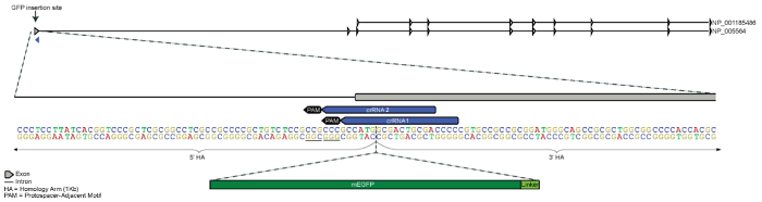
图1。LMNB1基因N端GFP标签的设计策略。 GFP标签设计为插入位于5号染色体上的LMNB1基因第一个外显子的5ʹ端上游。5ʹ和3ʹ同源臂各长1 kb,在起始密码子(ATG)与第二个密码子之间汇合(图中仅部分显示同源臂)。设计了两个候选crRNA,用于引导Cas9在尽可能接近目标插入位点的位置进行切割,同时确保在基因组中的唯一性。mEGFP序列及一个氨基酸连接序列被插入至起始密码子下游紧邻的3ʹ端(mEGFP与连接序列未按比例绘制)。请点击此处查看该图的放大版本。

图 2。生成内源性标记的 hiPSC 单克隆细胞系的工作流程。 转染组分包括 Cas9/crRNA/tracrRNA RNP 复合物(图示为红色 Cas9 蛋白、金色 crRNA 和紫色 tracrRNA)、含有同源臂(HAs,图示为金色)的供体质粒以及荧光蛋白+连接肽序列(FP+linker,图示为绿色),通过电穿孔导入细胞。转染 4 天后,通过 FACS 富集荧光蛋白阳性(FP-positive)的初步编辑细胞,并将所有分选后的细胞接种至 96 孔板的一个孔中(约 1,000 个细胞),扩增培养至获得数百万细胞的可用群体,作为“富集群体”进行后续检测(参见实验方案第 3.8 步)。由于不同实验中同源定向修复(HDR)效率存在差异,FP 阳性细胞的得率也有所不同12;在转染约 1.6 × 10⁶ 个 hiPS 细胞后,成功的富集通常可获得约 300–5,000 个阳性细胞。初步成像研究确认了富集群体中融合蛋白的信号及其亚细胞定位。随后人工挑选单克隆集落,转移至 96 孔板中进行扩增和冷冻保存。进一步采用微滴数字 PCR(ddPCR)及其他基于 PCR 的检测方法进行基因组质量控制分析,以鉴定正确编辑的单克隆,方法如前所述12。 请点击此处查看该图的放大版本。

图 3.富集假定编辑的细胞群体。(A)转染后第四天 LMNB1 编辑细胞的流式细胞术图谱。纵轴显示 GFP 强度,横轴显示前向散射。分选门控基于仅含缓冲液的对照组设定。由于 hiPSC 对扰动敏感,未使用活/死细胞染色,而是采用非常保守的 FSC/SSC 门控策略。(B)富集后,LMNB1 Cr1 编辑细胞群体中约 90% 的细胞显示 GFP 定位于核膜(符合预期的 LMNB1 定位)。该群体包含 GFP 强度不同的细胞,以及部分 GFP 阴性细胞。比例尺为 10 微米。(C)在获得单克隆细胞系后,细胞表现出均一的 GFP 强度,仅存在部分与细胞周期相关的差异。比例尺为 20 微米。
讨论
本文介绍的在hiPSCs中生成内源性调控的荧光蛋白融合体的方法,是一种多功能且强大的基因编辑细胞系构建策略,其应用范围广泛,包括活细胞成像、多种功能研究,以及利用患者来源的hiPSC系建立"疾病在培养皿中"模型13,14,15。尽管该方法已被用于在内源性蛋白的N端或C端引入较大的荧光蛋白标签,但其也有潜力用于引入其他标签或小的遗传改变,以模拟或纠正致病突变22,23。对于较小的插入片段,同源臂的长度可适当缩短,但本方法所描述的基因编辑总体策略仍然适用24,25。尽管强烈推荐使用hiPSCs以充分发挥其广泛的应用价值,但通过谨慎优化,本实验方案也可适用于其他哺乳动物细胞系的基因组编辑。
在确定用于荧光蛋白(FP)标记的目的基因时,转录本丰度估计值(来自芯片或RNA-Seq数据)是评估该基因或特定亚型是否表达的良好起点,尽管转录本水平并不总是与蛋白水平相关。本文所述的FACS富集策略最适合在目标细胞类型中至少中等表达水平的基因。该策略在筛选具有点状和/或明确定位模式的融合蛋白(如centrin、desmoplakin和paxillin)时也已取得成功,这些蛋白的信号与背景比非常低12,19。对于表达水平较低或仅在衍生细胞类型中表达的基因,可能需要额外的筛选策略。
在人类细胞系中设计crRNA和供体质粒时,应以人类参考基因组(GRCh38)为起点。由于同一种类的不同细胞系基因组可能存在差异,而CRISPR/Cas9具有序列特异性,因此明确与参考基因组不同的细胞系特异性变异(单核苷酸多态性或插入/缺失(indels))并将其纳入设计中极为重要。这可确保crRNA与宿主基因组兼容,并使供体质粒的同源臂保留细胞系特异性的变异。建议在设计过程中将纯合变异整合到crRNA和供体质粒的同源臂中;杂合变异的整合则为可选。下文将讨论大片段敲入实验所用的具体试剂及其他关键注意事项。
Cas9 蛋白
使用Cas9蛋白的主要优势在于,将Cas9和gRNA以核糖核蛋白(RNP)复合物形式导入细胞,其核酸酶活性持续时间较短,相比之下,基于质粒的方法中Cas9和gRNA的表达可能持续数天,从而导致更高的靶上和脱靶效应26,27。使用Cas9蛋白的另一个优势是,一旦进入细胞,该蛋白可立即发挥切割作用,而传统的Cas9 mRNA或Cas9/gRNA质粒方法则需要经过转录、翻译和蛋白质加工等过程才能产生成熟的功能蛋白26,28。目前,野生型S. pyogenes Cas9蛋白已可从多家商业来源获得。
向导RNA
有许多公开可用的工具可用于在目标荧光蛋白(FP)插入位点附近寻找 crRNA 靶点,这些靶点在宿主基因组中预测的脱靶效应为零或极少29,30,31,32。在特定基因位点使用不同 crRNA 靶点时,同源定向修复(HDR)的效率及修复结果的精确性存在广泛差异12。因此,建议每个位点测试多个 crRNA(2–4 个,最好位于目标插入位点 50 bp 范围内),以提高基因编辑实验成功的可能性。目前 gRNA 的递送方式包括:合成的双组分 crRNA 与 tracrRNA、合成的单体 gRNA(sgRNA)、in vitro 转录的 sgRNA,或通过质粒递送在细胞中由 U6 启动子驱动表达 sgRNA。本实验方案未针对高切割活性进行优化。本研究采用未经修饰的双组分 crRNA 与 tracrRNA(见材料表),旨在生成单等位基因荧光蛋白标记的细胞系,同时尽可能减少对细胞的潜在干扰。
供体模板质粒
由于供体质粒中提供的部分同源臂序列会在HDR过程中被整合到宿主基因组中,因此应针对crRNA识别位点引入点突变,以防止HDR完成后Cas9对修复位点的再次切割。通常最简单的破坏方法是突变PAM序列。由于某些非经典PAM序列仍可被野生型S. pyogenes Cas9识别,因此最好避免使用NGG、NAG或NGA33。在突变同源臂时,应避免引入非同义突变和稀有密码子。如果无法通过同义突变改变PAM序列,可考虑在crRNA结合位点的种子区域(靠近PAM的10 bp区域)引入三个同义点突变。在5ʹ非翻译区(UTR)进行此类修改时需格外谨慎,因为这些区域可能含有重要的调控序列。在此类情况下,可参考UCSC基因组浏览器的比较基因组学轨迹等遗传保守性数据库,以指导突变位点的选择,因为对非保守碱基的修改通常比对高度保守碱基的修改更易被耐受17。有时仅插入荧光蛋白(FP)序列就足以破坏crRNA结合位点(如图1所示);然而,仍应检查新插入的序列是否仍保留有crRNA结合位点或PAM序列。
建议在荧光蛋白(FP)与天然蛋白之间使用氨基酸连接序列,以保持融合蛋白的功能34。通常可根据连接序列的特定电荷或大小来选择合适的氨基酸连接序列。如果已有研究较为深入的cDNA融合设计,且其结构类似于目标内源性融合蛋白,则可采用相同的连接序列用于CRISPR/Cas9基因敲入实验12,19。若缺乏此类信息,也可成功使用如GTSGGS等较短的连接序列12。其他研究还表明,对于多种不同靶标,通用的三氨基酸小分子连接序列同样可实现有效融合35。
转染与FACS富集
许多市售转染试剂专为向细胞递送特定类型的分子而设计,而电穿孔系统则可用于递送在大小、电荷和组成上具有广泛差异的试剂。除了是转染难度较高的细胞(如人诱导多能干细胞,hiPSCs)常用的转染方法外,电穿孔还可同时递送本方法中所述CRISPR/Cas9介导的定点插入(FP knock-in)所需的全部三种组分。在本方法开发过程中,与其他市售试剂相比,电穿孔获得了最佳效果(数据未显示),且已有其他研究将其用于核糖核蛋白(RNP)的递送26,28,36。
在对hiPSCs进行基因编辑操作时,应特别注意在基因编辑前后轻柔处理细胞,以确保细胞存活率最佳,并最大限度减少自发性分化。具体而言,进行FACS富集时应针对干细胞分选进行优化:使用尽可能大的喷嘴(130 µm)、低流速(≤24 µL/min)、无防腐剂的鞘液(如生理盐水,参见材料表)以及低样本压力(10 psi)。为避免单细胞分选导致干细胞存活率不理想,建议将FACS富集后的hiPSCs以群体形式分选并扩增培养,以优化细胞存活率和干细胞完整性。然而,对于敏感性较低的细胞类型,单细胞分选可能是合适的。为促进细胞存活,细胞在用于FACS富集而收获后,应在一小时内重新接种回培养体系,并在整个分选过程中保持室温。对于某些细胞类型,在分选过程中将细胞置于冰上(4°C)孵育也可能有助于提高细胞存活率。
大量扩增FP阳性细胞可为在建立单克隆细胞系之前通过成像分析评估融合蛋白定位提供机会。尽管所获得的富集细胞群体可能已满足某些研究的需求,但这些群体通常表现出强度不一的FP信号。分离得到的单克隆细胞系则具有均一的信号(图3),因此更适用于功能实验12。
克隆细胞系的建立
在整个编辑和克隆系生成过程中,监测细胞形态非常重要。在无饲养层条件下培养的hiPSC集落应具有光滑的边缘以及均匀、紧密排列的中心12,18,19。分化细胞的比例应低于培养物的5%。在挑选单个集落时,应选择形态良好的集落。在96孔板传代过程中,需检查各克隆的形态,及时终止那些过度生长的克隆,因为这可能导致细胞分化或提示存在遗传不稳定性。
获得单克隆细胞系可用于精确编辑的遗传学验证,这一点非常重要,因为尽管在目标位点成功插入了标签,Cas9诱导的基因组双链断裂仍常常被非精确修复。先前报道的基于PCR的检测方法显示,在十个不同的基因组位点中,许多表达荧光蛋白(FP)的克隆(45%)均存在供体质粒骨架在靶向位点处的整合,或(较少见地)在基因组中随机整合12。此外,在十个不同位点中,23%的GFP阳性克隆(n=177)被发现其未标记等位基因的预期crRNA切割位点附近存在突变,这极有可能是由于NHEJ修复机制所致12。对大量单克隆细胞系(约100个克隆/编辑)的遗传学分析凸显了进行遗传学验证的重要性,而这种验证在混合细胞群体中无法实现,因为仅凭荧光蛋白表达和预期融合蛋白的定位并不能保证编辑的精确性12。此外,这些基于PCR的检测方法无法在富集的细胞群体中可靠地进行,因此在开展有意义的分析之前,必须首先建立单克隆细胞系。为了确保在靶向位点的精确编辑,不仅需要对插入的荧光蛋白标签进行遗传学确认,还需验证未编辑等位基因的遗传完整性(在单等位基因编辑的克隆中)。
迄今为止,使用该方法已观察到较低的双等位基因编辑率,并且未发现脱靶突变(通过Sanger测序和外显子组测序检测)(未发表数据)12。这与先前研究中描述的在CRISPR/Cas9实验中使用短暂存在的RNP的结果一致26,27。缺乏具有双等位基因编辑的克隆细胞系也可能具有位点特异性,或由于细胞无法耐受两个拷贝的标记必需蛋白所致;此前已发表的实验表明,对于某一基因位点(LMNB1)可观察到推测的双等位基因编辑细胞,而在另一基因位点(TUBA1B)则未能观察到12。利用该方法已成功生成经过完全验证的双等位基因编辑克隆细胞系,用于将ST6 β-半乳糖苷α-2,6-唾液酸转移酶1(ST6GAL1)和RAS癌基因家族成员RAB5A(RAB5A)与mEGFP进行标记19。
除了确认基因组编辑的精确性之外,还可采用多种质量控制检测方法,进一步表征单克隆细胞系,并筛选出符合未来研究所需的所有干细胞特性、基因组特征及细胞生物学标准的克隆。可通过细胞生物学和功能实验,验证融合蛋白的表达水平、亚细胞定位及功能是否正常12。与未经编辑的亲本对照细胞进行比较,有助于评估编辑过程对蛋白定位、动态行为及功能的影响。其他检测方法,如细胞生长分析和基因组稳定性检测,也有助于判断标记蛋白是否对细胞产生干扰。在本方案中使用人诱导多能干细胞(hiPSC)时,对多能性标志物的检测以及分化潜能的评估,对于筛选适用于后续研究的优质克隆至关重要12。由于长期培养hiPSC已被证实可能导致遗传不稳定性,因此监测单克隆细胞系的生长速率和核型同样重要12,37。然而,编辑后细胞的最终应用目的将决定所需质量控制分析的深度和广度,并因具体应用而异。
披露
作者无任何利益冲突需要披露。
致谢
我们感谢 Daphne Dambournet 在基因编辑方面的诸多有益讨论和建议,感谢 Thao Do 提供插图,感谢 Angelique Nelson 对手稿的审阅,以及 Andrew Tucker 生成了 mEGFP 标记的 Lamin B1 细胞系。我们谨向艾伦细胞科学研究所的干细胞与基因编辑及检测开发团队致以谢意,感谢他们在基因编辑和质量控制过程中的贡献。我们所使用的用于构建基因编辑细胞系的 WTC 细胞系由 Gladstone 研究所和加州大学旧金山分校(UCSF)的 Bruce R. Conklin 实验室提供。我们感谢艾伦细胞科学研究所创始人 Paul G. Allen 的远见卓识、鼓励与支持。
材料
| 姓名 | 公司 | 目录编号 | 评论 |
|---|---|---|---|
| Geneious R9 | Biomatters,或类似产品 | 用于in silico供体质粒设计的生物信息学软件 | |
| TE缓冲液 pH 8.0 | IDT,或类似产品 | 11-01-02-05 | |
| HERAcell VIOS 160i CO2培养箱,或类似设备 | ThermoFisher Scientific,或类似产品 | 51030408 | |
| 移液器(1000 µL、200 µL、20 µL、10 µL、2 µL) | Rainin,或类似产品 | ||
| 移液器吸头(1000 µL、200 µL、20 µL) | Rainin,或类似产品 | ||
| 多通道移液器(200 µL) | Rainin,或类似产品 | ||
| 血清移液管(25 mL、10 mL、5 mL) | Costar,或类似产品 | ||
| BRAND 8通道手动分液器(Quiksip型,可高压灭菌) | Millipore Sigma,或类似产品 | BR704526-1EA | 需与非滤芯吸头配合使用,例如下方列出的Molecular BioProducts低吸附移液吸头Pure 10 |
| Molecular BioProducts低吸附移液吸头,Pure 10 | Thermo Fisher,或类似产品 | 3501-05 | |
| XP2移液控制器 | Drummond,或类似产品 | 4-000-501 | |
| 一次性巴斯德吸管 | VWR,或类似产品 | 53300-567 | |
| II级A2型生物安全柜 | CELLGARD,或类似产品 | NU-481 | |
| Matrigel基质(生长因子减少型) | Corning | 354230 | 使用前需对hiPSC进行批次检测 |
| DMEM/F12(无酚红) | Gibco | 11039-021 | 冷藏,用于以1:30比例稀释Matrigel |
| mTeSR1完全培养基 | StemCell Technologies | 85850 | 推荐用于WTC hiPSC细胞系的生长培养基 |
| 青霉素-链霉素 | Gibco | 15070-063 | |
| WTC hiPSC细胞系 | Coriell | GM25256 | 本实验方案中使用的hiPSC细胞系可通过Coriell获取 |
| 组织培养皿(100 mm) | Falcon | 353003 | |
| 6孔细胞培养板 | CELLSTAR | 657160 | |
| StemPro Accutase | Gibco | A11105-01 | |
| 杜氏磷酸盐缓冲液(DPBS,无钙、无镁) | Gibco | 14190144 | |
| 15 mL聚苯乙烯锥形离心管 | Sarstedt | 62.554.100 | |
| Y-27632(ROCK抑制剂) | StemCell Technologies | 72308 | |
| Edit-R CRISPR-Cas9合成crRNA(未修饰,定制序列) | Dharmacon | Custom0247 | |
| Edit-R CRISPR-Cas9合成tracrRNA | Dharmacon | U-002005-05 | |
| 重组野生型 Streptococcus pyogenes Cas9-NLS纯化蛋白,40 µM | 加州大学伯克利分校QB3 Macrolab | ||
| 定制供体质粒(PriorityGENE) | Genewiz | 供体插入序列由Genewiz合成并克隆至pUC57骨架中 | |
| DNA LoBind管(1.5 mL) | Eppendorf | 22431021 | |
| NucleoBond Xtra Maxi EF | Clontech | 740424.50 | |
| Neon转染系统 | ThermoFisher Scientific | MPK5000 | |
| Neon转染系统100 µL试剂盒 | ThermoFisher Scientific | MPK10096 | |
| 5 mL聚苯乙烯圆底试管(带细胞筛网盖) | Falcon | 352235 | |
| 15 mL高透明度聚丙烯锥形离心管 | Falcon | 352196 | |
| FACSAriaIII Fusion流式细胞仪 | BD Biosciences | 656700 | |
| FACSDiva软件 | BD Biosciences | ||
| FlowJo 10.2版 | TreeStar | ||
| NERL血库生理盐水 | ThermoFisher Scientific | 8504 | 用作无防腐剂的FACS缓冲液 |
| Olympus SZX7体视显微镜,或类似设备 | Olympus,或类似产品 | ||
| 组织培养板(96孔) | Falcon | 353072 | |
| 96孔细胞培养板(V型底) | CELLSTAR | 351180 | |
| CryoStor CS10 | Sigma | C2874-100ML | 用作96孔板格式细胞的冷冻保存缓冲液 |
| Parafilm封口膜 | Bemis | PM-996 | |
| 24孔细胞培养板 | CELLSTAR | 662160 |
参考文献
- Wood, A. J., et al. Targeted genome editing across species using ZFNs and TALENs. Science. 333 (6040), 307(2011).
- Jinek, M., et al. A programmable dual-RNA-guided DNA endonuclease in adaptive bacterial immunity. Science. 337 (6096), 816-821 (2012).
- Cong, L., et al. Multiplex genome engineering using CRISPR/Cas systems. Science. 339 (6121), 819-823 (2013).
- Mali, P., et al. RNA-guided human genome engineering via Cas9. Science. 339 (6121), 823-826 (2013).
- Dambournet, D., Hong, S. H., Grassart, A., Drubin, D. G. Tagging endogenous loci for live-cell fluorescence imaging and molecule counting using ZFNs, TALENs, and Cas9. Methods in Enzymology. , 139-160 (2014).
- Ratz, M., Testa, I., Hell, S. W., Jakobs, S. CRISPR/Cas9-mediated endogenous protein tagging for RESOLFT super-resolution microscopy of living human cells. Sci Rep. 5, 9592(2015).
- Hendriks, W. T., Warren, C. R., Cowan, C. A. Genome Editing in Human Pluripotent Stem Cells: Approaches, Pitfalls, and Solutions. Cell Stem Cell. 18 (1), 53-65 (2016).
- Hockemeyer, D., Jaenisch, R. Induced Pluripotent Stem Cells Meet Genome Editing. Cell Stem Cell. 18 (5), 573-586 (2016).
- Doyon, J. B., et al. Rapid and efficient clathrin-mediated endocytosis revealed in genome-edited mammalian cells. Nature Cell Biology. 13 (3), 331-337 (2011).
- Cho, W. K., et al. Super-resolution imaging of fluorescently labeled, endogenous RNA Polymerase II in living cells with CRISPR/Cas9-mediated gene editing. Sci Rep. 6, 35949(2016).
- White, C. W., Vanyai, H. K., See, H. B., Johnstone, E. K. M., Pfleger, K. D. G. Using nanoBRET and CRISPR/Cas9 to monitor proximity to a genome-edited protein in real-time. Sci Rep. 7 (1), 3187(2017).
- Roberts, B., et al. Systematic gene tagging using CRISPR/Cas9 in human stem cells to illuminate cell organization. Mol Biol Cell. 28 (21), 2854-2874 (2017).
- Soldner, F., et al. Generation of isogenic pluripotent stem cells differing exclusively at two early onset Parkinson point mutations. Cell. 146 (2), 318-331 (2011).
- Young, J. E., Goldstein, L. S. Alzheimer's disease in a dish: promises and challenges of human stem cell models. Human Molecular Genetics. 21 (R1), R82-R89 (2012).
- Soares, F. A., Sheldon, M., Rao, M., Mummery, C., Vallier, L. International coordination of large-scale human induced pluripotent stem cell initiatives: Wellcome Trust and ISSCR workshops white paper. Stem Cell Reports. 3 (6), 931-939 (2014).
- Gene, NCBI- National Center for Biotechnology Information. , U.S. National Library of Medicine. https://www.ncbi.nlm.nih.gov/gene (2017).
- Gene, NCBI- National Center for Biotechnology Information. , UCSC Genome Browser. https://genome.ucsc.edu/ (2017).
- Allen Cell Methods: Single cell passaging human iPS cells. , https://www.youtube.com/watch?v=wao2UcMFPMc (2018).
- Allen Cell Explorer. , http://www.allencell.org/ (2017).
- Lingeman, E., Jeans, C., Corn, J. E. Production of Purified CasRNPs for Efficacious Genome Editing. Curr Protoc Mol Biol. 120, 31-31 (2017).
- Michael Davidson Fluorescent Protein Collection. , https://www.addgene.org/fluorescent-proteins/davidson/ (2017).
- Cox, D. B., Platt, R. J., Zhang, F. Therapeutic genome editing: prospects and challenges. Nature Medicine. 21 (2), 121-131 (2015).
- Haas, S. A., Dettmer, V., Cathomen, T. Therapeutic genome editing with engineered nucleases. Hamostaseologie. 37 (1), 45-52 (2017).
- Beumer, K. J., Trautman, J. K., Mukherjee, K., Carroll, D. Donor DNA Utilization During Gene Targeting with Zinc-Finger Nucleases. G3 (Bethesda). 3 (4), 657-664 (2013).
- Orlando, S. J., et al. Zinc-finger nuclease-driven targeted integration into mammalian genomes using donors with limited chromosomal homology. Nucleic Acids Research. 38 (15), e152(2010).
- DeWitt, M. A., Corn, J. E., Carroll, D. Genome editing via delivery of Cas9 ribonucleoprotein. Methods. 121-122, 9-15 (2017).
- Kim, S., Kim, D., Cho, S. W., Kim, J., Kim, J. S. Highly efficient RNA-guided genome editing in human cells via delivery of purified Cas9 ribonucleoproteins. Genome Research. 24 (6), 1012-1019 (2014).
- Lin, S., Staahl, B. T., Alla, R. K., Doudna, J. A. Enhanced homology-directed human genome engineering by controlled timing of CRISPR/Cas9 delivery. Elife. 3, e04766(2014).
- Labun, K., Montague, T. G., Gagnon, J. A., Thyme, S. B., Valen, E. CHOPCHOP v2: a web tool for the next generation of CRISPR genome engineering. Nucleic Acids Research. 44 (W1), W272-W276 (2016).
- Montague, T. G., Cruz, J. M., Gagnon, J. A., Church, G. M., Valen, E. CHOPCHOP: a CRISPR/Cas9 and TALEN web tool for genome editing. Nucleic Acids Research. 42 (Web Server issue), W401-W407 (2014).
- Bae, S., Park, J., Kim, J. S. Cas-OFFinder: a fast and versatile algorithm that searches for potential off-target sites of Cas9 RNA-guided endonucleases. Bioinformatics. 30 (10), 1473-1475 (2014).
- CRISPOR V4.3. , http://crispor.tefor.net/ (2017).
- Zhang, Y., et al. Comparison of non-canonical PAMs for CRISPR/Cas9-mediated DNA cleavage in human cells. Sci Rep. 4, 5405(2014).
- Chen, X., Zaro, J. L., Shen, W. C. Fusion protein linkers: property, design and functionality. Adv Drug Deliv Rev. 65 (10), 1357-1369 (2013).
- Leonetti, M. D., Sekine, S., Kamiyama, D., Weissman, J. S., Huang, B. A scalable strategy for high-throughput GFP tagging of endogenous human proteins. Proc Natl Acad Sci U S A. 113 (25), E3501-E3508 (2016).
- Liang, X., et al. Rapid and highly efficient mammalian cell engineering via Cas9 protein transfection. Journal of Biotechnology. 208, 44-53 (2015).
- Baker, D., et al. Detecting Genetic Mosaicism in Cultures of Human Pluripotent Stem Cells. Stem Cell Reports. 7 (5), 998-1012 (2016).
重印与许可
