我们介绍一种利用标准经食管超声心动图技术测量涡流形成时间(反映左心室充盈效率的指标)的方案,适用于接受心脏手术的患者。我们应用该技术分析了若干具有不同心脏病理状况的患者群体中的涡流形成时间。
需要JoVE订阅才能观看此内容。 请登录或开始免费试用
本文内容
摘要
摘要
跨瓣血流产生一种三维旋转流体结构,称为涡环,与连续的线性射流相比,该结构可提高左心室(LV)充盈的效率。涡环形成的程度通常用涡环形成时间(VFT)来量化,VFT 是一个基于刚性管道中流体射出的无量纲参数。本研究团队关注影响心脏手术期间左心室充盈效率的因素。在本报告中,我们描述了如何利用标准二维(2D)和多普勒经食管超声心动图(TEE)无创获取计算 VFT 所需的变量。我们通过在食管中段四腔心 TEE 切面测量的跨瓣早期左心室充盈和心房收缩期血流速度波形的速度-时间积分,计算心房充盈分数(β)。每搏输出量(SV)通过食管中段长轴 TEE 切面测得的左心室流出道直径与脉冲波多普勒在深胃底切面测定的流出道血流速度-时间积分的乘积计算得出。最后,二尖瓣直径(D)分别在正交的食管中段双交界和长轴成像平面上测得的长轴和短轴长度的平均值确定。随后,VFT 按公式 4 × (1-β) × SV/(πD3) 计算。我们已应用该技术分析了多组具有不同心脏异常患者的 VFT。本文讨论了该技术的应用及其潜在局限性,并回顾了迄今为止的研究结果。在接受心脏手术的麻醉患者中,利用 TEE 无创测量 VFT 操作简便。该技术可能使心脏麻醉医师和外科医生能够实时评估病理状态和外科干预对左心室充盈效率的影响。
引言
流体力学是左心室(LV)充盈的关键因素,却常常被低估。当流体通过一个孔口时,会形成一种被称为涡环的三维旋转流体结构1,2,3。与连续的线性射流相比,这种涡环可提高流体输送的效率4。在早期左心室充盈期间,血液通过二尖瓣流动从而形成涡环5,6,7,8,并通过保持流体的动量和动能促进其在心腔内的传播9。这些作用增强了左心室的充盈效率4,10,11,12,13。该涡环不仅抑制左心室心尖部的血流淤滞14,15,16,17,还优先引导血流经过二尖瓣前叶下方7,18,分别降低了心尖部血栓形成的风险,并促进左心室流出道的充盈19。对比超声心动图17、多普勒矢量血流成像6,20,21、磁共振成像7以及粒子图像测速技术9,22,23,24已被用于展示跨二尖瓣涡环在正常和病理状态下的形成与行为特征。左心房-左心室压力梯度、舒张期二尖瓣环位移程度、舒张期达到的最低左心室压力,以及左心室舒张的速度和程度,是决定跨二尖瓣涡环持续时间、大小、流速强度和位置的四个主要因素2,12,25,26,27,28,29。
涡环发展的量化通常采用一个无量纲参数(涡流形成时间;VFT),该参数基于流体从刚性管中喷射的情况3,其中形成时间(VFT)定义为时间平均流体速度与射流持续时间的乘积除以孔径直径。当VFT为4时,可获得最优尺寸的涡环。 体外 因为尾随射流和能量限制使其无法达到更大的尺寸3,4二尖瓣VFT已通过经胸超声心动图在临床上进行近似评估8,30,31基于对二尖瓣血流速度和二尖瓣直径(D)的分析,可轻松证明8 VFT = 4 × (1-β) × EF × α3,其中 β = 心房充盈分数,EF = 左心室射血分数,α = 舒张末期容积1/3/D,其中EDV为舒张末期容积。射血分数是每搏输出量(SV)与EDV的比值,因此该公式可简化为VFT = 4 × (1-β) × SV/(πD3由于VFT是无量纲的(体积/体积),该指数允许在不同体型的患者之间直接进行比较,而无需根据体重或体表面积进行校正。8健康受试者的最佳VFT范围在3.3至5.5之间8,结果与流体动力学模型中获得的结果一致3,32VFT在左心室收缩功能减退的患者中显示≤2.0,该结果亦得到理论预测的支持8. VFT 的降低可独立预测心力衰竭患者的发病率和死亡率30左心室后负荷升高33阿尔茨海默病34,舒张功能异常19,以及用人工瓣膜置换原生二尖瓣35 还发现可降低VFT。VFT的测量可能有助于识别急性心肌梗死患者的血流停滞或血栓形成。36,37.
本课题组关注影响心脏手术期间左心室充盈效率的因素38,39,40,41。我们采用标准的二维和多普勒经食管超声心动图(TEE)无创获取计算VFT所需的变量。在本报告中,我们将详细描述该方法,并综述迄今的研究结果。
访问受限。请登录或开始试用以查看此内容。
方案
克莱门特·J·扎布洛基退伍军人事务医疗中心的机构审查委员会已批准本研究方案。由于有创心脏监测和经食管超声心动图(TEE)在本机构所有接受心脏手术的患者中均为常规使用,因此免除了书面知情同意的要求。存在经食管超声心动图相对或绝对禁忌证、需再次行胸骨正中切开术或急诊手术,以及伴有房性或室性快速性心律失常的患者被排除在研究之外。
1. 麻醉
- 术前为每位患者静脉注射咪达唑仑(1 至 3 mg)和芬太尼(50 至 150 µg),以实现清醒镇静。
- 在置入静脉和桡动脉导管时,使用局部麻醉(皮下注射1%利多卡因)。通过针刺测试局部麻醉效果。
- 确保患者通过鼻导管接受辅助氧气(2 至 4 L/min)。
- 根据适当的临床指征,在无菌条件下,于超声引导下经右侧或左侧颈内静脉置入中心静脉导管或肺动脉导管,并使用局部麻醉(皮下注射1%利多卡因)。
- 采用静脉注射芬太尼(5 mcg/kg)、丙泊酚(1 至 2 mg/kg)和罗库溴铵(0.1 mg/kg)诱导麻醉。维持麻醉时使用七氟烷(呼气末浓度1%)与空气-氧气混合气体吸入,并联合芬太尼(1 至 2 µg/kg/h)和罗库溴铵(0.05 mg/kg),根据神经肌肉监测结果滴定剂量以达到所需效应。
- 使用口胃管进行胃部吸引。
- 将超声耦合剂置于患者的下咽部。向前抬起下颌,轻柔施压推进经食管超声心动图(TEE)探头进入食管,以克服下咽肌的阻力。
2. 经食管超声心动图
- 根据美国超声心动图学会/心血管麻醉学会指南42,对每位患者进行完整的经食管超声心动图(TEE)检查。
- 在食管中段四腔心 TEE 成像切面中(图1),将脉冲波多普勒取样容积置于二尖瓣瓣叶尖端之间,记录跨二尖瓣血流速度。
- 识别跨二尖瓣血流速度中的早期左心室充盈波和心房收缩期血流波形,并使用超声心动图设备的集成软件测量其相应的峰值流速及速度-时间积分(分别为 VTIE 和 VTIA)(图1)。
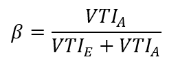
- 计算心房充盈分数(β),即心房充盈量与左心室总充盈量的比值:
- 在食管中段主动脉瓣长轴 TEE 视图中,于收缩中期测量主动脉瓣下方左心室流出道的最大直径(图2A)。
- 假设左心室流出道为圆形几何形状,其面积按 π/4 与直径平方的乘积计算(见上述步骤 2.5)。
- 获取深胃底长轴 TEE 视图,并在远端左心室流出道放置脉冲波多普勒取样容积,记录血流速度频谱包络(图2B),取样位置应与测量直径时相同(见上述步骤 2.5);使用超声心动图设备的软件对该波形进行积分,获得速度-时间积分(VTI)。
- 将左心室流出道血流速度波形的速度-时间积分(VTI)(图2B)乘以流出道面积(见步骤 2.6),计算每搏输出量(SV)。
- 分别记录食管中段双交界切面和左心室长轴 TEE 成像切面的视频片段42,确保每个片段包含多个心动周期。
- 在 ECG T 波之后,回放并目视检查视频片段(见上述步骤 2.9)的慢速图像,选择二尖瓣瓣叶最大开放时刻。
- 使用超声心动图设备的“卡尺”功能测量二尖瓣瓣叶之间的距离(图3A 和 3B)。
- 将二尖瓣直径(D)计算为长径(交界径,前外-后内方向)与短径(前后径)的平均值。
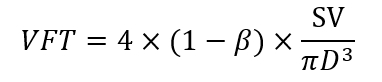
- 使用以下公式计算血流通过时间(VFT):
- 所有定量超声心动图测量均应在呼气末重复进行三次。
3. 实验设计
- 在10例接受冠状动脉手术且术前左心室射血分数正常的患者中,于体外循环(CPB)前30分钟及CPB后15、30和60分钟的稳态条件下,测定VFT、左心室舒张功能指数及血流动力学参数,以验证CPB会暂时降低VFT的假设39。
- 通过检测一组因主动脉瓣狭窄导致左心室压力超负荷肥厚的患者(共8例,接受主动脉瓣置换术),并与另一组左心室壁厚度正常的8例接受冠状动脉手术的患者进行比较,验证主动脉瓣狭窄引起的左心室压力超负荷肥厚会降低VFT的假设40。在CPB前30分钟的稳态条件下测定VFT、左心室舒张功能、血流动力学参数及舒张末期后壁厚度。
- 在8例伴有中度主动脉瓣反流的主动脉瓣狭窄患者与8例无反流瓣膜的主动脉瓣狭窄患者之间,检验异常的舒张期血流进入左心室是否影响经二尖瓣左心室充盈效率的假设38。VFT及其他参数的测定方法同上(步骤3.2)。
- 通过比较7例高龄患者(82 ± 2岁)与7例较年轻患者(55 ± 6岁)在接受冠状动脉手术时使用VFT量化评估的左心室充盈效率,检验高龄与左心室充盈效率下降相关的假设41。确保两组患者术前左心室射血分数均正常。VFT及其他参数的测定方法同上(步骤3.2)。
4. 统计学
- 以均值 ± 标准差形式表示数据。
- 使用方差分析(ANOVA)对数据进行评估,随后采用Student’s t检验的Bonferroni校正法进行多重比较 t测试。
- 采用线性回归分析确定VFT与舒张末期后壁厚度之间以及VFT与年龄之间的关系。
- 当 p 值小于显著性水平时,拒绝零假设。 < 0.05.
访问受限。请登录或开始试用以查看此内容。
结果
当前技术使我们能够通过在标准经食管超声心动图成像切面中获取血流和容积记录,从而在多种临床条件下可靠地测量心脏手术期间的瓣环位移(VFT)。在经食管中段四腔心切面中,将脉冲波多普勒取样容积置于二尖瓣瓣叶尖端,以获取计算心房充盈分数(β)所需的跨二尖瓣血流速度曲线; 图1)。通过连续性方程(左心室流出道血流速度时间积分乘以流出道面积)计算每搏输出量,左心室流出道直径在经食管中段左心室长轴切面测量。图2A),而流出道的血流则在深部经胃短轴成像平面测定(图2B)。最后,二尖瓣平均直径计算为食管中段双交界切面和左心室长轴切面测得的长轴与短轴直径的平均值(图3A 和 3B,分别)。VFT 测量的观察者内和观察者间变异分别为 5% 和 7%,与其他使用经食管超声心动图(TEE)测量的尺寸和血流指标相似(数据未显示)。...
访问受限。请登录或开始试用以查看此内容。
讨论
当前结果表明,使用本文所述的经食管超声心动图(TEE)技术,可在心脏手术期间可靠地测量左心室充盈分数(VFT)。既往对VFT的描述采用的是在清醒受试者中进行的经胸超声心动图检查,但当胸腔处于开放状态时,该方法无法应用。我们采用术中TEE来测定接受心脏手术的麻醉患者中的VFT,此类手术过程中常因缺血-再灌注损伤或外科干预而出现左心室(LV)充盈动力学的改变。我们的研究结果表明,VFT测量值能够反映由短暂体外循环(CPB)引起的舒张功能障碍(表现为松弛功能受损模式)、主动脉瓣疾病以及衰老所导致的左心室充盈效率变化。目前在心脏手术期间计算VFT的技术要求在稳定的血流动力学条件下获取高质量的TEE图像和视频片段,以确保对二尖瓣和左心室流出道的尺寸及血流进行精确测量(图1、图2 和 图3)。由于心脏存在偏轴旋转或心脏几何结构的病理性改变,并非所有患者都能获得理想的成像窗。尽管存在这些潜在限制,经验丰富的术中超声心动图医师在进行全面TEE检查时,应能够轻松获取必要的中食管...
访问受限。请登录或开始试用以查看此内容。
披露
作者声明,针对本研究不存在竞争性财务利益或其他利益冲突。
致谢
本材料是在威斯康星州密尔沃基市克莱门特·J·扎布洛基退伍军人事务医疗中心利用其资源和设施开展工作所取得的成果。
访问受限。请登录或开始试用以查看此内容。
材料
| 姓名 | 公司 | 目录编号 | 评论 |
|---|---|---|---|
| 超声心动图仪 | Philips Ultrasound, Bothall, WA | iE33 | |
| 经食管超声心动图探头 | Philips Ultrasound, Bothall, WA | X7-2t | |
| 统计分析软件 | AnalystSoft, Walnut, CA | StatPlus:mac Pro |
参考文献
- Collier, E., Hertzberg, J., Shandas, R. Regression analysis for vortex ring characteristics during left ventricular filling. Biomedical Sciences Instrumentation. 38 (2), 307-311 (2002).
- Kheradvar, A., Gharib, M. Influence of ventricular pressure drop on mitral annulus dynamics through the process of vortex ring formation. Annals of Biomedical Engineering. 35 (12), 2050-2064 (2007).
- Gharib, M., Rambod, E., Shariff, K. A universal time scale for vortex ring formation. Journal of Fluid Mechanics. 360 (1), 121-140 (1998).
- Krueger, P. S., Gharib, M. The significance of vortex ring formation to the impulse and thrust of a starting jet. Physics of Fluids. 15 (5), 1271-1281 (2003).
- Reul, H., Talukder, N., Muller, W. Fluid mechanics of the natural mitral valve. Journal of Biomechanics. 14 (5), 361-372 (1981).
- Kim, W. Y., et al. Two-dimensional mitral flow velocity profiles in pig models using epicardial Doppler echocardiography. Journal of the American College of Cardiology. 24 (2), 532-545 (1994).
- Kilner, P. J., et al. Asymmetic redirection of flow through the heart. Nature. 404 (6779), 759-761 (2000).
- Gharib, M., Rambod, E., Kheradvar, A., Sahn, D. J., Dabiri, J. O. Optimal vortex formation as an index of cardiac health. Proceedings of the National Academy of Sciences USA. 103 (16), 6305-6308 (2006).
- Rodriguez Munoz, D., et al. Intracardiac flow visualization: current status and future directions. European Heart Journal Cardiovascular Imaging. 14 (11), 1029-1038 (2013).
- Martinez-Legazpi, P., et al. Contribution of the diastolic vortex ring to left ventricular filling. Journal of the American College of Cardiology. 64 (16), 1711-1721 (2014).
- Dabiri, J. O., Gharib, M. The role of optimal vortex formation in biological fluid transport. Proceedings of the Royal Society B. 272 (1572), 1557-1560 (2003).
- Kheradvar, A., Gharib, M. On mitral valve dynamics and its connection to early diastolic flow. Annals of Biomedical Engineering. 37 (1), 1-13 (2009).
- Linden, P. F., Turner, J. S. The formation of "optimal" vortex rings, and the efficiency of propulsion devices. Journal of Fluid Mechanics. 427 (1), 61-72 (2001).
- Domenichini, F., Pedrizzetti, G., Baccani, B. Three-dimensional filling flow into a model left ventricle. Journal of Fluid Mechanics. 539 (1), 179-198 (2005).
- Sengupta, P. P., et al. Left ventricular isovolumic flow sequence during sinus and paced rhythms: new insights from use of high-resolution Doppler and ultrasonic digital particle imaging velocimetry. Journal of the American College of Cardiology. 49 (8), 899-908 (2007).
- Rodriguez Munoz, D., et al. Flow mapping inside a left ventricular aneurysm: a potential tool to demonstrate thrombogenicity. Echocardiography. 31 (1), E10-E12 (2014).
- Son, J. W., et al. Abnormal left ventricular vortex flow patterns in association with left ventricular apical thrombus formation in patients with anterior myocardial infarction: a quantitative analysis by contrast echocardiography. Circulation Journal. 76 (11), 2640-2646 (2012).
- Kheradvar, A., Falahatpisheh, A. The effects of dynamic saddle annulus and leaflet length on trans-mitral flow pattern and leaflet stress of a bileaflet bioprosthetic mitral valve. The Journal of Heart Valve Disease. 21 (2), 225-233 (2012).
- Kheradvar, A., Assadi, R., Falahatpisheh, A., Sengupta, P. P. Assessment of trans-mitral vortex formation in patients with diastolic dysfunction. Journal of the American Society of Echocardiography. 25 (2), 220-227 (2012).
- Chen, R., et al. Assessment of left ventricular hemodynamics and function of patients with uremia by vortex formation using vector flow mapping. Echocardiography. 29 (9), 1081-1090 (2012).
- Hendabadi, S., et al. Topology of blood transport in the human left ventricle by novel processing of Doppler echocardiography. Annals of Biomedical Engineering. 41 (12), 2603-2616 (2013).
- Sengupta, P. P., Pedrizetti, G., Narula, J. Multiplaner visualization of blood flow using echocardiographic particle imaging velocimetry. Journal of the American College of Cardiology Cardiovascular Imaging. 5 (5), 566-569 (2012).
- Sengupta, P. P., et al. Emerging trends in CV flow visualization. Journal of the American College of Cardiology Cardiovascular Imaging. 5 (3), 305-316 (2012).
- Hong, G. R., Kim, M., Pedrizzetti, G., Vannan, M. A. Current clinical application of intracardiac flow analysis using echocardiography. Journal of Cardiovascular Ultrasound. 21 (4), 155-162 (2013).
- Kheradvar, A., Milano, M., Gharib, M. Correlation between vortex ring formation and mitral annulus dynamics during ventricular rapid filling. American Society for Artificial Internal Organs Journal. 53 (1), 8-16 (2007).
- Hong, G. R., et al. Characterization and quantification of vortex flow in the human left ventricle by contrast echocardiography using vector particle image velocimetry. Journal of the American College of Cardiology Cardiovascular Imaging. 1 (6), 705-717 (2008).
- Zhang, H., et al. The evolution of intraventricular vortex during ejection studied by using vector flow mapping. Echocardiography. 30 (1), 27-36 (2013).
- Nogami, Y., et al. Abnormal early diastolic intraventricular flow 'kinetic energy index' assessed by vector flow mapping in patients with elevated filling pressure. European Heart Journal Cardiovascular Imaging. 14 (3), 253-260 (2013).
- Zhang, H., et al. The left ventricular intracavity vortex during the isovolumic contraction period as detected by vector flow mapping. Echocardiography. 29 (5), 579-587 (2012).
- Poh, K. K., et al. Left ventricular filling dynamics in heart failure: echocardiographic measurement and utilities of vortex formation time. European Heart Journal Cardiovascular Imaging. 13 (5), 385-393 (2012).
- Belohlavek, M. Vortex formation time: an emerging echocardiographic index of left ventricular filling efficiency? European Heart Journal Cardiovascular Imaging. 13 (5), 367-369 (2012).
- Dabiri, J. O., Gharib, M. Starting flow through nozzles with temporally variable exit diameter. Journal of Fluid Mechanics. 538 (1), 111-136 (2005).
- Jiamsripong, P., et al. Impact of acute moderate elevation in left ventricular afterload on diastolic trans-mitral flow efficiency: analysis by vortex formation time. Journal of the American Society of Echocardiography. 22 (4), 427-431 (2009).
- Belohlavek, M., et al. Patients with Alzheimer disease have altered trans-mitral flow: echocardiographic analysis of the vortex formation time. Journal of Ultrasound in Medicine. 28 (11), 1493-1500 (2009).
- Pedrizzetti, G., Domenichini, F., Tonti, G. On the left ventricular vortex reversal after mitral valve replacement. Annals of Biomedical Engineering. 38 (3), 769-773 (2010).
- Martinez-Legazpi, P., et al. Stasis mapping using ultrasound: a prospective study in acute myocardial infarction. Journal of the American College of Cardiology Cardiovascular Imaging. 11 (3), 514-515 (2018).
- Harfi, T. T., et al. The E-wave propagation index (EPI): a novel echocardiographic parameter for prediction of left ventricular thrombus. Derivation from computational fluid dynamic modeling and validation on human subjects. International Journal of Cardiology. 227 (1), 662-667 (2017).
- Pagel, P. S., Boettcher, B. T., De Vry, D. J., Freed, J. K., Iqbal, Z. Moderate aortic valvular insufficiency invalidates vortex formation time as an index of left ventricular filling efficiency in patients with severe degenerative calcific aortic stenosis undergoing aortic valve replacement. Journal of Cardiothoracic and Vascular Anesthesia. 30 (5), 1260-1265 (2016).
- Pagel, P. S., Gandhi, S. D., Iqbal, Z., Hudetz, J. A. Cardiopulmonary bypass transiently inhibits intraventricular vortex ring formation in patients undergoing coronary artery bypass graft surgery. Journal of Cardiothoracic and Vascular Anesthesia. 26 (3), 376-380 (2012).
- Pagel, P. S., Hudetz, J. A. Chronic pressure-overload hypertrophy attenuates vortex formation time in patients with severe aortic stenosis and preserved left ventricular systolic function undergoing aortic valve replacement. Journal of Cardiothoracic and Vascular Anesthesia. 27 (4), 660-664 (2013).
- Pagel, P. S., Dye, L., Boettcher, B. T., Freed, J. K. Advanced age attenuates left ventricular filling efficiency quantified using vortex formation time: a study of octogenarians with normal left ventricular systolic function undergoing coronary artery surgery. Journal of Cardiothoracic and Vascular Anesthesia. 32 (4), 1775-1779 (2018).
- Shanewise, J. S., et al. ASE/SCA guidelines for performing a comprehensive intraoperative multiplane transesophageal echocardiography examination: recommendations of the American Society of Echocardiography Council for Intraoperative Echocardiography and the Society of Cardiovascular Anesthesiologists Task Force for Certification in Perioperative Transesophageal Echocardiography. Journal of the American Society of Echocardiography. 12 (10), 884-900 (1999).
- Gaspar, T., et al. Three-dimensional imaging of the left ventricular outflow tract: impact on aortic valve area estimation by the continuity equation. Journal of the American Society of Echocardiography. 25 (7), 749-757 (2012).
- Karamnov, S., Burbano-Vera, N., Huang, C. C., Fox, J. A., Shernan, S. A. Echocardiographic assessment of mitral stenosis orifice area: a comparison of a novel three-dimensional method versus conventional techniques. Anesthesia and Analgesia. 125 (3), 774-780 (2017).
- Pagel, P. S., Kampine, J. P., Schmeling, W. T., Warltier, D. C. Comparison of end-systolic pressure-length relations and preload recruitable stroke work as indices of myocardial contractility in the conscious and anesthetized, chronically instrumented dog. Anesthesiology. 73 (2), 278-290 (1990).
- Pagel, P. S., Kampine, J. P., Schmeling, W. T., Warltier, D. C. Alteration of left ventricular diastolic function by desflurane, isoflurane, and halothane in the chronically instrumented dog with autonomic nervous system blockade. Anesthesiology. 74 (6), 1103-1114 (1991).
- De Hert, S. G., Rodrigus, I. E., Haenen, L. R., De Mulder, P. A., Gillebert, T. C. Recovery of systolic and diastolic left ventricular function early after cardiopulmonary bypass. Anesthesiology. 85 (5), 1063-1075 (1996).
- Gorcsan, J., Diana, P., Lee, J., Katz, W. E., Hattler, B. G. Reversible diastolic dysfunction after successful coronary artery bypass surgery. Assessment by transesophageal Doppler echocardiography. Chest. 106 (5), 1364-1369 (1994).
- Djaiani, G. N., et al. Mitral flow propagation velocity identifies patients with abnormal diastolic function during coronary artery bypass graft surgery. Anesthesia and Analgesia. 95 (3), 524-530 (2002).
- Casthely, P. A., et al. Left ventricular diastolic function after coronary artery bypass grafting: a correlative study with three different myocardial protection techniques. Journal of Thoracic and Cardiovascular Surgery. 114 (2), 254-260 (1997).
- Tulner, S. A., et al. Perioperative assessment of left ventricular function by pressure-volume loops using the conductance catheter method. Anesthesia and Analgesia. 97 (4), 950-957 (2003).
- Firstenberg, M. S., et al. Relationship between early diastolic intraventricular pressure gradients, an index of elastic recoil, and improvements in systolic and diastolic function. Circulation. 104 (12 Suppl 1), I330-I335 (2001).
- Cooke, J., Hertzberg, J., Boardman, M., Shandas, R. Characterizing vortex ring behavior during ventricular filling with Doppler echocardiography: an in vitro study. Annals of Biomedical Engineering. 32 (2), 245-256 (2004).
- Grossman, W., Jones, D., McLaurin, L. P. Wall stress and patterns of hypertrophy in the human left ventricle. Journal of Clinical Investigation. 56 (1), 56-64 (1975).
- Hess, O. M., et al. Diastolic function and myocardial structure in patients with myocardial hypertrophy. Special reference to normalized viscoelastic data. Circulation. 63 (2), 360-371 (1981).
- Hess, O. M., et al. Diastolic stiffness and myocardial structure in aortic valve disease before and after valve replacement. Circulation. 69 (5), 855-865 (1984).
- Sandstede, J. J. W., et al. Cardiac systolic rotation and contraction before and after valve replacement for aortic stenosis: a myocardial tagging study using MR imaging. American Journal of Roentgenology. 178 (4), 953-958 (2002).
- Stuber, M., et al. Alterations in the local myocardial motion pattern in patients suffering from pressure overload due to aortic stenosis. Circulation. 100 (4), 361-368 (1999).
- Nagel, E., et al. Cardiac rotation and relaxation in patients with aortic valve stenosis. European Heart Journal. 21 (7), 582-589 (2000).
- Rakowski, H., et al. Canadian consensus recommendations for the measurement and reporting of diastolic dysfunction by echocardiography: from the Investigators of Consensus on Diastolic Dysfunction by Echocardiography. Journal of the American Society of Echocardiography. 9 (5), 736-760 (1996).
- Homeyer, P., Oxorn, D. C. Aortic regurgitation: echocardiographic diagnosis. Anesthesia and Analgesia. 122 (1), 37-42 (2016).
- Landzberg, J. S., et al. Etiology of the Austin Flint murmur. Journal of the American College of Cardiology. 20 (2), 408-413 (1992).
- Flint, A. On cardiac murmurs. American Journal of Medical Sciences. 91 (1), 27(1886).
- Botvinick, E. H., Schiller, N. B., Wickramasekaran, R., Klausner, S. C., Gertz, E. Echocardiographic demonstration of early mitral valve closure in severe aortic insufficiency. Its clinical implications. Circulation. 51 (5), 836-847 (1975).
- Mann, T., McLaurin, L., Grossman, W., Craige, E. Assessing the hemodynamic severity of acute aortic regurgitation due to infective endocarditis. New England Journal of Medicine. 293 (3), 108-113 (1975).
- Borlaug, B. A., et al. Longitudinal changes in left ventricular stiffness: a community-based study. Circulation Heart Failure. 6 (5), 944-952 (2013).
- Wong, J., et al. Age-related changes in intraventricular kinetic energy: a physiological or pathological adaptation? American Journal of Physiology Heart Circulatory Physiology. 310 (6), H747-H755 (2016).
- Carrick-Ranson, G., et al. Effect of healthy aging on left ventricular relaxation and diastolic suction. American Journal of Physiology Heart Circulatory Physiology. 303 (3), H315-H322 (2012).
- Iskandrian, A. S., Hakki, A. H. Age-related changes in left ventricular diastolic performance. American Heart Journal. 112 (1), 75-78 (1986).
- Schulman, S. P., et al. Age-related decline in left ventricular filling at rest and exercise. American Journal of Physiology. 263 (6 Pt 2), H1932-H1938 (1992).
- Stork, M., et al. Age-related hemodynamic changes during diastole: a combined M-mode and Doppler echo study. Internal Journal of Cardiovascular Imaging. 6 (1), 23-30 (1991).
- Sanders, D., Dudley, M., Groban, L. Diastolic dysfunction, cardiovascular aging, and the anesthesiologist. Anesthesiology Clinics. 27 (3), 497-517 (2009).
访问受限。请登录或开始试用以查看此内容。
重印与许可
