本文介绍了用于诊断和评估系统性硬化症相关间质性肺疾病的胸部高分辨率计算机断层扫描的实用操作建议。
本文内容
摘要
摘要
系统性硬化症相关间质性肺病(SSc-ILD)的早期诊断至关重要,有助于尽早开始治疗,最大限度减少延误。然而,SSc-ILD的诊断具有挑战性,因其主要症状缺乏特异性。胸部高分辨率计算机断层扫描(HRCT)被公认为是一种诊断和评估SSc-ILD的敏感影像学方法。尽管患者暴露于电离辐射可能被视为一种局限性,但可通过采取适当的方法学措施加以控制。本文提出了HRCT扫描操作及结果解读的实用建议。SSc-ILD在HRCT上的主要特征包括非特异性间质性肺炎(NSIP)模式,表现为外周分布的磨玻璃样影和广泛的牵拉性支气管扩张。尽管SSc-ILD与特发性肺纤维化(IPF)存在相似之处,HRCT仍可用于区分这两种疾病:与IPF相比,SSc-ILD中磨玻璃样影的比例更高,纤维化表现较不粗糙。扩张、含气的食管,直径>10 mm,提示存在食管运动功能障碍,在SSc-ILD中常见。肺动脉直径大于相邻的升主动脉提示合并肺动脉高压。由于肺癌风险增加,需对肺内结节进行监测。HRCT上病变范围较大(≥20%)或纤维化评分较高提示死亡风险增加。HRCT在SSc-ILD的诊断中具有核心作用,连续的影像评估有助于监测疾病进展或治疗反应。
引言
系统性硬化症(SSc)是一种复杂的、异质性的自身免疫性疾病,可表现为血管病变、雷诺现象以及皮肤和内脏器官的纤维化1。SSc可分为以下亚型:局限性皮肤型、弥漫性皮肤型、无皮肤受累型(sine scleroderma)和重叠综合征型1。
系统性硬化症(SSc)并非以孟德尔方式遗传,但遗传因素似乎会影响个体对该疾病的易感性。不同种族群体之间的发病率存在差异,且有家族病史的个体发病率更高2,3。环境风险因素也可能存在,高浓度接触二氧化硅或有机溶剂似乎会增加SSc的发生风险4。全球范围内SSc的患病率约为万分之一1。女性患者多于男性,报道的女男比例在3:1至8:1之间,发病率最高的年龄组为45–54岁5。
肺是系统性硬化症(SSc)患者中第二常见的受累内脏器官6。SSc 的主要肺部表现有两种:间质性肺疾病(ILD)和肺动脉高压7。ILD 通常表现为纤维化,约80%的 SSc 患者会出现该病变,在弥漫性皮肤型硬皮病中的发生率高于局限性硬皮病1,8。肺动脉高压可表现为孤立性肺动脉高压(PAH,其在 SSc 患者中的患病率为13–35%),或由左心室受累/舒张功能障碍或 ILD/低氧血症引起的肺动脉高压7。SSc-ILD 患者与 SSc-PAH 患者的抗体谱存在差异。例如,抗 Scl-70 抗体的存在与 SSc-ILD 相关8,而抗着丝点抗体在合并 PAH 的 SSc 患者中比无 PAH 的患者更常见9。
系统性硬化症相关间质性肺病(SSc-ILD)的症状包括呼吸困难、咳嗽、胸痛和活动耐力下降。间质性肺病(ILD)是系统性硬化症患者发病的重要原因10,11,12。因此,据报道,SSc-ILD患者的年度全因医疗费用高于无ILD的SSc患者:分别为31,285–55,446美元与18,513–23,268美元13。
系统性硬化症相关间质性肺病(SSc-ILD)是系统性硬化症(SSc)患者死亡的首要原因,在该患者群体中占死亡原因的30–35%10,14。据报道,SSc-ILD患者的中位生存期为5–8年10,15;相比之下,约76%的SSc总体人群在疾病发病后可存活超过10年16。SSc-ILD死亡风险的重要预测因素包括年龄、用力肺活量(FVC)、一氧化碳弥散量(DLCO)基线值、高分辨率计算机断层扫描(HRCT)上肺部病变范围、肺动脉高压的存在以及肺部克雷布斯抗原6(Krebs von den Lungen 6, KL-6)的水平17,18。
早期诊断对于及时开展治疗至关重要,对于表型呈进展性的患者,还可能有助于延缓疾病进展。然而,系统性硬化症相关间质性肺病(SSc-ILD)的诊断具有挑战性,因为咳嗽、呼吸困难和疲劳等非特异性症状可能与其他系统性硬化症(SSc)表现(如心脏疾病和肌肉骨骼受累)相混淆。用于诊断间质性肺病(ILD)的评估手段包括:临床表现、病史、吸烟状况、肺功能、影像学检查,以及在某些情况下的肺活检。确诊SSc-ILD通常需要多项检查,并常采用多种方法联合评估19。其中最常用的评估方法包括肺功能检测和高分辨率计算机断层扫描(HRCT)20,21,22,23。其他影像学方法,如胸部X线摄影和减少辐射暴露的影像技术(例如磁共振成像[MRI]、肺部超声)也可能被采用22。肺功能检测可用于评估ILD的严重程度并监测其病程变化。然而,单独使用肺功能检测对诊断SSc-ILD的价值有限24,25。胸部高分辨率CT(HRCT)被认为是辅助SSc-ILD鉴别诊断最敏感的无创方法19。基线HRCT结果以及随时间的变化可用于预测肺部疾病的未来进展趋势及对治疗的潜在反应26。
高分辨率计算机断层扫描(HRCT)的辐射暴露有时被认为是常规筛查的一个限制因素27,28;减少扫描层数是降低辐射风险的一种潜在方法,此外还可通过降低电压或电流来减少辐射剂量29,30,31。也可考虑采用其他评估方法。例如,磁共振成像(MRI)在间质性肺疾病(ILD)患者的评估与随访中显示出一定潜力22。在一项使用呼吸同步T2加权MRI图像的研究中,同时以HRCT作为“金标准”进行评估,报道MRI在判断ILD是否存在时的敏感性为100%,特异性为60%32。另一项研究也报告了MRI与HRCT在ILD的检出和分类方面具有相似的一致性33。尽管结果令人鼓舞,但目前MRI仍处于研究阶段,尚未准备好广泛用于临床。
本文对影像学结果的解读提供了实用的概述,重点是高分辨率计算机断层扫描(HRCT),用于诊断系统性硬化症(SSc)中的肺部受累情况、判断预后,并探讨可能改进影像学方法及结果解读的未来发展方向。文中包含了代表性病例的HRCT图像。
方案
1. HRCT 扫描
- 进行胸部容积式高分辨率CT(HRCT)扫描36。无需使用对比剂36,37:
- 按照表136,37中所示参数获取以下扫描图像:
- 从肺尖至肺底进行仰卧位吸气相扫描(容积式)。
- 从肺尖下方2 cm处至肺底进行仰卧位呼气相扫描(序列式,层间距10–20 mm)。
- 可选俯卧位吸气相扫描(序列式,层间距10–20 mm),范围从隆突至肺底。
- 每次扫描前向患者说明呼吸指令36,37。进行吸气相扫描时,指导语为:“深吸一口气……然后呼出。再深吸一口气……然后呼出。再深吸一口气,屏住呼吸。继续保持屏气状态”37。
- 在完全吸气末获取吸气相扫描图像35,36。
- 采用最薄准直、最短旋转时间和最高螺距,以确保获得无运动伪影的图像36。推荐的扫描参数详见表137。
- 为获得最佳质量的容积扫描图像,应使用高空间分辨率重建方法获取薄层(<2 mm)图像35,36。
- 扫描完成后立即查看图像,若存在运动伪影或吸气不足,则需重新扫描37。
2. 报告
- 编写一份解读报告。
- 将报告和高分辨率CT图像与患者的医疗团队共享,并将其添加到患者的病历中。
结果
诊断
HRCT 上 SSc-ILD 的关键特征通常包括非特异性间质性肺炎(NSIP)模式,表现为外周分布的磨玻璃样影和广泛的牵拉性支气管扩张(图 1 和 图 2)。磨玻璃样影的病因广泛,常无特异性40,41,42。病变以中央为主,或呈外周分布但胸膜下区域相对保留,高度提示 NSIP(图 3)。
通常,HRCT图像中的ILD表现为网状影伴结构扭曲,导致牵拉性支气管扩张/细支气管扩张(符合纤维化型NSIP)。事实上,牵拉性支气管扩张和牵拉性细支气管扩张常为SSc-ILD的主要特征图4)43。其他可能的发现包括蜂窝样改变(图5;在局限性 SSc 中更常见),小叶间隔增厚和小叶内线状影,以及微小结节40,44蜂窝状改变是指成簇分布的囊性含气腔隙,其直径通常较为均一(约3–10 mm),囊壁增厚且边界清晰。31蜂窝状改变和牵拉性支气管扩张是普通型间质性肺炎(UIP)在高分辨率CT(HRCT)上的关键特征。尽管该影像模式最常与特发性肺纤维化(IPF)相关——IPF是具有进行性表型的典型纤维化性间质性肺病——但在系统性硬化相关间质性肺病(SSc-ILD)患者中有时也可观察到此类表现。10最近,在结缔组织病相关性间质性肺病(包括系统性硬化症相关性间质性肺病,SSc-ILD)且高分辨率CT(HRCT)表现为普通型间质性肺炎(UIP)模式的患者中,已识别出若干影像学征象,而这些征象在特发性肺纤维化(IPF)患者中未见出现。这些征象包括:直线边缘征(即肺纤维化局限于肺底部,在冠状位图像上沿头尾方向呈现清晰的边界,且在肺外侧缘无明显延伸)、蜂窝样改变为主(或显著)征>肺部70%的纤维化区域),以及上叶前部征象(即上叶前部区域纤维化集中,而上叶其他部分相对保留,同时伴有下叶受累)45直线征也与非特异性间质性肺炎(NSIP)的病理特征相关46,这是 SSc-ILD 的主要 CT 表现模式10.
食管扩张并充气在系统性硬化症(SSc)患者中常见(图6)47,48,49,也见于SSc-ILD患者47,48。尽管目前尚无公认的年龄上限用于判断食管扩张是否仍有助于区分SSc-ILD与特发性肺纤维化(IPF),但在65岁以上患者中,由于食管运动功能障碍的发生率逐渐增加,食管扩张的解读可能更为困难。纵隔淋巴结肿大(通常为反应性),即淋巴结短轴超过10 mm,在SSc-ILD患者中也常被观察到47,50。肺动脉直径大于邻近升主动脉提示可能合并肺动脉高压(图6),即使在无纤维化性肺部疾病的患者中亦如此51,52,53。实变区域提示可能存在重叠感染、误吸、机化性肺炎、出血或恶性肿瘤。由于SSc-ILD患者发生肺癌的风险增加,肺结节需密切监测7;在SSc-ILD患者中最常见的原发性肺癌类型为腺癌7,54。
系统性硬化症相关间质性肺病(SSc-ILD)在临床表现、发病机制和病理特征方面与特发性肺纤维化(IPF)存在诸多相似之处15,55。然而,某些影像学特征可用于区分这两种间质性肺病15,45。与IPF相比,SSc-ILD中磨玻璃样阴影的比例更高,且纤维化程度较轻、结构更细。在SSc患者出现普通型间质性肺炎(UIP)模式时,蜂窝状改变可能累及超过70%的纤维化肺组织——即显著蜂窝征56,57。此外,四角征(又称前上肺叶征)在SSc-ILD中的发生率显著高于IPF;该征象表现为炎症和/或纤维化病灶性或不成比例地累及双侧前外侧上叶及后上部下叶58。
胸部X线片可能初步发现间质性肺病(ILD);然而,其对比度分辨率不足以进行可靠诊断。在系统性硬化症相关间质性肺病(SSc-ILD)患者的胸部X线片中,最常见的表现为以肺基底部为主的网状阴影59。其他可能的影像特征还包括可见的支气管扩张、肺容积缩小和蜂窝样改变。与高分辨率CT(HRCT)类似,若发现扩张且含气的食管,可能有助于支持SSc-ILD的诊断47。
预后
已有多种不同的影像学表现被证实与系统性硬化症相关间质性肺病(SSc-ILD)的预后相关。在高分辨率CT(HRCT)上病变范围≥20%的患者,其死亡风险更高(病变范围高于与低于20%阈值的患者10年生存率分别为43%和67%)60。类似地,HRCT上的高纤维化评分(基于网状影和蜂窝样改变的范围)也与死亡率升高相关61。食管直径较大与间质性肺病(ILD)严重程度增加及一氧化碳弥散量(DLCO)降低相关48。肺密度和肺动脉直径可能用于预测肺动脉高压的风险62。计算机化的定量CT参数也可用于识别患者肺功能下降或死亡的风险。一项研究表明,通过HRCT定量评估的ILD范围可用于预测12个月内用力肺活量(FVC)的下降63。另一项研究发现,定量胸部CT参数提供的死亡风险评估结果与临床预测模型一致64。尽管这些影像学生物标志物显示出明显潜力,但目前其应用仍主要限于人群层面,因其在个体患者中的临床实用性尚未确立。
治疗反应
环磷酰胺和吗替麦考酚酯对 SSc-ILD 患者具有一定的治疗益处。在标志性的硬皮病肺研究 I(Scleroderma Lung Study I)中,与安慰剂相比,环磷酰胺治疗可延缓肺纤维化的进展速度65。最近,硬皮病肺研究 II(Scleroderma Lung Study II)报告表明,吗替麦考酚酯相较于环磷酰胺具有相似的疗效且耐受性更佳66。基于积极的 SENSCIS® 试验结果67,尼达尼布成为首个获得美国食品药品监督管理局(US Food and Drug Administration)批准用于减缓 SSc-ILD 患者肺功能下降速率的治疗药物。然而,SSc-ILD 患者的治疗选择仍有待进一步改善。目前正在研究的治疗方法包括单克隆抗体(如利妥昔单抗、abituzumab)、抗纤维化药物(如吡非尼酮)、直接凝血酶抑制剂达比加群、蛋白酶体抑制剂硼替佐米以及造血干细胞移植19,68。
连续高分辨率CT扫描显示系统性硬化症相关间质性肺病患者病情进展
在不同时间点进行的高分辨率CT评估可用于研究疾病的进展。 图7 显示一位 SSc-ILD 患者相隔 10 年的两组胸部高分辨率 CT 轴向和冠状位图像。初始的轴向和冠状位图像(图7A 和 B胸部高分辨率CT显示以肺基底部为主的磨玻璃影和网状影,伴有轻度牵拉性支气管扩张,胸膜下区相对 spared,符合该系统性硬化症(SSc)患者非特异性间质性肺炎(NSIP)的表现。后一组图像(图7C 和 D10 年后拍摄的图像显示,肺基底部网状结构增多及牵拉性支气管扩张,同时在轴位和冠状位上磨玻璃样阴影减少图7C 和 D胸部CT图像显示肺纤维化轻度加重。连续的高分辨率CT(HRCT)扫描也可用于监测治疗反应。69,70,71这在硬皮病肺研究II(Scleroderma Lung Study II)中得到了证实,该研究利用基于高分辨率CT(HRCT)扫描的计算机辅助诊断评分,比较了环磷酰胺与吗替麦考酚酯在系统性硬化症相关间质性肺病(SSc-ILD)患者中的疗效69.
| 相位 | 探测器 准直 | 电压 (kVp) | 电流 (mAs) | 扫描 间隔 | 螺距 | 旋转时间 | 管电流 调制 |
| 仰卧吸气相 | 螺旋 1.2 mm | 120(可降低) | 230(可降低) | 不适用 | ~1.0 | 0.5 秒或更快 | 开启 |
| 仰卧呼气相 | 轴向 2 × 1.0 mm | 120 | 150 | 20 mm | 不适用 | 1.0 秒 | 开启 |
| 俯卧吸气相 | 轴向 2 × 1.0 mm | 120 | 150 | 20 mm | 不适用 | 1.0 秒 | 开启 |
表1:计算机断层扫描采集参数37。 kVp = 千伏峰值;N/A = 不适用。
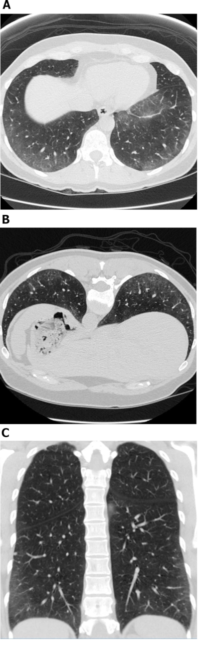
图1:系统性硬化症伴细胞性非特异性间质性肺炎(NSIP)模式。 轴向(A)、俯卧位(B)和冠状位(C)高分辨率计算机断层扫描图像均显示广泛的外周和以肺底部为主的磨玻璃样混浊影;这些表现是非特异性间质性肺炎(NSIP)的典型特征。缺乏牵拉性支气管扩张提示为细胞性NSIP模式。NSIP = 非特异性间质性肺炎。 请点击此处查看该图的放大版本。

图2:系统性硬化症伴纤维化型非特异性间质性肺炎表现。 轴位计算机断层扫描图像显示广泛、以肺底部为主的磨玻璃样影,并伴有牵拉性支气管扩张。值得注意的是,食管明显扩张;此表现为硬皮病的典型特征。请点击此处查看该图的放大版本。
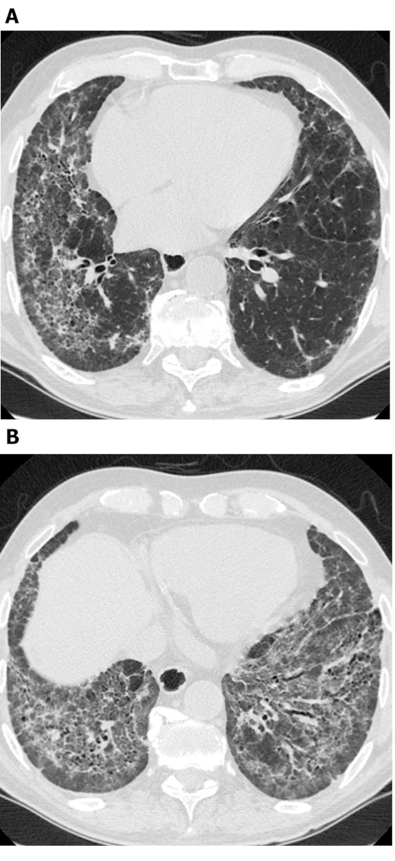
图3:系统性硬化症伴纤维化型非特异性间质性肺炎(NSIP)表现。 轴向高分辨率计算机断层扫描图像(A 和 B)显示广泛磨玻璃样影、网状影、结构扭曲及牵拉性支气管扩张。值得注意的是,可见胸膜下保留区域;此征象为非特异性间质性肺炎(NSIP)的典型特征,在约50%的病例中可见。NSIP = 非特异性间质性肺炎。 请点击此处查看该图的高清版本。
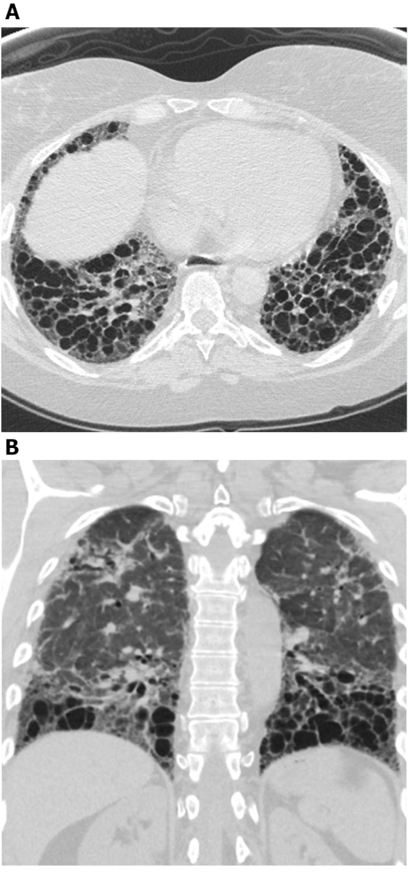
图4:伴有显著牵拉性支气管扩张的系统性硬化症。 轴向(A)和冠状面(B)高分辨率计算机断层扫描图像显示中肺和下肺区域为主的广泛性牵拉性支气管扩张。尽管此类表现可能被误认为蜂窝肺,但这些囊性区域相互连通,且未累及紧邻胸膜下的肺组织,此特征符合支气管扩张的表现。请点击此处查看该图的放大版本。

图5:系统性硬化症伴肺纤维化UIP型表现。 轴向(A)和冠状面(B)计算机断层扫描图像显示以外周和肺底分布为主的蜂窝样改变及牵拉性支气管扩张,符合典型的寻常型间质性肺炎(UIP)型肺纤维化表现。请注意扩张的食管(继发于硬皮病)以及“显著”的蜂窝样改变(提示间质性肺病与结缔组织病相关,而非特发性肺纤维化)。UIP = 寻常型间质性肺炎。 请点击此处查看该图的放大版本。
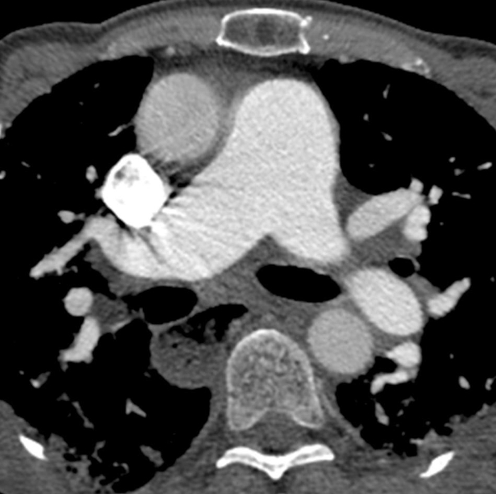
图6:系统性硬化症合并肺动脉高压及食管扩张。增强胸部计算机断层扫描显示肺动脉干明显增宽,其直径大于邻近的升主动脉,提示存在基础肺动脉高压。食管显著扩张,此表现归因于硬皮病。请点击此处查看该图的放大版本。
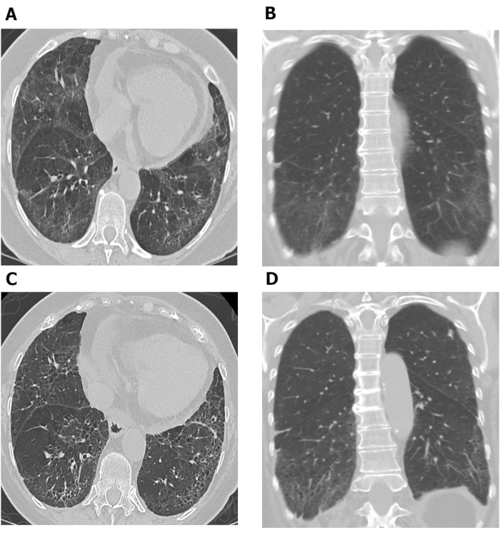
图7:系统性硬化症相关间质性肺病(SSc-ILD)患者肺纤维化进展的系列胸部高分辨率CT(HRCT)图像。 该SSc患者胸部高分辨率CT的轴向(A)和冠状面(B)图像显示以肺基底部为主的磨玻璃样影和网状影,伴有轻度牵拉性支气管扩张,胸膜下区域相对保留,符合非特异性间质性肺炎的表现。10年后,轴向(C)和冠状面(D)胸部高分辨率CT图像显示肺基底部网状影和牵拉性支气管扩张加重,磨玻璃样影减少,提示肺纤维化轻度进展。HRCT = high-resolution computed tomography;SSc-ILD = systemic scleroderma-associated interstitial lung disease。请点击此处查看本图的高清版本。
讨论
尽管高分辨率计算机断层扫描(HRCT)目前是诊断和评估系统性硬化症相关间质性肺病(SSc-ILD)的确定性影像学方法,但它使用电离辐射且相对昂贵。可改用胸部X线摄影,但其在鉴别诊断方面的效能不如HRCT,且正常的胸部X线结果并不能排除间质性肺病(ILD)的可能性。胸部X线摄影最适宜的应用可能是用于在HRCT检查间隔期间监测疾病是否进展,以及在症状急性加重时排除其他并发症(如感染性肺炎)。
HRCT 的一个被认为的局限性是辐射暴露。如前所述,新的CT扫描方法可能有助于降低辐射暴露31;此外,目前的CT扫描仪提供了一系列先进技术,未来有可能将辐射暴露降至接近胸部X线摄影的水平。 alternatively,未来的影像学方法如MRI或肺部超声可能被用于避免使患者暴露于辐射32,72,73,74。我们认为,尽管影像学应用存在风险与获益的权衡,但CT在诊断和患者管理方面的优势远远超过其潜在风险。
影像学数据,尤其是高分辨率计算机断层扫描(HRCT),可提供用于诊断系统性硬化症相关间质性肺病(SSc-ILD)的最重要信息。对HRCT扫描图像模式和特征的详细分析通常足以将SSc-ILD与其他肺部疾病区分开来,从而避免进行有创活检的需要。
高分辨率CT扫描的视觉评估具有一定的主观性,并可能导致观察者间变异。基于计算机的高分辨率CT扫描解读方法已被研究作为提高准确性的可能途径63,75。例如,可采用定量方法评估肺纤维化程度或疾病累及范围,以判断治疗反应69,71,76。然而,目前这些方法尚未在日常临床实践中广泛应用。
我们希望本文提供的信息能够作为实用指南,帮助医生利用高分辨率CT扫描诊断系统性硬化症相关间质性肺病(SSc-ILD)并判断预后。改进的影像获取方法和扫描解读方法有望减少患者接受的辐射暴露,并提高诊断和预后判断的准确性。
披露
Jonathan Chung 对本文章所述的研究、作者身份和/或出版不存在任何潜在的利益冲突;Christopher Walker 宣布从 Amirsys(Elsevier)获得版税;Stephen Hobbs 宣布从 Elsevier 和 Wolters Kluwer Health 获得版税。本文的开放获取出版由 Boehringer Ingelheim Pharmaceuticals, Inc. 赞助。
致谢
作者符合国际医学期刊编辑委员会(ICMJE)推荐的作者署名标准。作者未因本稿件的撰写而获得任何直接报酬。稿件的撰写协助由UDG Healthcare plc旗下的Ashfield公司GeoMed提供,由理学学士Ken Sutor负责,该公司由勃林格殷格翰制药有限公司(BIPI)签约并资助。BIPI有权对稿件进行审阅,以确保其医学和科学准确性,并考虑知识产权相关事宜。
作者在视频制作过程中未获得任何直接补偿。视频脚本的医学写作支持由Ashfield公司旗下的GeoMed公司提供,由Leon Newman博士负责,该公司由勃林格殷格翰制药有限公司(BIPI)签约并资助。BIPI有权对最终视频进行审阅,以确保其医学和科学准确性,并考虑知识产权相关事宜。
材料
| 姓名 | 公司 | 目录编号 | 评论 |
|---|---|---|---|
| CT 扫描仪 | Philips | NA | 多种 |
参考文献
- Denton, C. P., Khanna, D. Systemic sclerosis. Lancet. 390 (10103), 1685-1699 (2017).
- Arnett, F. C., et al. Familial occurrence frequencies and relative risks for systemic sclerosis (scleroderma) in three United States cohorts. Arthritis & Rheumatism. 44 (6), 1359-1362 (2001).
- Barnes, J., Mayes, M. D. Epidemiology of systemic sclerosis: incidence, prevalence, survival, risk factors, malignancy, and environmental triggers. Current Opinion in Rheumatology. 24 (2), 165-170 (2012).
- Marie, I., Gehanno, J. F. Environmental risk factors of systemic sclerosis. Seminars in Immunopathology. 37 (5), 463-473 (2015).
- Silman, A. J. Epidemiology of scleroderma. Annals of the Rheumatic Diseases. 50, Suppl 4 846-853 (1991).
- Scholand, M. B., et al. Interstitial lung disease in systemic sclerosis: diagnosis and management. Rheumatology. 1, 008(2012).
- Solomon, J. J., et al. Scleroderma lung disease. European Respiratory Review. 22 (127), 6-19 (2013).
- Walker, U. A., et al. Clinical risk assessment of organ manifestations in systemic sclerosis: a report from the EULAR Scleroderma Trials And Research group database. Annals of the Rheumatic Diseases. 66 (6), 754-763 (2007).
- Hinchcliff, M., Fischer, A., Schiopu, E., Steen, V. D., Investigators, P. Pulmonary hypertension assessment and recognition of outcomes in scleroderma (PHAROS): baseline characteristics and description of study population. The Journal of Rheumatology. 38 (10), 2172-2179 (2011).
- Giacomelli, R., et al. Interstitial lung disease in systemic sclerosis: current and future treatment. Rheumatology International. 37 (6), 853-863 (2017).
- Sanchez-Cano, D., et al. Interstitial lung disease in systemic sclerosis: data from the spanish scleroderma study group. Rheumatology International. 38 (3), 363-374 (2018).
- Silver, K. C., Silver, R. M. Management of systemic-sclerosis-associated interstitial lung disease. Rheumatic Diseases Clinics of North America. 41 (3), 439-457 (2015).
- Fischer, A., Kong, A. M., Swigris, J. J., Cole, A. L., Raimundo, K. All-cause healthcare costs and mortality in patients with systemic sclerosis with lung involvement. The Journal of Rheumatology. 45 (2), 235-241 (2018).
- Tyndall, A. J., et al. Causes and risk factors for death in systemic sclerosis: a study from the EULAR Scleroderma Trials and Research (EUSTAR) database. Annals of the Rheumatic Diseases. 69 (10), 1809-1815 (2010).
- Herzog, E. L., et al. Review: interstitial lung disease associated with systemic sclerosis and idiopathic pulmonary fibrosis: how similar and distinct. Arthritis & Rheumatology. 66 (8), 1967-1978 (2014).
- Rubio-Rivas, M., Royo, C., Simeon, C. P., Corbella, X., Fonollosa, V. Mortality and survival in systemic sclerosis: systematic review and meta-analysis. Seminars in Arthritis and Rheumatism. 44 (2), 208-219 (2014).
- Stock, C., et al. Serum KL-6 as a marker of disease progression in SSc-ILD. European Respiratory Journal. 52, Suppl 62, abstract PA3664 (2018).
- Winstone, T. A., et al. Predictors of mortality and progression in scleroderma-associated interstitial lung disease: a systematic review. Chest. 146 (2), 422-436 (2014).
- Khanna, D., et al. Ongoing clinical trials and treatment options for patients with systemic sclerosis-associated interstitial lung disease. Rheumatology (Oxford). 58 (4), 567-579 (2019).
- Behr, J., Furst, D. E. Pulmonary function tests. Rheumatology (Oxford). 47, Suppl 5 65-67 (2008).
- Hax, V., et al. Clinical algorithms for the diagnosis and prognosis of interstitial lung disease in systemic sclerosis. Seminars in Arthritis and Rheumatism. 47 (2), 228-234 (2017).
- Molberg, O., Hoffmann-Vold, A. M. Interstitial lung disease in systemic sclerosis: progress in screening and early diagnosis. Current Opinion in Rheumatology. 28 (6), 613-618 (2016).
- Raghu, G. Interstital lung disease. Goldman-Cecil Medicine. Goldman, L., Schafer, A. I. , Elsevier Science. Philadelphia, USA. 575-588 (2016).
- Showalter, K., et al. Performance of forced vital capacity and lung diffusion cutpoints for associated radiographic interstitial lung disease in systemic sclerosis. The Journal of Rheumatology. 45 (11), 1572-1576 (2018).
- Suliman, Y. A., et al. Brief report: pulmonary function tests: high rate of false-negative results in the early detection and screening of scleroderma-related interstitial lung disease. Arthritis & Rheumatology. 67 (12), 3256-3261 (2015).
- Roth, M. D., et al. Predicting treatment outcomes and responder subsets in scleroderma-related interstitial lung disease. Arthritis & Rheumatology. 63 (9), 2797-2808 (2011).
- Kalra, M. K., Maher, M. M., Rizzo, S., Kanarek, D., Shepard, J. A. Radiation exposure from chest CT: issues and strategies. Journal of Korean Medical Science. 19 (2), 159-166 (2004).
- Siegel, J. A., Pennington, C. W., Sacks, B., Welsh, J. S. The birth of the illegitimate linear no-threshold model: an invalid paradigm for estimating risk following low-dose radiation exposure. American Journal of Clinical Oncology. 41 (2), 173-177 (2018).
- Frauenfelder, T., et al. Screening for interstitial lung disease in systemic sclerosis: performance of high-resolution CT with limited number of slices: a prospective study. Annals of the Rheumatic Diseases. 73 (12), 2069-2073 (2014).
- Kubo, T., et al. Radiation dose reduction in chest CT: a review. American Journal of Roentgenology. 190 (2), 335-343 (2008).
- Nguyen-Kim, T. D. L., et al. The impact of slice-reduced computed tomography on histogram-based densitometry assessment of lung fibrosis in patients with systemic sclerosis. Journal of Thoracic Disease. 10 (4), 2142-2152 (2018).
- Muller, C. S., Warszawiak, D., Paiva, E. D. S., Escuissato, D. L. Pulmonary magnetic resonance imaging is similar to chest tomography in detecting inflammation in patients with systemic sclerosis. Revista Brasileira de Reumatologia English Edition. 57 (5), 419-424 (2017).
- Pinal-Fernandez, I., et al. Fast 1.5 T chest MRI for the assessment of interstitial lung disease extent secondary to systemic sclerosis. Clinical Rheumatology. 35 (9), 2339-2345 (2016).
- Sverzellati, N. Highlights of HRCT imaging in IPF. Respiratory Research. 14, Suppl 1 3(2013).
- Lynch, D. A., et al. Diagnostic criteria for idiopathic pulmonary fibrosis: a Fleischner Society White Paper. The Lancet Respiratory Medicine. 6 (2), 138-153 (2018).
- Raghu, G., et al. Diagnosis of idiopathic pulmonary fibrosis. An official ATS/ERS/JRS/ALAT clinical practice guideline. American Journal of Respiratory and Critical Care Medicine. 198 (5), 44-68 (2018).
- Exam: CT thorax for interstitial lung disease. , Available from: https://www.pulmonaryfibrosis.org/docs/default-source/medical-community-documents/ct-chest-for-ild-protocol.pdf?sfvrsn=0 (2019).
- Doyle, T. J., Hunninghake, G. M., Rosas, I. O. Subclinical interstitial lung disease: why you should care. American Journal of Respiratory and Critical Care Medicine. 185 (11), 1147-1153 (2012).
- Peroni, D. G., Boner, A. L. Atelectasis: mechanisms, diagnosis and management. Paediatric Respiratory Reviews. 1 (3), 274-278 (2000).
- Branley, H. M. Pulmonary fibrosis in systemic sclerosis: diagnosis and management. Respiratory Medicine CME. 3, 10-14 (2010).
- Engeler, C. E., Tashjian, J. H., Trenkner, S. W., Walsh, J. W. Ground-glass opacity of the lung parenchyma: a guide to analysis with high-resolution CT. American Journal of Roentgenology. 160 (2), 249-251 (1993).
- Goldin, J. G., et al. High-resolution CT scan findings in patients with symptomatic scleroderma-related interstitial lung disease. Chest. 134 (2), 358-367 (2008).
- Strollo, D., Goldin, J. Imaging lung disease in systemic sclerosis. Current Rheumatology Reports. 12 (2), 156-161 (2010).
- Pandey, A. K., et al. Significance of various pulmonary and extrapulmonary abnormalities on HRCT of the chest in scleroderma lung. Indian Journal of Radiology and Imaging. 23 (4), 304-307 (2013).
- Chung, J. H., et al. CT Features of the usual interstitial pneumonia pattern: differentiating connective tissue disease-associated interstitial lung disease from idiopathic pulmonary fibrosis. American Journal of Roentgenology. 210 (2), 307-313 (2018).
- Zhan, X., et al. Differentiating usual interstitial pneumonia from nonspecific interstitial pneumonia using high-resolution computed tomography: the "Straight-edge Sign". Journal of Thoracic Imaging. 33 (4), 266-270 (2018).
- Farrokh, D., Abbasi, B., Fallah-Rastegar, Y., Mirfeizi, Z. The extrapulmonary manifestations of systemic sclerosis on chest high resolution computed tomography. Tanaffos. 14 (3), 193-200 (2015).
- Salaffi, F., et al. Relationship between interstitial lung disease and oesophageal dilatation on chest high-resolution computed tomography in patients with systemic sclerosis: a cross-sectional study. La Radiologia Medica. 123 (9), 655-663 (2018).
- Vonk, M. C., et al. Oesophageal dilatation on high-resolution computed tomography scan of the lungs as a sign of scleroderma. Annals of the Rheumatic Diseases. 67 (9), 1317-1321 (2008).
- Chowaniec, M., Skoczynska, M., Sokolik, R., Wiland, P. Interstitial lung disease in systemic sclerosis: challenges in early diagnosis and management. Reumatologia. 56 (4), 249-254 (2018).
- McCall, R. K., Ravenel, J. G., Nietert, P. J., Granath, A., Silver, R. M. Relationship of main pulmonary artery diameter to pulmonary arterial pressure in scleroderma patients with and without interstitial fibrosis. Journal of Computer Assisted Tomography. 38 (2), 163-168 (2014).
- Pandey, A. K., et al. Predictors of pulmonary hypertension on high-resolution computed tomography of the chest in systemic sclerosis: a retrospective analysis. Canadian Association of Radiologists Journal. 61 (5), 291-296 (2010).
- Raymond, T. E., Khabbaza, J. E., Yadav, R., Tonelli, A. R. Significance of main pulmonary artery dilation on imaging studies. Annals of the American Thoracic Society. 11 (10), 1623-1632 (2014).
- Colaci, M., et al. Lung cancer in scleroderma: results from an Italian rheumatologic center and review of the literature. Autoimmunity Reviews. 12 (3), 374-379 (2013).
- Distler, O., et al. Design of a randomised, placebo-controlled clinical trial of nintedanib in patients with systemic sclerosis-associated interstitial lung disease (SENSCIS). Clinical and Experimental Rheumatology. 35 (4), Suppl 106 75-81 (2017).
- Desai, S. R., et al. CT features of lung disease in patients with systemic sclerosis: comparison with idiopathic pulmonary fibrosis and nonspecific interstitial pneumonia. Radiology. 232 (2), 560-567 (2004).
- Mira-Avendano, I., et al. Interstitial lung disease and other pulmonary manifestations in connective tissue diseases. Mayo Clinic Proceedings. 94 (2), 309-325 (2019).
- Walkoff, L., White, D. B., Chung, J. H., Asante, D., Cox, C. W. The four corners sign: a specific imaging feature in differentiating systemic sclerosis-related interstitial lung disease from idiopathic pulmonary fibrosis. Journal of Thoracic Imaging. 33 (3), 197-203 (2018).
- Kotnur, M. R., Suresh, P., Reddy, V. S., Sharma, T., Salim, N. A. Systemic sclerosis with multiple pulmonary manifestations. Journal of Clinical & Diagnostic Research. 10 (6), 16-17 (2016).
- Goh, N. S., et al. Interstitial lung disease in systemic sclerosis: a simple staging system. American Journal of Respiratory and Critical Care Medicine. 177 (11), 1248-1254 (2008).
- Takei, R., et al. Radiographic fibrosis score predicts survival in systemic sclerosis-associated interstitial lung disease. Respirology. 23 (4), 385-391 (2018).
- Bakker, M. E., et al. Lung density and pulmonary artery diameter are predictors of pulmonary hypertension in systemic sclerosis. Journal of Thoracic Imaging. 32 (6), 391-397 (2017).
- Khanna, D., et al. Predictors of lung function decline in scleroderma-related interstitial lung disease based on high-resolution computed tomography: implications for cohort enrichment in systemic sclerosis-associated interstitial lung disease trials. Arthritis Research & Therapy. 17, 372(2015).
- Ariani, A., et al. Quantitative chest computed tomography is associated with two prediction models of mortality in interstitial lung disease related to systemic sclerosis. Rheumatology (Oxford). 56 (6), 922-927 (2017).
- Goldin, J., et al. Treatment of scleroderma-interstitial lung disease with cyclophosphamide is associated with less progressive fibrosis on serial thoracic high-resolution CT scan than placebo: findings from the scleroderma lung study. Chest. 136 (5), 1333-1340 (2009).
- Tashkin, D. P., et al. Mycophenolate mofetil versus oral cyclophosphamide in scleroderma-related interstitial lung disease (SLS II): a randomised controlled, double-blind, parallel group trial. The Lancet Respiratory Medicine. 4 (9), 708-719 (2016).
- Distler, O., et al. Nintedanib for Systemic Sclerosis-Associated Interstitial Lung Disease. New England Journal of Medicine. 380, 2518-2528 (2019).
- Cappelli, S., et al. Interstitial lung disease in systemic sclerosis: where do we stand. European Respiratory Review. 24 (137), 411-419 (2015).
- Goldin, J. G., et al. Longitudinal changes in quantitative interstitial lung disease on CT after immunosuppression in the Scleroderma Lung Study II. Annals of the American Thoracic Society. 5 (11), 1286-1295 (2018).
- Wangkaew, S., Euathrongchit, J., Wattanawittawas, P., Kasitanon, N. Correlation of delta high-resolution computed tomography (HRCT) score with delta clinical variables in early systemic sclerosis (SSc) patients. Quantitative Imaging in Medicine and Surgery. 6 (4), 381-390 (2016).
- Kim, H. J., et al. Transitions to different patterns of interstitial lung disease in scleroderma with and without treatment. Annals of the Rheumatic Diseases. 75 (7), 1367-1371 (2016).
- Tardella, M., et al. Ultrasound B-lines in the evaluation of interstitial lung disease in patients with systemic sclerosis: cut-off point definition for the presence of significant pulmonary fibrosis. Medicine (Baltimore). 97 (18), 0566(2018).
- Hassan, R. I., et al. Lung ultrasound as a screening method for interstitial lung disease in patients with systemic sclerosis. Journal of Clinical Rheumatology. , (2018).
- Wang, Y., Gargani, L., Barskova, T., Furst, D. E., Cerinic, M. M. Usefulness of lung ultrasound B-lines in connective tissue disease-associated interstitial lung disease: a literature review. Arthritis Research & Therapy. 19 (1), 206(2017).
- Ariani, A., et al. Quantitative CT indexes are significantly associated with exercise oxygen desaturation in interstitial lung disease related to systemic sclerosis. The Clinical Respiratory Journal. 11 (6), 983-989 (2017).
- Kim, H. J., et al. Quantitative texture-based assessment of one-year changes in fibrotic reticular patterns on HRCT in scleroderma lung disease treated with oral cyclophosphamide. European Radiology. 21 (12), 2455-2465 (2011).
重印与许可
