我们开发了一种简单且低成本的方案,用于将Cas9/单导向RNA(sgRNA)核糖核蛋白复合物装载到病毒样颗粒中。这些被称为“Nanoblades”的颗粒能够高效地将Cas9/sgRNA复合物递送至永生化细胞、原代细胞以及体内环境。
本文内容
摘要
摘要
成簇规律间隔短回文重复序列(CRISPR)-Cas系统已使真核细胞的基因组编辑实现普及化,并推动了众多创新应用的发展。然而,将Cas9蛋白和单导向RNA(sgRNA)递送至靶细胞在技术上仍具挑战性。经典的病毒载体,例如源自慢病毒(LVs)或腺相关病毒(AAVs)的载体,能够在多种原代细胞及体内实现编码Cas9蛋白及其相关sgRNA的转基因的高效递送。尽管如此,这些载体仍存在一些缺陷,例如转基因在靶细胞基因组中的整合、装载容量有限,以及Cas9蛋白和导向RNA在靶细胞中长期表达等问题。
为克服上述部分问题,研究人员开发了一种基于小鼠白血病病毒(MLV)的递送载体,可在不包含任何编码转基因的情况下,将Cas9蛋白及其相关的向导RNA包装递送。通过将Cas9蛋白与MLV结构蛋白Gag的C末端融合,形成了装载Cas9蛋白和sgRNA的病毒样颗粒(VLPs),被称为"Nanoblades"。Nanoblades可从生产细胞的培养基中收集,经纯化、定量后用于转导靶细胞,递送具有活性的Cas9/sgRNA复合物。Nanoblades能够在多种原代细胞和永生化细胞中瞬时且快速地递送其核糖核蛋白(RNP)货物,并可通过使用修饰的Cas9蛋白,编程实现其他应用,例如靶向基因的瞬时转录激活。Nanoblades能够在注射后的成年小鼠肝脏以及卵母细胞中实现体内基因组编辑,从而生成转基因动物。最后,它们还可与供体DNA形成复合物,实现"无需转染"的同源定向修复。Nanoblades的制备过程简单、成本相对较低,可在任何细胞生物学实验室中轻松完成。
引言
与其他可编程核酸酶相比,CRISPR-Cas 系统极大地简化并普及了真核细胞中序列特异性的基因组靶向与切割操作。通过简单表达一条单导向RNA(sgRNA),用户即可将Cas9蛋白(或其优化变体)编程至几乎任意的细胞基因位点1。在此情况下,Cas9蛋白与sgRNA的递送成为实现定点诱变的主要限制因素。在永生化细胞中,sgRNA和Cas蛋白可通过转染质粒实现高效表达,从而在大多数细胞中完成有效的基因组靶向。然而,Cas9/sgRNA复合物的持续表达可能增加Cas9蛋白的脱靶活性,导致非特异性位点发生非预期的遗传改变2。在原代细胞中,DNA转染在技术上往往难以实现,可能导致表达水平低下或转染细胞比例过低。除经典DNA转染外,替代方法包括使用病毒载体递送编码Cas9和sgRNA的转基因,或通过电穿孔将重组Cas9蛋白与合成sgRNA共同导入细胞。然而,这些方法可能导致转基因整合至宿主细胞基因组(如经典的逆转录病毒和慢病毒表达载体),受到细胞内因子的限制,并引发Cas9蛋白和sgRNA的持续表达。
通过电穿孔将Cas9/sgRNA核糖核蛋白复合物导入细胞,可克服上述大多数问题,实现原代细胞及体内高效且瞬时的递送,并允许产生剂量依赖性反应。然而,该方法通常依赖昂贵的设备和试剂,且当需要处理大量细胞时,难以实现规模化扩增。作为上述技术的替代方案,这些研究人员开发了"Nanoblades"——一种用于递送Cas9蛋白和sgRNA的逆转录病毒载体3,其概念与其他基于病毒的衣壳蛋白递送系统相似4,5,6,7,8。Nanoblades利用逆转录病毒Gag多蛋白在培养细胞中单独表达时,能够自发形成并释放至细胞外培养基中的病毒样颗粒(VLPs)这一天然特性9。通过将Cas9蛋白与小鼠白血病病毒(MLV)Gag多蛋白的C末端融合,并共表达sgRNA和病毒包膜糖蛋白,可将Cas9蛋白包裹于释放的VLPs(即Nanoblades)中。经纯化后,Nanoblades可与靶细胞共孵育,或直接注射至体内,从而实现Cas9/sgRNA核糖核蛋白复合物的快速、瞬时且具有剂量依赖性的递送3。
Nanoblade 可通过多个 sgRNA 进行编程,以实现不同位点的同步编辑,或利用 Cas9 变体执行其他应用,例如靶向特异性的转录激活或抑制3。与依赖重组表达的蛋白质电穿孔技术相比,文献中新描述的 Cas 变体可轻松克隆至 Gag 融合表达载体中,使其成为一个多功能的平台。Nanoblade 还可进一步与单链或双链寡脱氧核苷酸(ssODN)形成复合物或负载此类分子,以实现同源定向修复3。Nanoblade 的制备相对简单且成本较低。此外,Nanoblade 可在 4 °C 下保存多天,或在 -80 °C 下长期保存。通常情况下,Nanoblade 可在大多数永生化细胞和原代培养细胞中实现高效、无转基因的基因组编辑。然而,某些原代细胞可能对病毒颗粒的存在较为敏感,导致细胞死亡率升高。先天免疫系统的细胞也可能因 Nanoblade 具有病毒来源而对其产生反应并被激活。在此类情况下,必须优化转导方案,以缩短细胞与 Nanoblade 的接触时间,并将非特异性效应降至最低。Nanoblade 是现有 CRISPR 递送方法的一种切实可行且易于实施的替代方案。
方案
1. sgRNA 设计与克隆
注意:sgRNA 的设计指南可从多个来源获取,例如 https://blog.addgene.org/how-to-design-your-grna-for-crispr-genome-editing 或来自 Hanna 和 Doench10.
- 设计好20个核苷酸的sgRNA序列后,订购以下单链DNA寡核苷酸:
- 正向:5' caccgNNNNNNNNNNNNNNNNNNNN 3'(N对应靶向位点序列,不含原间隔序列邻近基序(PAM)序列)
- 反向:5' aaacNNNNNNNNNNNNNNNNNNNNc 3'(N对应靶向位点反向互补序列,不含PAM序列)
注意:订购寡核苷酸时无需特殊修饰(无需5'磷酸化)。
- 在0.2 mL聚合酶链式反应(PCR)管中,将两条DNA寡核苷酸退火,反应体系包括:5 µL退火缓冲液(500 mM NaCl;100 mM Tris-HCl;100 mM MgCl₂;10 mM DTT;pH 7.9,25 °C),每条DNA寡核苷酸各1 µL(100 µM水溶液),以及42 µL水。
- 在PCR仪中,将样品于95 °C孵育15秒,然后以0.5 °C/s的降温速率降至20 °C。孵育完成后可置于室温或-20 °C保存。
注意:本步骤可暂停操作。 - 在50 µL总反应体系中,使用10单位BsmBI-v2限制性内切酶在55 °C消化10 µg BLADE或SUPERBLADE sgRNA表达质粒,反应时间3小时。
注意:经酶切的载体应释放出约1.9 kb的DNA插入片段和另一段约3.3 kb的DNA片段。 - 将限制性酶切产物上样至含5 µg/mL溴化乙锭(或更安全的替代性DNA凝胶染料)的1%琼脂糖凝胶中。
注意:操作溴化乙锭时应佩戴适当的防护装备,因其可能引起遗传缺陷。- 在波长312 nm的紫外(UV)灯台上(以避免DNA损伤),切下凝胶中的3.3 kb DNA片段,并将其放入1.5 mL微量离心管中。
注意:操作溴化乙锭及在紫外灯台上工作时,应佩戴适当的防护装备(手套和防紫外线护目镜)。 - 使用专用的DNA凝胶回收试剂盒(见材料表)从含有3.3 kb DNA片段的凝胶切片中提取DNA。使用分光光度计测定纯化后DNA的浓度。
注意:本步骤可暂停操作。
- 在波长312 nm的紫外(UV)灯台上(以避免DNA损伤),切下凝胶中的3.3 kb DNA片段,并将其放入1.5 mL微量离心管中。
- 将步骤1.2中退火形成的正向与反向DNA寡核苷酸双链连接至经BsmBI酶切并经凝胶纯化的BLADE或SUPERBLADE载体(来自步骤1.5.2)。连接反应体系如下:加入2 µL T4 DNA连接酶缓冲液、50 ng凝胶纯化的载体(来自步骤1.5.2)、1 µL退火后的DNA寡核苷酸(来自步骤1.2)、加水至总体积19 µL,最后加入1 µL T4 DNA连接酶。于25 °C孵育反应10分钟。
- 将连接产物转化至感受态细菌中(见材料表),具体操作参见文献11。将转化后的细菌涂布于氨苄青霉素Luria Bertani琼脂平板,37 °C过夜培养。
- 从琼脂平板上挑选数个独立菌落进行质粒小提11(见材料表),并使用U6正向引物(5' GACTATCATATGCTTACCGT 3')进行Sanger测序,以确认sgRNA可变序列的正确连接。
注意:可使用其他不编码Cas9蛋白的sgRNA表达质粒,以避免干扰Nanoblade的制备。
2. 质粒制备
- 对所有必需的质粒进行大提(参见材料表),并将其以1 µg/mL的浓度分装为10 µg每份,于-20 °C保存。避免质粒反复冻融;每份分装物最多使用两次后即弃用。
3. Nanoblade 的制备
- 第1天,在10 cm细胞培养皿中接种3.5至4 × 10⁶个HEK293T细胞(参见材料表),加入10 mL含高糖、丙酮酸钠、L-谷氨酰胺、10%胎牛血清(FBS)及青霉素/链霉素的Dulbecco改良Eagle培养基(DMEM)。轻轻前后移动培养皿,再左右移动(重复此操作5次),使细胞在培养皿内均匀分布。将细胞置于37 °C、5% CO2的细胞培养箱中孵育。
注意:所有涉及培养细胞和Nanoblades的操作均应在细胞培养超净工作台内进行,以避免污染。 - 第2天:质粒转染
- 接种24小时后,细胞应达到70–80%融合度(图1A)。转染前,更换为10 mL新鲜DMEM培养基(含高糖、丙酮酸钠、L-谷氨酰胺和10% FBS;青霉素和链霉素可省略,但非必须)。
注意:此步骤中细胞不应完全融合,否则可能降低转染效率和颗粒产量。 - 为每个10 cm培养皿,在1.5 mL离心管中准备以下质粒量:0.3 µg pCMV-VSV-G、0.7 µg pBaEVRless、2.7 µg MLV Gag/Pol、1.7 µg BIC-Gag-Cas9、4.4 µg编码克隆sgRNA的BLADES或SUPERBLADES质粒(若使用两个sgRNA,则各2.2 µg)。
- 加入500 µL转染缓冲液(参见材料表),涡旋10秒,然后离心1秒。加入20 µL转染试剂(参见材料表),涡旋1秒,再离心1秒。
- 室温孵育10分钟,使用P1000移液器将全部溶液逐滴加入含DMEM的细胞中。轻轻前后移动10 cm培养皿,再左右移动(重复5次),使转染试剂均匀分布于细胞表面。将细胞在37 °C、5% CO2的细胞培养箱中孵育至少40小时。
注意:如有需要,可在转染后4小时更换培养基。
- 接种24小时后,细胞应达到70–80%融合度(图1A)。转染前,更换为10 mL新鲜DMEM培养基(含高糖、丙酮酸钠、L-谷氨酰胺和10% FBS;青霉素和链霉素可省略,但非必须)。
- 第3天,在显微镜下观察转染细胞的形态。
注意:生产细胞将开始发生融合。这是由于融合性病毒包膜蛋白表达所致,属于正常现象(图1B、C)。 - 第4天:收集Nanoblades
注意:转染至少40小时后,细胞会因融合性病毒包膜蛋白的表达而聚集融合,有时甚至完全从培养皿表面脱落(图1D)。- 使用10 mL移液管收集9 mL培养上清液。
注意:Nanoblades是病毒样颗粒(VLPs),可将Cas9蛋白及其相关sgRNA递送至原代细胞及体内。尽管其不含遗传物质,不被视为转基因生物,但仍可诱导基因改变。因此,操作时必须谨慎,避免与操作者接触(尤其是当其被设计用于靶向抑癌基因时)。建议遵循当地关于逆转录病毒载体操作的安全规范,并在生物安全二级(BSL-2)实验室中进行VLP制备和转导实验。Nanoblades可用70%乙醇或0.5%次氯酸钠灭活。建议将所有塑料废弃物(移液枪头、组织培养皿、离心管等)用0.5%次氯酸钠处理至少10分钟,以灭活Nanoblades。 - 将收集的上清液在500 × g条件下离心5分钟,去除细胞碎片,并小心回收上清液,避免扰动细胞沉淀。
注意:若Nanoblades将用于原代细胞,可使用0.45 µm或0.8 µm滤膜过滤上清液。需注意,此步骤会显著降低Nanoblades滴度,因大量颗粒会被滤膜截留。 - 在4 °C条件下,使用摆桶转子以4,300 × g离心过夜(12–16小时),或使用超速离心机以209,490 × g离心75分钟,沉淀Nanoblades(参见材料表)。
注意:若靶细胞可在DMEM中生长,则可在步骤3.4.2后直接使用上清液进行孵育,无需浓缩Nanoblades。
- 使用10 mL移液管收集9 mL培养上清液。
- 第5天:Nanoblades的重悬与保存
- 离心后,缓慢吸除上清液,用100 µL预冷的1×磷酸盐缓冲液(PBS)重悬白色沉淀。用封口膜封住管口,在4 °C轻柔振荡孵育1小时,然后通过上下吹打使沉淀充分重悬。
注意:重悬时可能出现白色粘稠物质,此为正常现象,不会显著影响转导效率。 - 若计划在四周内使用Nanoblades,可将其保存于4 °C。否则,应使用液氮速冻后于-80 °C保存。
注意:操作液氮时需佩戴护目镜和低温手套。速冻及-80 °C保存会导致Nanoblades活性显著下降。此外,解冻后的Nanoblades不应再次冷冻。本方案可在此处暂停。
- 离心后,缓慢吸除上清液,用100 µL预冷的1×磷酸盐缓冲液(PBS)重悬白色沉淀。用封口膜封住管口,在4 °C轻柔振荡孵育1小时,然后通过上下吹打使沉淀充分重悬。
4. 在蔗糖垫上浓缩Nanoblades
注意:作为过夜离心或超速离心(步骤 3.4.3)的替代方法,可使用蔗糖垫对Nanoblades进行浓缩。该方法可获得纯度更高的Nanoblades组分,但回收总量会较低。
- 在1× PBS中配制10%(质量/体积)的蔗糖溶液,并通过0.2 µm针头滤器过滤(参见材料表)。
- 开始在蔗糖垫上浓缩Nanoblades。
- 将9 mL含病毒样颗粒(VLP)的样品(来自步骤3.4.3)加入超速离心管中(参见材料表)。使用3 mL注射器和套管,将2.5 mL 10%蔗糖溶液缓慢铺加在样品下方,注意避免混合含VLP的样品与蔗糖溶液。
- 或者,先将2.5 mL 10%蔗糖溶液加入超速离心管中(参见材料表)。倾斜离心管,使用低速移液器缓慢加入9 mL含VLP的样品(来自步骤3.4.3),并在操作过程中逐渐将离心管扶至垂直位置。
- 在4 °C条件下,于超速离心机中以209,490 × g离心90分钟。
注:该技术可根据文献12所述,改为在4,300 × g条件下低速离心过夜。 - 离心后,小心移除上清液,将离心管倒置在纸巾上以去除残留液体。1分钟后,加入100 µL 1× PBS,将离心管置于4 °C,在振荡摇床上的离心管架中加盖封口膜,孵育1小时(参见材料表),然后通过上下吹打重悬沉淀。
注:本实验流程可在此处暂停。
5. 通过点印迹法监测Nanoblade中的Cas9装载情况
- 通过将1体积含有非离子型表面活性剂的裂解缓冲液(参见材料表)加入4体积的1×PBS中,配制稀释缓冲液。取2 µL浓缩的Nanoblades加入50 µL稀释缓冲液中,短暂涡旋混匀,然后将25 µL该混合液转移至另一新管中,其中已含有25 µL稀释缓冲液。重复此操作,共制备4管Nanoblades稀释液(每步为2倍稀释)。
- 对于标准对照,将2 µL重组Cas9核酸酶(参见 材料表)加入50 µL稀释缓冲液中,短暂涡旋混匀,随后进行八次连续稀释(每步为2倍稀释)。
- 使用多通道移液器小心地将每种VLP稀释液各2.5 µL以及每种标准品各2.5 µL点样于硝酸纤维素膜上(体积过大可能导致斑点重叠)。
注意:也可使用经甲醇处理的聚偏二氟乙烯膜(PVDF膜)。 - 待颗粒完全吸附至膜上后,用含非离子型表面活性剂(TBS-T)并补充5%(w/v)脱脂奶粉的1×Tris缓冲盐水在室温下封闭膜45分钟。
注意:此处可暂停实验流程,将膜置于4 °C的1×TBS-T中保存。 - 弃去含脱脂奶粉的1×TBST,将膜在4 °C下与Cas9-辣根过氧化物酶抗体(按1/1000比例溶于1×TBST并含5%脱脂奶粉)孵育过夜。用TBS-T洗涤膜3次,然后使用增强型化学发光底物试剂盒显影信号。
- 使用凝胶成像系统配套的专用软件或ImageJ13对Nanoblades及重组Cas9标准品稀释系列的斑点强度进行定量分析。建立斑点强度与Cas9浓度之间的线性曲线关系。根据所得曲线的函数关系,推算各制备样品中的Cas9含量。
注意:重组Cas9蛋白对照在标准稀释系列中最浓样品中可能导致信号饱和(图2)。因此,在定义线性曲线时,若未稀释样品(有时还包括前几步稀释样品)的读数不在已知Cas9浓度的线性范围内,则应将其剔除。同样,在推算Nanoblades样品中Cas9含量时,仅使用处于标准曲线线性范围内的读数。
6. 使用Nanoblades转导靶细胞(12孔板转导操作步骤)
- 在12孔板中,每孔接种100,000–200,000个细胞(原代细胞或永生化贴壁细胞),加入1 mL适当的细胞培养基。在转导前,让细胞充分贴附于孔板表面。
- 在1.5 mL微量离心管中,加入5–20 µL浓缩的Nanoblades(来自步骤3.5.1或4.4),与500 µL细胞培养基混合,使用P1000移液器上下吹打混匀。吸除细胞培养液,替换为500 µL上述Nanoblades混合液。
注意:每种细胞类型的转导条件均需优化。应使用尽可能小的培养基体积(同时避免目标细胞干燥),以保持Nanoblades的高浓度。贴壁细胞必须在贴附于孔板的状态下直接进行转导(切勿在悬浮状态下转导,否则将显著降低转导效率)。某些细胞可耐受较长时间的Nanoblades暴露(24–48小时),而另一些细胞则较为敏感,可能出现小合胞体。在此情况下,Nanoblades与细胞共孵育时间应控制在4–6小时,随后更换培养基。对于悬浮培养的细胞,离心增强转导(spinoculation)14也可提高转导效率。某些细胞类型中,阳离子聚合物等辅助剂(参见材料表)亦可提升转导效率。 - 细胞在含Nanoblades的少量培养基中孵育4–6小时后,将培养基体积补充至正常量(若使用12孔板,则补至1 mL),或在细胞对病毒样颗粒(VLPs)敏感时更换为新鲜培养基。
注意:含Nanoblades的细胞培养基在丢弃前必须用0.5%次氯酸钠处理10分钟以灭活。操作次氯酸钠时应佩戴手套和护目镜。若Nanoblades引起细胞死亡,应调整使用量及总暴露时间以降低细胞死亡率。
7. 通过T7核酸内切酶实验测定靶向位点的CRISPR编辑效率
- 设计PCR引物以扩增包含CRISPR切割位点的400-700个碱基对(bp)区域。
注意:切割位点应距离扩增子边缘至少200 bp,并略微偏离扩增子中心,以便在T7核酸内切酶切割后产生两个不同大小的片段。 - 从用靶向目的基因的Nanoblades处理的细胞以及用携带对照sgRNA的Nanoblades处理的对照细胞中提取基因组DNA(参见材料表)。
注意:此步骤可暂停。 - 以150 ng基因组DNA为模板,按照试剂生产商的说明书配制总体积为30 µL的PCR反应体系。通过电泳分析2%琼脂糖凝胶(含5 µg/mL溴化乙锭或更安全的替代染料)来确认PCR扩增产物为单一且大小符合预期的扩增子。
注意:操作可能引起遗传缺陷的溴化乙锭时,请穿戴适当的防护装备。此步骤可暂停。 - 异源双链生成与消化
- 在0.2 mL PCR管中加入5 µL酶缓冲液(随T7核酸内切酶I提供)、20 µL水和24 µL第7.3步的PCR产物。将样品加热至94 °C持续3分钟,随后以每分钟降低2 °C的速率降温至40 °C,以促进异源双链的形成。
- 向每个含有异源双链的PCR管(包括对照管)中加入0.5 µL室温的T7核酸内切酶I,37 °C孵育15分钟。将反应产物上样至含溴化乙锭的2.5%(质量/体积)琼脂糖凝胶中,电泳后在紫外透射仪上成像。
注意:操作可能引起遗传缺陷的溴化乙锭时,请穿戴适当的防护装备;使用紫外透射仪时请佩戴防紫外线护目镜。 - 通过分析消化反应后的凝胶图像,使用适当的软件(参见材料表)量化各条带的强度,从而测定切割效率。
8. 通过Sanger测序和TIDE分析测定靶位点的CRISPR编辑效率
注意:除了T7核酸酶实验外,还可以通过基于TIDE方案的Sanger测序结果分析与解卷积来监测CRISPR效率15。
- 使用正向或反向PCR引物,对步骤7.3中的PCR扩增产物(包括未处理细胞的对照条件)进行Sanger测序。
- 利用TIDE服务器(https://tide.nki.nl)并按照其分析指南,分析对照条件(未处理细胞)和Nanoblade处理样本的Sanger测序结果。
9. 用于同源定向修复的 Nanoblade 复合物与 ssODN 供体的形成(12 孔板转导操作步骤)
注意:关于高效介导同源定向修复编辑的ssODN设计指南此前已有描述16。
- 在12孔板中,每孔接种100,000–200,000个细胞,加入1 mL适当的细胞培养基。在转导前,让细胞贴壁附着于孔板表面。
- 准备100 µL阳离子聚合物溶液(参见材料表),用1×PBS配制成8 µg/mL的浓度。
- 将19 µL阳离子聚合物溶液与100 pmol的ssODN模板混合。加入20 µL浓缩的Nanoblades(来自步骤3.5.1或4.4),冰上孵育15分钟。
- 将冰上孵育后的Nanoblades/ssODN复合物取出,加入500 µL细胞培养基(37 °C)。吸去靶细胞(来自步骤9.1)中的培养基,加入含Nanoblades/ssODN复合物的500 µL培养基。细胞继续培养48小时后进行基因分型。
- 使用专用基因组DNA提取试剂盒(参见材料表)从部分细胞群体中提取基因组DNA。
- 设计PCR引物,扩增包含敲入位点的400–700 bp区域。
注意:PCR引物不应与ssODN的同源臂重叠,以避免因靶细胞内残留的ssODN被PCR扩增而导致假阳性结果。 - 以150 ng来自对照细胞(未处理)或Nanoblade处理细胞的基因组DNA为模板,按照试剂制造商的说明程序设置30 µL的PCR反应。
注意:转导复合物后数天,细胞培养基中仍可能存在ssODN残留。该残留ssODN可能作为PCR检测的不完全模板,导致假阳性结果。因此,建议在转导后至少传代两次细胞,以避免出现假阳性检测结果。 - 将对照组和Nanoblade处理组的PCR产物各取5 µL,上样至含溴化乙锭的1%(质量/体积)琼脂糖凝胶中。电泳结束后,在紫外透射仪上成像。
注意:若同源重组成功且插入的遗传物质超过1 bp,则对照组与Nanoblade处理组的PCR扩增产物分子量应存在差异。由于HDR效率无法达到100%,在Nanoblade处理组的样品中应可见两条条带(一条与对照组PCR扩增产物大小相近,对应未编辑等位基因;另一条分子量更高,对应敲入等位基因,见图3B 中间图)。
- 设计PCR引物,扩增包含敲入位点的400–700 bp区域。
- 对对照组和Nanoblade处理组的PCR扩增产物进行Sanger测序。
- 使用TIDER方法17定量分析敲入效率。
10. 体内 Nanoblade 递送
- 若操作对象为小鼠,可通过眶后注射递送最多 25 µL 来自步骤 3.5.1 的浓缩 Nanoblades,或通过尾静脉注射递送最多 100 µL,具体操作如文献18所述。
注意:所有涉及动物实验的操作(包括用于基因组编辑的 Nanoblades 注射)均需获得本地伦理委员会批准的实验方案。 - 为制备转基因小鼠,使用显微注射仪将 1 pL 至 10 pL 来自步骤 4.4 的浓缩 Nanoblades 注入小鼠卵母细胞的受精膜下腔隙,操作方法参见先前研究18。
注意:进行受精膜下腔隙注射时,必须通过蔗糖垫对 Nanoblades 进行纯化和浓缩,以避免显微注射仪针头堵塞。
结果
Nanoblade 制备的实验方案相对简单,除需使用细胞培养罩、CO2 培养箱以及摆动桶式离心机或超速离心机外,仅需常规实验室设备。然而,某些步骤需特别注意,例如生产细胞的来源与处理方式,以及转导条件。如图 1A所示,接种细胞时应确保其在培养板中均匀分布,并在转染当天达到约 70–80% 的融合度(避免细胞成团)。转染后 24 小时(图 1B、C),生产细胞将形成合胞体,表现为体积更大、含多个细胞核的细胞。转染后 40 小时(图 1D),培养板中大多数细胞将形成合胞体并开始从培养板脱落。
这种情况完全正常,是由包膜糖蛋白的表达引起的,该蛋白可诱导相邻细胞之间的融合。通过离心浓缩(或直接取自生产细胞的上清液)后,可通过点印迹法在硝酸纤维素膜上使用重组Cas9作为参照,对Nanoblades内装载的Cas9量进行绝对定量(图2)。此步骤对于确定转导靶细胞时所需Nanoblades的正确用量至关重要。进行点印迹实验时,应仅考虑落在标准曲线线性范围内的读数。然而,无论Nanoblades中Cas9的含量如何,都必须通过T7核酸内切酶实验(图3)或Sanger测序在靶细胞上直接检测基因组编辑效率。
如图3所示,Nanoblades的效率可能因批次不同而有所差异,尽管其通常与Cas9的含量相关。在图3所示的示例中,第1泳道的批次导致总体编辑效率为20%,而第3泳道的批次则达到60%的效率。在此情况下,可通过增加第1批次Nanoblades的使用体积,以达到与第3批次相似的编辑效率。图4展示了在不同类型的原代细胞中使用Nanoblades所获得的最大编辑效率。需要注意的是,编辑效率可能因所用sgRNA的序列及靶标可及性而有所不同。
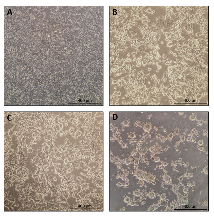
图1:Nanoblade生产过程中生产细胞的形态学特征。(A)铺板后24小时,汇合度为70-80%的HEK293T细胞。(B和C)转染后24小时HEK293T细胞的形态。(D)转染后40小时HEK293T细胞的形态。比例尺 = 400 µm。请点击此处查看该图的放大版本。
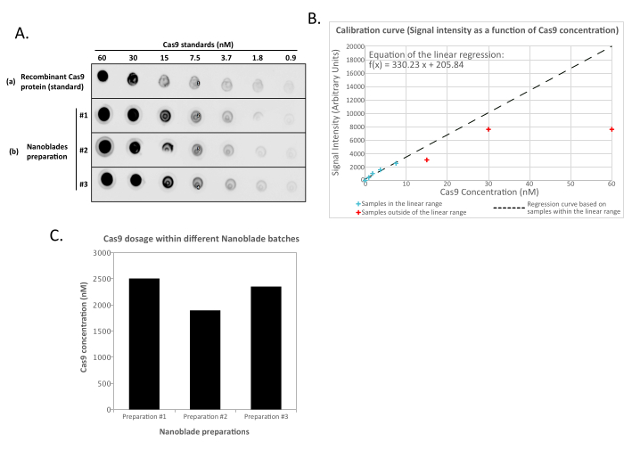
图 2:通过点印迹法测定纳米刀具中Cas9的载量。(A)将重组Cas9蛋白或经超速离心浓缩100倍的纳米刀具样品(#1、#2和#3)进行连续2倍梯度稀释后,点样于硝酸纤维素膜上,随后与HRP偶联的抗Cas9抗体孵育。通过增强型化学发光法显色。(B)采集并量化重组Cas9稀释样品的化学发光信号,将信号强度对已知点样量作图。根据处于线性范围内的稀释点(见蓝色叉形标记)计算回归曲线,排除所有超出线性范围的浓度点(见红色叉形标记)。(C)利用(B)中所得线性回归方程推算每种纳米刀具制备物中的Cas9浓度(nM)。为此,应仅使用落在回归曲线线性范围内的纳米刀具稀释样品的定量信号进行计算。请点击此处查看该图的放大版本。
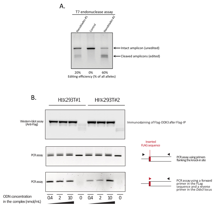
图 3:转导后编辑效率的监测。(A)T7 核酸内切酶检测用于测定 Nanoblade 处理细胞中的切割效率。使用靶向 EMX1 基因的 Nanoblade 转导的细胞通过 T7 核酸内切酶检测进行分析。泳道 1:Nanoblade 制备批次 #1(切割效率为 20%);泳道 2:对照细胞;泳道 3:Nanoblade 制备批次 #2(切割效率为 60%)。(B)在 DDX3 开放阅读框中敲入 Flag 标签序列。使用靶向 DDX3 位点的 sgRNA 编程的浓缩 Nanoblade 从不同的 HEK293T 克隆(#1、#2)中制备,并与剂量递增的 Flag-DDX3 ssODN 模板形成复合物,所得复合物用于转导 HEK293T 靶细胞。转导后,细胞培养三天,随后收集细胞以提取基因组 DNA 和总蛋白。使用抗 Flag 琼脂糖磁珠对 Flag-DDX3 蛋白进行免疫沉淀,再用抗 Flag 抗体对回收的蛋白进行蛋白质印迹分析(上图)。通过 PCR 检测 Ddx3 位点中 Flag 标签的定点插入,PCR 引物分别采用跨越插入位点的引物(中图),或采用识别 Flag 标签序列的正向引物与位于 Flag 插入位点下游的 Ddx3 位点特异性反向引物(下图)。缩写:EMX1 = Empty Spiracles Homeobox 1;DDX3 = DEAD-box RNA 解旋酶 3;PCR = 聚合酶链式反应;ODN = 脱氧寡核苷酸;ssODN = 单链 ODN;sgRNA = 单导向 RNA;IP = 免疫沉淀。 请点击此处查看该图的放大版本。
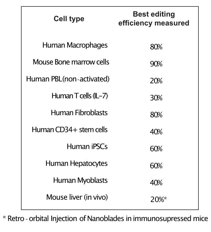
图 4:使用纳米刀在不同原代细胞类型中实现的编辑效率。 缩写:PBL = 外周血淋巴细胞;IL = 白细胞介素;CD = 分化群;iPSC = 诱导性多能干细胞。 请点击此处查看此图的放大版本。
讨论
Nanoblades 可实现 Cas9/sgRNA RNP 复合物在细胞系和原代细胞中的快速且剂量依赖性递送。与传统的转染方法和其他病毒载体递送方式不同,Nanoblades 与蛋白质电穿孔类似,具有以无转基因方式瞬时递送 Cas9/sgRNA RNP 的优势。Nanoblades 提供了一个高度通用、简便且经济的蛋白质递送平台,可轻松快速地适配于不断扩展的 CRISPR 变体家族。Nanoblades 可在 HEK293T 细胞系或其衍生细胞系中生产。本研究中使用的 HEK293T 细胞系经过优化,以最大化逆转录病毒和慢病毒颗粒的产量(参见材料表)。然而,尽管其他来源的 HEK293T 细胞也可能适用,用户必须对来自不同来源的 HEK293T 细胞进行测试和比较,因为不同来源的 HEK293T 细胞在病毒颗粒产量上已观察到显著差异。此外,细胞还需频繁检测支原体污染,并每三天传代一次(通常按 ⅛ 稀释),以避免过度汇合,后者会对病毒颗粒的产量产生不利影响。
细胞传代次数不应超过20代。细胞培养使用添加了葡萄糖、青霉素/链霉素、谷氨酰胺和10%灭活胎牛血清的DMEM培养基。由于血清来源可能影响Nanoblade制备的质量,因此在大规模生产前应测试不同批次的血清。转染后的第二天,Nanoblade也可在其他培养基中高效生产,例如RPMI或可替代DMEM的无血清最低必需培养基改良配方。如下所述,尽管使用某些DNA转染试剂进行转染后更换培养基是可选步骤,但调整病毒样颗粒(VLPs)释放所处的培养基可能具有优势,特别是有助于减少颗粒制备中的血清残留。然而,目前尚未尝试在转染前一天将细胞培养于低血清最低必需培养基中。
如前所述,Nanoblades 是通过在生产细胞中过表达一组质粒混合物而产生的。过表达似乎是实现高效生产所必需的。事实上,本实验室曾构建了一株生产细胞系,其中携带 Gag-Pol 表达结构的质粒通过抗生素筛选得以稳定存在;然而,该系统未能产生可观测量的 Nanoblades。当将编码 sgRNA 的结构稳定整合至生产细胞基因组时,也观察到了类似结果。正如其他颗粒生产系统所报道的那样,构建一株稳定表达至少部分参与 Nanoblades 生成的组分的细胞系可能是可行的;但这种方法必然需要处理大量上清液,并采用适当的技术来纯化颗粒。上述方案描述了生产 Nanoblades 的首选方法,该方法利用了特定的转染试剂(参见材料表)。
尽管其他厂商的转染试剂也已成功测试使用,但本课题组绝大多数关于Nanoblade的实验结果均遵循本文所述的操作流程。使用磷酸钙试剂可实现低成本转染,并获得良好的生产效率;然而,该方法必须在转染后第二天更换培养基,且可能在沉淀的病毒颗粒制备物中残留磷酸钙。与生产细胞中Nanoblade组分需要高水平表达的要求一致,已有观察表明,与Cas9蛋白结合的sgRNA数量可能是高效基因组编辑的限制因素。为提高sgRNA的装载效率,近期已有两个独立研究团队利用类似于Nanoblade的蛋白递送载体开发了两种技术方法。这些方法依赖于T7聚合酶介导的sgRNA在细胞质中的表达6,或在sgRNA序列中引入逆转录病毒包装信号以介导其与Gag多蛋白的结合6。这些策略确实有可能改善Nanoblade中sgRNA的装载,但目前尚未经过验证。
靶细胞的转导是该实验流程中的关键步骤。在大多数永生化细胞系中,使用Nanoblade进行转导几乎不会产生细胞病变效应。然而,在原代细胞中,可能存在细胞毒性问题。因此,必须针对每种细胞类型优化转导条件。具体而言,在优化转导方案时,Nanoblade的处理时间是一个需要调整的重要因素。对于原代神经元或骨髓细胞等敏感细胞,使用Nanoblade孵育4–6小时后更换培养基,可在最大限度降低细胞毒性的同时实现Cas9蛋白的有效递送。此外,某些佐剂(如阳离子聚合物等)可显著提高部分细胞的转导效率(参见材料表)。需要注意的是,Nanoblade属于病毒样颗粒(VLPs),可能引发免疫原性反应。当使用某些类型的原代细胞(例如巨噬细胞或树突状细胞)时,这一特性可能成为限制因素,因为与Nanoblade共孵育可能引起细胞基因表达和表型的显著改变。如果巨噬细胞和树突状细胞来源于造血干细胞前体(如小鼠骨髓细胞),建议在细胞完全分化之前进行Nanoblade转导,以避免诱导针对Nanoblade的细胞免疫反应。否则,在处理已分化的免疫细胞时,Cas9蛋白的电穿孔递送可作为一种可行的替代方案。
Nanoblade 可用于体内转导小鼠受精卵或胚胎,以生成转基因动物。与经典的逆转录病毒或慢病毒载体类似,它们也可直接注射到成年动物的组织中。然而,Nanoblade(与逆转录病毒和慢病毒载体相似)可能被宿主动物的免疫反应灭活;因此,注射剂量需针对每种应用进行优化。这种免疫反应还可能限制功能性病毒样颗粒(VLPs)向注射部位附近组织的分布。最后,与慢病毒载体不同,Nanoblade 不含外源转基因,且 Cas9 的递送具有时间限制性。因此,它们无法用于需要在细胞筛选后对 sgRNA 进行高通量测序的全基因组功能筛选。当需要快速、剂量依赖且无外源转基因的基因组编辑时,Nanoblade 具有重要应用价值20。此外,与蛋白质电穿孔类似,相较于通过 DNA 转染或经典病毒载体实现的 Cas9/sgRNA 长期表达,Nanoblade 引起的脱靶效应更少3。未来对 Nanoblade 的开发将聚焦于整合适用于不同技术应用的 Cas9 变体,例如碱基编辑和 RNA 靶向。
披露
Philippe E. Mangeot 和 Emiliano P. Ricci 被列为与 Nanoblades 技术相关的一项专利的发明人(专利申请人:Institut National de la Santé et de la Recherche Médicale (INSERM)、Centre National de la Recherche Scientifique (CNRS)、Ecole Normale Superieure de Lyon、Université Claude Bernard Lyon 1、Villeurbanne Cedex;申请号:WO 2017/068077 Al;专利状态:已公布,2017年4月27日;本稿件的所有内容均涵盖在该专利申请范围内。其余作者声明不存在利益冲突。
致谢
本工作由里昂大学的Labex Ecofect(ANR-11-LABX-0048)资助,该项目隶属于法国国家科研署(ANR)实施的“未来投资计划”(ANR-11-IDEX-0007);同时获得FINOVI基金会、法国国家艾滋病与病毒性肝炎研究署(ANRS-ECTZ3306)以及欧洲研究理事会(ERC-StG-LS6-805500,授予E.P.R.)在欧盟“地平线2020”科研与创新计划下的资助。
材料
| 姓名 | 公司 | 目录编号 | 评论 |
|---|---|---|---|
| 13.2 mL,薄壁聚丙烯管,14 x 89 mm - 50支装 | Beckman Coulter Life Sciences | 331372 | 用于Nanoblades纯化的超速离心管 |
| Amersham Protran Premium 蛋白印迹膜,硝酸纤维素膜 | Merck | GE10600004 | 用于定量纯化Nanoblades中Cas9水平的硝酸纤维素膜 |
| BIC-Gag-CAS9 | Addgene | 119942 | 编码GAG(F-MLV)-CAS9(sp)融合蛋白。可在生产细胞中与过表达的gRNA(s)及适当的包膜蛋白共同作用,产生GAG-CAS9类病毒颗粒 |
| BICstim-Gag-dCAS9-VPR | Addgene | 120922 | 编码GAG-dCAS9-VPR融合蛋白,用于靶向转录激活 |
| BLADE | Addgene | 134912 | 用于在Nanoblades系统中克隆sgRNA序列的空骨架载体 |
| BsmBI-v2 | New England Biolabs | R0739S | 限制性内切酶,用于消化BLADE和SUPERBLADES载体以进行sgRNA克隆 |
| Cas9 (7A9-3A3) 小鼠单克隆抗体(HRP偶联)#97982 | Cell Signaling Technology | 97982S | 用于点印迹法定量Cas9的抗Cas9抗体 |
| Cas9 核酸酶,S. pyogenes | New England Biolabs | M0386T | 重组Cas9蛋白,用作Nanoblades中Cas9载量绝对定量的参考标准 |
| 溴化乙锭溶液(10 mg/mL,溶于H2O) | Sigma-Aldrich | E1510-10ML | 用于琼脂糖凝胶染色及DNA可视化 |
| Fisherbrand Wave Motion 摇床 | Fisher Scientific | 88-861-028 | 离心后重悬Nanoblades的振荡平台 |
| gelAnalyzer | http://www.gelanalyzer.com;用于酶切后条带强度的定量分析 | ||
| Gesicle Producer 293T | Takara | 632617 | Nanoblades生产细胞系 |
| Gibco DMEM,高糖,含丙酮酸钠 | ThermoFisher Scientific | 41966052 | Gesicle Producer 293T细胞的培养基 |
| GoTaq G2 DNA聚合酶 | Promega | M7848 | T7核酸酶检测前用于基因组DNA扩增的Taq聚合酶 |
| jetPRIME 转染试剂盒(用于DNA及DNA/siRNA) | Polyplus | POL114-15 | 用于在Gesicle Producer 293T细胞中生产Nanoblades的转染试剂 |
| Millex-AA,0.80 µm,针头滤器 | Millipore | SLAA033SS | 在浓缩Nanoblades前去除细胞碎片的针头滤器 |
| Millex-GS,0.22 µm,针头滤器 | Millipore | SLGS033SS | 用于蔗糖垫溶液灭菌的针头滤器 |
| Millex-HP,0.45 µm,聚醚砜,针头滤器 | Millipore | SLHP033RS | 在浓缩Nanoblades前去除细胞碎片的针头滤器 |
| Monarch DNA凝胶回收试剂盒 | New England Biolabs | T1020L | 用于BsmBI酶切后纯化pBLADES或pSUPERBLADES质粒片段的DNA凝胶回收试剂盒 |
| NEB Stable 感受态E. coli(高效率) | New England Biolabs | C3040I | 用于质粒转化与扩增的感受态细菌 |
| NucleoBond Xtra Midi 试剂盒(用于转染级质粒DNA) | Macherey-Nagel | 740410.50 | 从培养细菌中纯化质粒DNA的大提试剂盒 |
| Nucleospin 基因组DNA提取试剂盒 | Macherey-Nagel | 740952.50 | 从转导细胞中提取基因组DNA |
| NucleoSpin 质粒DNA小提试剂盒 | Macherey-Nagel | 740588.50 | 从培养细菌中纯化质粒DNA的小提试剂盒 |
| NucleoSpin 组织DNA小提试剂盒(适用于细胞与组织) | Macherey-Nagel | 740952.5 | 基因组DNA提取试剂盒 |
| Optima XE-90 | Beckman Coulter Life Sciences | A94471 | 超速离心机 |
| pBaEVRless | Els Verhoeyen (Inserm U1111) | 个人请求需发送至:els.verhoyen@ens-lyon.fr | 见Girard-Gagnepain A. 等人发表于Blood 124, 1221–1231 (2014) 的研究:狒狒内源性逆转录病毒Rless包膜蛋白。狒狒包膜假型慢病毒在向早期细胞因子刺激及静息状态造血干细胞进行基因转移时优于VSV-G假型慢病毒 |
| pBS-CMV-gagpol | Addgene | 35614 | 编码小鼠白血病病毒(Murine Leukemia Virus)的gag和pol基因 |
| pCMV-VSV-G | Addgene | 8454 | 用于生产慢病毒和MuLV逆转录病毒颗粒的包膜蛋白 |
| 磷酸盐缓冲液(PBS) | ThermoFisher Scientific | 14200091 | 10X PBS,需用Millipore水稀释 |
| Polybrene 转染试剂 | Millipore Sigma | TR-1003-G | 阳离子聚合物,可增强特定哺乳动物细胞中逆转录病毒转导效率。也可促进寡脱氧核苷酸(ODN)通过病毒依赖性方式进入细胞,实现同源定向修复 |
| 蔗糖,分子生物学级,≥99.5%(GC) | Sigma-Aldrich | S0389-5KG | 用于制备超速离心纯化Nanoblades时的蔗糖垫 |
| SUPERBLADE5 | Addgene | 134913 | 用于在Nanoblades系统中克隆sgRNA序列的空骨架载体(经Chen B等人,2013年优化,以提高基因组编辑效率) |
| SuperSignal West Dura 增强型化学发光底物 | ThermoFisher Scientific | 34076 | 用于Cas9点印迹检测的增强型化学发光(ECL)HRP底物 |
| SW 41 Ti 摆动桶转子 | Beckman Coulter Life Sciences | 331362 | 超速离心用转子 |
| SYBR Safe DNA凝胶染料 | ThermoFisher Scientific | S33102 | 溴化乙锭的替代品,用于琼脂糖凝胶染色及DNA可视化 |
| T4 DNA连接酶 | New England Biolabs | M0202S | 用于将BLADE或SUPERBLADES载体与对应sgRNA可变区的双链DNA寡核苷酸连接的DNA连接酶 |
| T7 内切酶 I | New England Biolabs | M0302S | T7内切酶I可识别并切割不完全匹配的DNA,用于监测特定基因座上的基因组编辑程度 |
| 含Triton的裂解缓冲液 | Promega | E291A | 用于裂解Nanoblades并实现Cas9定量的裂解缓冲液 |
| TWEEN 20 | Sigma-Aldrich | P9416 | 用于TBST缓冲液的配制 |
参考文献
- Jinek, M., et al. A programmable dual-RNA-guided DNA endonuclease in adaptive bacterial immunity. Science. 337 (6096), 816-821 (2012).
- Zuris, J. A., et al. Cationic lipid-mediated delivery of proteins enables efficient protein-based genome-editing in vitro and in vivo. Nature Biotechnology. 33, 73-80 (2015).
- Mangeot, P. E., et al. Genome editing in primary cells and in vivo using viral-derived Nanoblades loaded with Cas9-sgRNA ribonucleoproteins. Nature Communications. 10 (1), 45(2019).
- Cai, Y., Bak, R. O., Mikkelsen, J. G. Targeted genome editing by lentiviral protein transduction of zinc-finger and TAL-effector nucleases. eLife. 3, 01911(2014).
- Choi, J. G., et al. Lentivirus pre-packed with Cas9 protein for safer gene editing. Gene Therapy. 23 (7), 627-633 (2016).
- Gee, P., et al. Extracellular nanovesicles for packaging of CRISPR-Cas9 protein and sgRNA to induce therapeutic exon skipping. Nature Communications. 11, 1334(2020).
- Indikova, I., Indik, S. Highly efficient 'hit-and-run' genome editing with unconcentrated lentivectors carrying Vpr.Prot.Cas9 protein produced from RRE-containing transcripts. Nucleic Acids Research. 48 (14), 8178-8187 (2020).
- Lyu, P., Javidi-Parsijani, P., Atala, A., Lu, B. Delivering Cas9/sgRNA ribonucleoprotein (RNP) by lentiviral capsid-based bionanoparticles for efficient 'hit-and-run' genome editing. Nucleic Acids Research. 47 (17), 99(2019).
- Gheysen, D., Jacobs, E., de Foresta, F., Thiriart, C. Assembly and release of HIV-1 precursor Pr55gag virus-like particles from recombinant baculovirus-infected insect cells. Cell. 59 (1), 103-112 (1989).
- Hanna, R. E., Doench, J. G. Design and analysis of CRISPR-Cas experiments. Nature Biotechnology. 38 (7), 813-823 (2020).
- Sambrook, J. Molecular cloning a laboratory manual. Third edition. , Cold Spring Harbor Laboratory Press. Cold Spring Harbor, N.Y. (2001).
- Jiang, W., et al. An optimized method for high-titer lentivirus preparations without ultracentrifugation. Scientific Reports. 5, 13875(2015).
- Rueden, C. T., et al. ImageJ2: ImageJ for the next generation of scientific image data. BMC Bioinformatics. 18 (1), 529(2017).
- O'Doherty, U., Swiggard, W. J., Malim, M. H. Human immunodeficiency virus type 1 spinoculation enhances infection through virus binding. Journal of Virology. 74 (21), 10074-10080 (2000).
- Brinkman, E. K., Chen, T., Amendola, M., van Steensel, B. Easy quantitative assessment of genome editing by sequence trace decomposition. Nucleic Acids Research. 42 (22), 168(2014).
- Bollen, Y., Post, J., Koo, B. -K., Snippert, H. J. G. How to create state-of-the-art genetic model systems: strategies for optimal CRISPR-mediated genome editing. Nucleic Acids Research. 46 (13), 6435-6454 (2018).
- Brinkman, E. K., et al. Easy quantification of template-directed CRISPR/Cas9 editing. Nucleic Acids Research. 46 (10), 58(2018).
- Dussaud, S., Pardanaud-Glavieux, C., Sauty-Colace, C., Ravassard, P. Lentiviral mediated production of transgenic mice: a simple and highly efficient method for direct study of founders. Journal of Visualized Experiments. (140), e57609(2018).
- Montagna, C., et al. VSV-G-enveloped vesicles for traceless delivery of CRISPR-Cas9. Molecular Therapy. Nucleic Acids. 12, 453-462 (2018).
- Marnef, A., et al. A cohesin/HUSH- and LINC-dependent pathway controls ribosomal DNA double-strand break repair. Genes & Development. 33 (17-18), 1175-1190 (2019).
重印与许可
