我们介绍一种利用甲醛交联在活酵母和哺乳动物细胞中快速稳定翻译(蛋白质生物合成)复合物的技术。该方法能够解析瞬时中间产物以及动态的RNA:蛋白质相互作用。交联后的复合物可用于多种下游应用,例如基于深度测序的分析方法、显微镜观察和质谱分析。
本文内容
摘要
摘要
涉及信使RNA(mRNA)快速重分布及mRNA翻译改变的快速响应,与细胞正在进行的稳态调节密切相关。这些调节对于真核细胞在营养和盐度波动、温度变化以及各种化学和辐射胁迫条件下的存活能力和“损伤控制”至关重要。由于RNA水平响应具有高度动态性,且许多RNA:RNA和RNA:蛋白质中间体不稳定,因此仅通过有限的几种方法才能获得有意义的细胞质RNA状态快照。全转录组范围、基于RNA测序的核糖体谱分析(ribosome profiling)类实验是研究翻译调控最具信息量的数据来源之一。然而,若缺乏对RNA及RNA:蛋白质中间体的统一稳定化处理,则可能导致不同的偏差,尤其是在快速响应的细胞通路中。本文提供了一种适用于不同通透性真核细胞的快速固定详细方案,以辅助稳定RNA及RNA:蛋白质中间体。我们进一步提供了基于核糖体及多(核糖)体组分共沉降方法分离稳定化RNA:蛋白质复合物的示例。所分离的稳定化材料可后续用于核糖体谱分析类实验,例如翻译复合物谱分析(Translation Complex Profile sequencing, TCP-seq)及其衍生技术。目前,TCP-seq类方法的通用性已在多种生物体和细胞类型的应用中得到验证。由于交联易于逆转,这些稳定化的复合物还可进一步进行亲和纯化,并通过电子显微镜成像,或分离至不同的多(核糖)体组分后进行RNA测序。因此,基于瞬时冷冻和甲醛固定的处理方法,结合基于沉降或其他类型的RNA:蛋白质复合物富集技术,特别适用于在活细胞中深入探究快速RNA:蛋白质复合物动态的精细机制。
引言
生物体在其生命周期中会经历动态的细胞内和细胞外变化,需要快速响应以维持稳态并确保生存。为实现环境适应,真核细胞会调节其代谢活动 通过 基因表达调控。基因表达调控可在转录和/或翻译过程中施加影响;其中翻译水平的响应通常发生得更为迅速1,2,3,4例如,翻译水平的变化通常在应激开始后1-30分钟内出现,而转录水平的改变则在应激暴露数小时后发生3,4,5由于细胞质中信使(m)RNA分子持续存在,翻译水平的调控可更快速地实现对基因表达的改变。相反,在转录水平,必须先合成新的mRNA分子,并在真核生物中经过加工并从细胞核输出,导致响应时间显著延迟。2,4,6,7,8.
急性应激的翻译响应通常表现为整体翻译产出的下降,同时选择性上调对细胞存活至关重要的蛋白质1,3,4,9。由于翻译过程消耗大量能量,降低蛋白质合成输出被认为至关重要3,7。为实现选择性抑制与上调,翻译响应依赖于一系列复杂的调控机制。这些调控可作用于翻译的各个阶段:多肽生物合成的起始、延伸、终止以及核糖体的再循环10,11,12,13,但最显著的作用发生在起始阶段5,7,9,10,13。在起始阶段,小核糖体亚基(SSU)在真核起始因子(eIFs)的协助下结合到mRNA的5'非翻译区(UTR),并沿其扫描,直至识别起始密码子2,5,6,8,11,12,13。调控机制通常靶向影响结合、扫描及起始密码子识别的eIFs。例如,起始因子eIF2是一种关键的翻译因子,可协助将起始Met-tRNAiMet招募至SSU,在应激条件下常成为调控靶点4,6,11。在酵母中,营养缺乏和渗透压应激可诱导该因子的磷酸化1,4,11,14,15;而在哺乳动物细胞中,氨基酸饥饿、内质网(ER)应激、紫外线应激、病毒感染以及氧浓度变化均可触发这一响应8,9,11。在哺乳动物细胞对缺氧的响应中,特定mRNA翻译的快速上调尤为明显,表现为全局性的快速翻译抑制,同时选择性上调缺氧诱导因子(HIFs)的生物合成。HIFs作为转录因子,进一步在DNA转录水平引发长期的细胞重编程8,9,16。在酵母中,热应激也观察到类似响应:热休克蛋白(HSPs)的翻译迅速表达,随后出现延迟的转录水平响应17,18。除营养缺乏和热休克外,酵母在不同氧气8,19、盐度5、磷酸盐、硫20,21及氮22,23水平下的翻译响应也已被广泛研究。这些研究对酵母在烘焙和发酵等工业应用中具有广泛意义24,25。翻译响应还有助于深入理解神经退行性疾病和心脏病等疾病,这些疾病常伴随氧化应激等细胞内应激状态。总体而言,翻译响应是真核生物基因表达调控的重要组成部分,有助于快速适应多种应激条件。
为了研究翻译响应,需要采用能够提供翻译景观最小失真快照的方法。多聚核糖体分析是一种用于研究mRNA翻译的经典方法,涉及分离mRNA的多聚(核糖)体组分 通过 蔗糖梯度超速离心26,27该方法可用于探究单个mRNA的翻译水平(通过逆转录聚合酶链反应等检测方法,RT-PCR)26),或与高通量技术(微阵列或RNA-seq)结合进行全局分析28,29)。更进一步的方法是核糖体谱分析,该方法能够在全基因组范围内研究延伸中的核糖体在mRNA分子上的位置,同时推断翻译效率在转录组中的分布以及主要和替代起始位点的利用情况30,31核糖体谱分析涉及分离并测序由核糖体占据而受到保护的 mRNA 片段。核糖体谱分析已在多种条件下为翻译动态提供了深入见解,包括缺氧应激、热休克和氧化应激。31,32该技术已适用于多种来源材料,包括酵母和哺乳动物细胞。
尽管多核糖体分析和核糖体图谱技术在拓展翻译研究能力方面发挥了基础性作用,但翻译过程包含多种翻译中间体和复合物,这些方法难以捕获。11,13另一个局限性源于无法研究快速响应类型,因为翻译复合物要么被稳定化 体内 通过添加特异性翻译抑制剂(抗生素),导致特定的核糖体分布伪影,或 离体 细胞裂解时特异性地(抗生素)或非特异性地(高盐或镁离子)剥夺寿命较短或稳定性较差的中间产物33,34,35.
甲醛被广泛用于交联核酸与蛋白质,例如在染色质免疫沉淀(ChIP)和交联免疫沉淀(CLIP)研究中。由于其分子尺寸小且细胞通透性良好,可实现快速的体内作用36。基于甲醛快速交联的特性,核糖体谱分析方法已扩展为翻译复合物谱测序(TCP-seq)10,36,37,38,39,40。TCP-seq 最初在酵母中建立,能够捕获所有翻译中间体,包括扫描或翻译终止后的SSU复合物以及多种核糖体构型37,38,41,42。该方法已在多项研究中得到应用10,38,39,41,42,其中一些研究采用翻译抑制剂与甲醛交联相结合的方法,以促进翻译过程的阻滞。该技术的一个进一步改进版本——选择性 TCP-seq39,最近已被用于对交联复合物进行免疫纯化,从而拓宽了 TCP-seq 的应用范围。甲醛交联具有快速、高效且可逆的特性,使这些方法适用于研究瞬时的 mRNA:翻译复合物相互作用,特别是在高度动态的翻译水平响应通路中的研究。
本文详细阐述了用于全面稳定和分离翻译复合物的 体内 甲醛交联过程。我们分别提供了适用于酵母和哺乳动物细胞的精细实验方案(图1)。此外,我们还概述了交联稳定化材料的后续应用示例(图1),例如通过免疫印迹(western blotting)检测共纯化的蛋白质因子、免疫辅助纯化(或“免疫沉淀”;IP)以及富集含有特定目标因子的翻译复合物、电子显微镜分析和RNA测序。
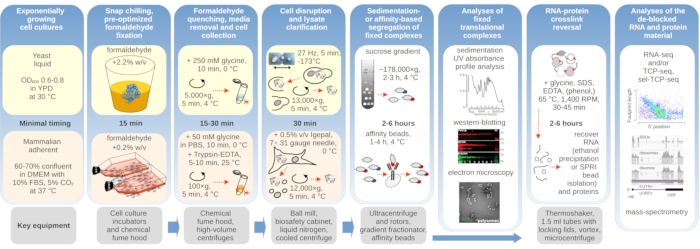
图1示意图展示了典型实验装置的概览。 主要步骤 体内 翻译复合物的甲醛稳定化流程以流程图形式展示,并补充了关键所需仪器的相关信息。文中概述了交联材料可能的下游应用,包括已成功使用但本方案未直接涵盖的方法实例,如RNA的SPRI磁珠纯化、RNA测序和质谱分析。 请点击此处查看此图的放大版本。
方案
1. 酵母细胞实验方案
- 酵母细胞培养与固定
注意:细胞固定与收获步骤改编自10,38 并加以改进。- 设置1 L酵母细胞培养液(以野生型(WT)BY4741为例),在摇床中培养,起始光密度在600 nm处不超过0.05 AU(OD600)置于合适的培养基中(以含1% w/v酵母提取物、2% w/v蛋白胨、2% w/v右旋糖(葡萄糖)、40 mg/L硫酸腺嘌呤的YPD培养基为例),在所需条件(30 °C)下培养 °本实验中使用的 C。
- 设置一台制备型离心机,配备适用的转子和离心瓶,用于沉淀酵母细胞的液体悬浮培养物。对于葡萄糖饥饿实验,在600 nm波长下细胞培养液的光密度达到0.6–0.8 AU(OD600) 时,使用30 °C,5,000 × g 1 分钟。
注意:记录正在生长的细胞的 OD 值,并让细胞继续生长至 OD600 当对数生长期是研究重点时,OD值达到0.6–0.8 AU。 - 立即将沉淀重悬于预热(30℃)的培养基中 °C)YP 培养基,不添加或低浓度(0.25% w/v)添加葡萄糖,将培养物在 30 ℃下继续孵育 10 分钟 °在轨道摇床培养箱中。
注意:培养基成分可能影响后续交联效率。本实验方案仅在YPD培养基中进行过测试。进行饥饿实验时,必须严格遵守时间安排,并尽量减少各步骤之间的延迟。 - 细胞准备好后,在通风橱内准备一个冰盒,并放入一个装有250 g洁净碎冰的烧杯。确保通风橱内备有25 mL移液管以及新购入的甲醇稳定37%(w/v)甲醛溶液。将1 L培养液倒入盛有25%(w/v)碎冰的烧杯中。
注意:除非另有说明,在细胞冻结之前的后续所有操作中,均需将细胞置于冰上。 - 加入75 mL 37% w/v的甲醛溶液,使其终浓度为2.2% w/v,并剧烈搅拌混合物直至冰完全融化。
- 冰融化后,设置10分钟的计时器。
注意:请遵循推荐的时间和温度方案,以获得可重复的固定结果。 - 孵育10分钟后,将培养物转移至预冷的离心瓶中,于4℃下离心收集细胞 °C,5,000 × g 5分钟。在此离心过程中,预先冷却一个50 mL离心管,并将新配制的缓冲液A(含甘氨酸以中和残留的甲醛)置于冰上。
注:有关确切的缓冲液组成,请参阅随附的表格。 - 离心后,将离心管置于冰上,使沉淀一侧与冰接触。将离心管移入通风橱,将上清液弃入甲醛废液容器中。
- 将所有离心管中的细胞沉淀用 25 mL 移液管加入 20 mL 缓冲液 A 重悬,并转移至 50 mL 离心管中。
注意:此洗涤步骤对于避免不可重复的交联至关重要,从细胞开始处理至缓冲液加入的时间不得超过 20 分钟' 收获 - 用缓冲液 A 定容至 40 mL,并在 4 °C 下离心收集洗涤后的细胞 °C,5,000 × g 5 分钟。
- 弃去上清液,将细胞沉淀重悬于40 mL缓冲液A1中, 不含甘氨酸的缓冲液A,以去除任何甘氨酸污染。
- 再次通过在 4 ℃ 下离心沉淀细胞 °C,5,000 × g 5 分钟。
- 用缓冲液 A1 再重复洗涤一次。弃去上清液,将细胞沉淀置于冰上。称量含有沉淀的离心管(每 1 L 细胞培养液对应的湿细胞质量应约为 1 g)。
- 酵母细胞破碎与细胞质提取
- 在聚苯乙烯泡沫箱内衬铝箔,注入液氮至深度约3厘米。将一个50 mL离心管直立放入箱中。
- 将沉淀物(约1 g湿细胞质量)重悬于550 µ通过移液并涡旋震荡10秒,将样品与10 µL缓冲液A2混合。加入10 µ40 U/升µ加入 RNase 抑制剂 1 L,再次涡旋振荡 10 秒。
警告:操作液氮时需佩戴适当的防护装备,例如隔热手套。确保盛装液氮的容器无泄漏,且内部试管架不会浮起或倾倒。应在通风良好的区域操作,以避免缺氧。 - 使用1 mL移液器,将细胞悬液逐滴加入含有液氮的50 mL离心管中。
注意:滴加过程必须缓慢且小心,以避免液滴聚集。确保在加入新液滴之前,原有液滴已完全冻结。 - 将装有冷冻细胞悬液液滴的50 mL离心管转移至室温,待液氮完全蒸发后,盖紧管盖,并将细胞沉淀物于-80℃保存。 °C 或立即继续下一步。
警告:在密封试管前,务必确保液氮已完全蒸发。密封试管中残留的液氮可能导致危险的压强积聚。 - 为准备下一步,将1.5 mL无核酸酶离心管和10 mL不锈钢研磨罐置于干冰上预冷。
- 使用洁净无菌的刮勺将冷冻的细胞悬液液滴转移至罐中。
警告:确保研磨罐已紧密密封。 - 将研磨罐浸入液氮中1分钟,确保液相保持在接头下方。设置冷冻混合球磨机在27 Hz频率下振荡1分钟。
注意:研磨罐必须始终用相同型号的另一个研磨罐配重平衡 即使样品处理仅需一个罐体。 - 以27 Hz的频率振荡密封的研磨罐1分钟 混合研磨仪
- 将研磨罐如前所述在液氮中重新冷却,然后在混合研磨仪中以27 Hz频率继续振荡1分钟。
- 将罐子转移至装有干冰的冰盒中,同时放入1.5 mL无核酸酶的离心管。使用小型钢制刮勺将所得粉末样品分装至离心管中,每份约100 mg,并将离心管于-80 ℃保存。 °C.
注意:建议每次实验使用约 600 mg 的样品,用于多聚核糖体沉降谱分析,将胞质溶胶分离为翻译和非翻译组分,并在 RNase 消化后进一步将翻译组分分离为 SSU、核糖体和双体(disome)组分。
- 从固定化的(多)核糖体复合物中分离非翻译区的细胞溶质组分
注意:此前建立的步骤10,38 通常通过翻译中RNA与(多)核糖体共沉降的方法来富集翻译中的RNA。本文介绍了一种更精细的分离翻译中与非翻译细胞质组分的方法,无需对材料进行沉淀及后续再溶解。- 配制 2.5 mL 线性 10%-20% (w/v) 使用冻融法以缓冲液B制备蔗糖梯度43 在超薄壁超速离心管(5 mL,13 x 51 mm)中。
注意:冻融方法是通过依次添加浓度呈线性递减的缓冲蔗糖溶液层,并逐层冷冻完成的。参见 补充表1 详情请见。 - 为制备不连续的50%(w/v)蔗糖垫,在线性梯度解冻并稳定后,使用连接19 G × 1.5针头的1 mL注射器,将0.5 mL含缓冲液B的50%蔗糖溶液缓慢加入离心管底部。" 针头或尺寸相似/合适的玻璃毛细管。在加样前,从预形成的蔗糖梯度顶部到底部,小心缓慢地将针头或毛细管插入,避免扰动,直至到达管底。
注意:参见 补充表1 有关缓冲液B的制备说明。 - 通过移除顶部部分或在缓冲液 B 中叠加更多 10 w/v 的蔗糖仔细平衡梯度,并保持其冰上或 4 ℃低温环境 °C.
注意:需要使用底部为50%蔗糖层的不连续梯度,以收集沉降速率较高的物质,同时防止其在离心管壁上沉淀。 - 将约100 mg冷冻的细胞粉末样品在室温下解冻,立即置于冰上。加入150 µ通过移液将缓冲液A2加至L,加入RNase抑制剂至1 U/µL,并涡旋混匀10秒(避免过度起泡及与气相混合)。
注意:除非另有说明,所有操作均需在冰上进行。 - 通过在4℃下离心试管,使细胞碎片沉淀 °C,13,000 × g 5 分钟,收集澄清的上清液(约 150 µL) 置于新的 1.5 mL 低蛋白结合离心管中。
- 将所得澄清混合物上样至1.3.3步骤制备的不连续蔗糖梯度管中,并仔细平衡。
- 使用中等容量的摆动桶转子在超速离心机中离心试管,转速为 4 °C,平均相对离心力为 287,980 × g (k因子49)作用1小时30分钟。
注:这些条件已预先优化(通过超速离心后梯度紫外吸光度轨迹分析),可在保留游离(非(多)核糖体)小亚基(SSUs)和大亚基(LSUs,large ribosomal subunit)位于梯度上部(10%–20%蔗糖)的同时,将(多)核糖体组分富集于底部(50%蔗糖垫)而不形成沉淀。 - 使用一支新的无菌1 mL注射器,配备19 G × 1.5" 用针头收集翻译后的细胞质组分。将5 mL梯度置于稳定的支架上,确保可见管底。
- 从管子的顶部将针头垂直插入梯度底部(切勿刺穿管子),轻轻吸取含有翻译后RNA池的底部溶液,吸取体积精确为0.5 mL,过程中避免产生气泡。
注意:确保此步骤在低温室中进行,并牢固握住离心管。建议将全部0.5 mL液体一次性向上抽吸,以避免扰动梯度。 - 通过超速离心后测定蔗糖梯度的吸光度,确认所得混合物中(多)核糖体的存在以及小亚基、大亚基和较轻组分的耗尽。
- 将上一步收集的翻译后RNA混合物浓缩至100 µ使用截留分子量为10 kDa的再生纤维素膜微型浓缩装置通过超滤进行浓缩。
注意:用 0.5 mL 缓冲液 1 预清洗微型浓缩装置的膜(参见 图2a)并使用离心条件(g)由制造商推荐。 - 将上一步骤的材料进一步稀释五倍(加入400) µL)用缓冲液1洗涤并浓缩回200 µL,以实现较小体积及部分去除蔗糖。
注意:建议将所得混合物在 -80 ℃ 保存 °可于 -80 °C 保存长达 6 个月,并作为“总翻译 RNA”RNA-seq 文库构建或 TCP-seq 文库构建中 RNase 消化步骤的输入材料。“非翻译”细胞质组分可通过类似方法从梯度顶部回收,并于 -80 °C 保存 °C.
- 配制 2.5 mL 线性 10%-20% (w/v) 使用冻融法以缓冲液B制备蔗糖梯度43 在超薄壁超速离心管(5 mL,13 x 51 mm)中。
- 固定后的(多)核糖体复合物的RNase消化及消化产物的小核糖体亚基(SSU)、单核糖体(核糖体,RS)和双核糖体(二聚体,DS)组分分离
注意:该步骤通常遵循先前描述的方法10,38 但采用了改良的梯度类型、分离时间、加速度及RNase消化条件,以在所有三个分离组分中实现最佳分辨率。- 使用冻融法,在 13 mL 薄壁聚丙烯管(14 × 89 mm)中,用缓冲液 1 制备精确配平的 12.5 mL 线性 10%–40% w/v 蔗糖梯度43 如步骤1.3.1及其中注释所述。
- 在室温下解冻,并立即将样品置于冰上,或取自步骤1.3.12的浓缩且已去除蔗糖的翻译后胞质组分。
注意:除非另有说明,所有操作均需在冰上进行。 - 通过加入4.5 U的消化酶消化翻译后的细胞质组分 E. coli 每1 OD单位的RNase I260 30 分钟内 23 个分数单位 °C. 立即加入RNase I失活型RNase抑制剂,通过移液混匀,使其终浓度为0.25 U/μLµ向混合物中加入 L,以灭活 RNase I。
注意:使用能够抑制RNase I的RNase抑制剂。测定AU260 通过使用AU260 =(260 nm 处的吸光度标准化为相当于 1 cm 光程 × 裂解物体积的光密度单位) µL) / 1,000 - 立即将样品转移至冰上。
注意:必须严格遵守推荐的消化条件,并准确测量所添加的 RNase I 用量。此处提到的 RNase I 活性单位定义为在规定条件下,每分钟水解 1 μg 酵母 RNA 所需的酶量。 µ37℃下30分钟内从鼠肝RNA中提取的酸溶性物质克数 °C. RNase I 不同批次的酶活性可能存在未记录的差异,可能需要通过实验确定最佳消化条件。如果酶原液浓度过高,建议用缓冲液1稀释,以避免移取过小体积的溶液。 - 将反应混合物上样至步骤1.4.1制备的10%-40%(w/v)蔗糖梯度中。
注:使用终体积为150–300的范围 µ每梯度升。每次纯化至少需要两个梯度。使用不同的上样体积(较低AU)260,10-11 AU260,用于DS和相对较高的AU260,13-14 AU260,以实现最佳的SSU或RS分离效果。 - 使用中等容量的水平转子在超速离心机中离心试管,4 °C,平均相对离心力 178,305 × g (k因子143.9)离心3小时30分钟。
警告:如果需要备用平衡管,请使用与含样品管质量及质量分布相等的管子。应使用备用的蔗糖梯度管,并在其上层覆盖与样品管上层体积相当的缓冲液,而不是使用均一蔗糖浓度的管子。 - 至少在超速离心结束前30分钟,安装好梯度分馏装置,包括向0.2 µ将重追加溶液(例如,此处使用的去离子水中60%蔗糖)经0.45 μm滤膜过滤后注入置换泵中。
注意:建议在每次运行前后,使用去离子水对分级仪的管路和管道进行去污染,随后依次使用含1%-2% SDS的去离子水溶液、去离子水以及含80%乙醇的去离子水溶液进行清洗。 - 通过先用去离子水填充系统,并根据制造商的建议将光学读数调零,然后使用与样品管缓冲液相同(例如缓冲液1)的空白14 x 89 mm蔗糖梯度进行基线漂移校正,来调整吸光度读数的基线。
注意:进行调整时应使用与样品读数相同的位移速度,例如 1.5 mL/min。 - 通过精确计时溶液首次进入检测器光路至首次出现在组分收集器出口之间的时间,测量位移系统的死体积。
注意:以推荐的1.5 mL/min流速进行分馏时,可在室温下操作。收集到的组分应立即置于冰上。 - 使用254 nm实时吸光度读数进行组分分离,流速为1.5 mL/min,并根据样品预期的沉降位置和吸光度谱进行在线组分检测。采用收集管切换,其时间延迟应与此前测定的死体积相对应。
- 分离对应于SSU、RS和DS复合物位置及迁移率的组分,并收集到新的低蛋白结合1.5 mL微量离心管中;立即将分离的组分置于冰上,若不能立即进一步处理,则应冷冻保存。
注意:建议将收集的组分立即在干冰或液氮中速冻,并于 -80 ℃ 保存 °C 或更低温度下保存长达 6 个月。
- 核糖体复合物的去交联及RNA的分离以构建RNA-seq文库
- 为解除交联并使RNA从结合的蛋白质中释放出来,将约一半的蔗糖梯度组分转移至新的低核酸吸附、无核酸酶的聚丙烯1.5 mL微量离心管中(350 µ每管 L,带盖安全/锁定装置。
- 向混合物中补充 40 µ100% 终止溶液(10% SDS w/v 和 100 mM EDTA)的 L,4 µ1 M Tris-HCl pH 2(25 ℃)的溶液 °C(至10 mM),1.6 µ2.5 M 甘氨酸溶液(至终浓度 10 mM)和去离子无核酸酶水,定容至 400 mL µL.
- 用移液器混匀各管中的内容物,并将试管置于室温下。
- 向每管中加入等体积的酸性酚:氯仿:异戊醇 125:24:1(pH 4.0–5.0)混合液。使用涡旋振荡器以最大速度剧烈振荡混合物2分钟。
警告:苯酚和氯仿具有腐蚀性和毒性。避免与液体直接接触,应在通风良好的区域或通风橱中操作。使用苯酚或氯仿时,务必佩戴手套、实验服以及护目镜或面罩。 - 将试管放入恒温振荡器中,在65℃下持续振荡 °4 ℃,1 400 rpm 离心 30 min。
- 通过在 12,000 x g 条件下离心混合物以促进相聚集 g 室温下孵育10分钟。
- 收集上层水相,并转移至新的低核酸结合性1.5 mL离心管中。
注意:为避免交叉污染,不要试图完全回收水相。合理的回收体积为 300–350 µL. - 向收集的水相中加入0.1体积的3 M乙酸钠(pH 5,25℃) °℃, 20 µ克糖原(使用5 µ克/µL 储液)和 2.5 体积的无水乙醇。将溶液在涡旋振荡器上振荡 1 分钟,充分混匀。
- 通过在 -20 ℃ 下孵育样品使 RNA 沉淀 °至少 2 小时(建议过夜)。
- 将离心管 warming 至室温,并通过涡旋振荡混匀。
注意:将离心管预热并在室温下(无需强制冷却)进行后续离心,有助于减少盐和苯酚的共沉淀及残留。若按照所述操作并使用足够纯净的乙醇,则这些条件不会导致材料损失或RNA回收效率降低。 - 通过在 12,000 x g 条件下离心试管,使 RNA 沉淀成团 g 室温下孵育30分钟。
- 弃去上清液,用80% v/v乙醇将沉淀洗涤两次,每次均在12,000 x g条件下离心收集沉淀。 g 室温下孵育10分钟。
- 将RNA沉淀的管盖打开,把开盖的离心管置于45℃的干式加热块中,使RNA沉淀干燥。 °℃下10分钟。将所得干燥沉淀物溶解于20 µ1x HE 缓冲液的 L
- 使用紫外吸收光谱法测定估算所得RNA浓度。
注意:可使用变性凝胶电泳进一步评估RNA片段长度和总量,例如在基于荧光的自动化毛细管凝胶电泳装置中进行。
- 通过带标签的eIFs选择性共免疫纯化SSU,并对SSU的选择性富集进行蛋白质印迹分析
注意:使用步骤 1.4.11 中获得的约 15 AU(260 nm)经消化并经沉降分离的小亚基(SSU)组分,利用磁性 IgG 微珠进行亲和纯化。保留约 5% 的 SSU 组分作为输入对照(输入组分,I)。实验采用带有 eIF4A 标签的(TIF1-TAP;串联亲和纯化标签)酵母菌株,该菌株还可通过抗-TAP 抗体检测 TAP 标签以实现对 eIF4A 的检测。- 转移 100 µ将磁性IgG珠悬浮液(每15 AU(260 nm)裂解液或组分使用1 mg珠)加入新的低蛋白结合1.5 mL离心管中;用磁力架收集珠子并吸除上清。
- 用移液枪重悬磁珠,并用磁力架收集,以 1 mL 缓冲液 1 洗涤磁珠两次。
- 洗涤后,将磁珠保留在磁力架上,收集并弃去上清液。
- 将SSU组分加入经洗涤的磁珠中,4℃下旋转孵育4小时 °在约 20 rpm 的旋涡混合器中。
- 使用磁力架在 4 ℃ 收集磁珠 °C,并保存上清液(流穿组分,FT)。
- 4°C下用缓冲液洗涤磁珠两次 °用含4 mM DTT的缓冲液1洗涤C,每次在旋转混合器中旋转10分钟,然后在磁力架上收集并倾去珠子。保留洗涤液(W1和W2组分)。
- 对于分析应用(如蛋白质印迹),通过加入pH 8.5的LDS(十二烷基硫酸锂)聚丙烯酰胺凝胶电泳(PAGE)样品缓冲液至1x浓度以及DTT至2 mM,可在变性与还原条件下洗脱结合的物质。
- 在95℃下加热混合物 °在温控仪中于 C 反应 5 分钟以完成洗脱。
- 使用磁力架收集磁珠,并将变性洗脱液(E组分)转移至新的低蛋白吸附性1.5 mL微量离心管中。
- 使用上一步骤获得的E组分立即进行变性十二烷基硫酸钠(SDS)聚丙烯酰胺凝胶电泳(PAGE),或保存E组分 在 -20 °C.
注意:若需为后续任何应用制备性收集经TAP标签富集的翻译复合物,请采用烟草蚀纹病毒(TEV)蛋白酶进行洗脱的替代方法。参见 补充表1 如需更多详情。 - 为浓缩稀释的 FT、W1 和 W2 组分,加入 3 倍体积的预冷丙酮沉淀其中物质。将样品与丙酮混合物在 -20 °C 下孵育。 °3小时,4℃
- 通过在 13,000 x g 条件下离心试管,使沉淀物沉淀。 g 4℃下孵育10分钟 °C.
- 弃去上清液,将沉淀物置于敞口离心管中,室温下晾干30分钟。
- 将沉淀溶解于 7 µL 1× LDS 上样缓冲液,其中添加 2 mM DTT。将样品置于设定为 95 ℃ 的热块中加热 °5 分钟内降至 C。
- 将所有 I、FT、W1、W2 和 E 样品上样至 4%–12%(w/v)丙烯酰胺梯度 Bis-Tris 变性聚丙烯酰胺凝胶。使用 1× MES SDS(2-[N-吗啉代]乙磺酸,十二烷基硫酸钠)电泳缓冲液,以 80 V 电压运行电泳,直至蛋白 Marker(10–250 kDa)条带清晰分离,且前沿染料迁移至凝胶底部。
注意:建议加载WCL(全细胞裂解液)的系列稀释液(2-10 µg) 作为对照上样至凝胶。要使各组分材料在凝胶上的上样量达到可比水平,可能需要多次尝试。 - 按照蛋白质印迹设备制造商的建议,在冷室中以100 V电压通过湿法转移方式将凝胶中的蛋白质转移至聚偏二氟乙烯(PVDF)膜上,持续1小时。
- 使用适当的封闭缓冲液(基于磷酸盐缓冲液)在室温下孵育膜 1 小时,并持续振荡以封闭膜。
- 根据抗体稀释的制造商说明,使用抗TAP抗体探测膜以检测标记的eIF4A蛋白,或使用抗Pab1p抗体β-肌动蛋白抗体(或其他任何理想靶标抗体),将膜置于冷室中,用封闭缓冲液(PBS)按1:1,000比例稀释的抗体溶液在旋转混合仪上孵育过夜。
注:1:1,000 的抗体稀释比例是一个良好的起始点。 - 用 1× 磷酸盐缓冲液(含 0.2% v/v Tween 20,PBST)洗涤膜 3 次,每次 10 分钟。
- 按照制造商说明,使用荧光标记的二抗在室温下于旋涡混合器中孵育1小时,对膜进行杂交。
注:1:20,000 的抗体稀释比例是一个合适的起始点。 - 用1×PBST洗涤膜三次,每次10分钟。用去离子水短暂冲洗膜,再用纯甲醇冲洗。根据制造商说明书,在荧光成像系统中干燥并成像膜。
注意:通过使用与不同荧光通道相匹配的染料标记的二抗,可实现对其他蛋白质的染色(例如在 eIF4A-TAP 中) 与 β-肌动蛋白配对在此处使用),通过在同一张膜上依次进行染色、剥离再染色,或将在加样模式为重复分馏的凝胶中切割出的膜条分别用相应的抗体探针检测(如此处使用的 Pab1p 示例所示)。
2. 哺乳动物细胞实验方案
- 哺乳动物细胞培养与固定
- 在2个T-175培养瓶中,使用含10% v/v胎牛血清的杜氏改良伊格尔培养基(DMEM),于37 °C、5% v/v二氧化碳条件下培养HEK293细胞至60%-70%汇合度。
注:完全培养基的配制方法为:将55 mL市售胎牛血清(FBS)加入500 mL市售高糖DMEM中,该DMEM含有L-谷氨酰胺、酚红和碳酸氢钠,但不含HEPES或丙酮酸钠。每个T-175培养瓶在70%汇合度时的细胞数量应在1.7–2.0 × 10⁷范围内。 - 在预定固定时间前至少3小时,将T-175培养瓶中的培养基更换为精确30 mL预热的完全培养基,并将培养瓶放回细胞培养箱。
注:确保将新鲜培养基加至培养瓶中细胞单层的对侧,以避免细胞脱落。尽量快速完成换液操作,以最小化气体和温度平衡的扰动。 - 更换细胞培养基后,准备固定所需的缓冲液和试剂。通过向500 mL瓶装DPBS中加入10.2 mL 2.5 M甘氨酸储备液并混匀,配制含50 mM甘氨酸的杜氏磷酸盐缓冲盐水(DPBS)。
- 按照步骤2.1.1的方法配制一瓶含10% FBS的DMEM,用于非无菌条件操作,并准备100 mL 0.25%胰蛋白酶-EDTA溶液。另准备一瓶商业预配含氯化钙(CaCl2)和氯化镁(MgCl2)的DPBS。
注:上述溶液可在4 °C保存最多2周。 - 准备一个装满碎冰的冰盒,使T-175培养瓶能平稳放置其上,并将已准备好的缓冲液一同置于冰上,放置于通风橱内。
注:由于翻译过程对任何环境变化均具有快速响应,因此从培养瓶离开培养箱到加入甲醛溶液之间的所有操作时间必须尽可能缩短。 - 为实现快速冷却,将T-175培养瓶从培养箱中取出后,紧压于冰上以确保最大接触面积。在化学通风橱内,将培养瓶倾斜至一侧,使培养基聚集于细胞对侧。用移液器将168 µL 37% w/v甲醛直接加入聚集的培养基中(终浓度为0.2% w/v)。立即轻轻前后摇动培养瓶混匀,盖紧后重新水平放置于冰上,确保细胞被均匀覆盖。
警告:甲醛是一种有害物质,可能具有长期不良影响,且对呼吸系统和皮肤具有刺激性。必须仅在合适的化学通风橱中操作。当不在通风橱内时,甲醛容器必须始终密封。
注:确保甲醛直接加入细胞培养基中,而非培养瓶壁。步骤2.1.6应在1分钟内完成。 - 继续将培养瓶置于冰上孵育10分钟。通过培养瓶细胞对侧倾倒去除培养基至适当的废液容器中。
- 使用移液管沿细胞对侧加入30 mL不含钙、镁离子且额外含50 mM甘氨酸的杜氏磷酸盐缓冲盐水(DPBS),轻轻摇动混匀;将培养瓶恢复水平位置,继续在冰上孵育10分钟。
- 通过培养瓶细胞对侧倾倒去除溶液,轻轻加入7 mL标准0.25% w/v胰蛋白酶-EDTA溶液以解离并重悬细胞。室温孵育5–10分钟。
注:确保胰蛋白酶-EDTA溶液均匀覆盖所有细胞。可周期性轻柔倾斜和摇动以促进细胞脱落。 - 将培养瓶竖直放置,使用移液管轻柔冲洗瓶壁上残留的细胞以收集脱落的细胞。将悬液转移至置于冰上的50 mL离心管中。
注:固定后的细胞可能更脆弱;避免剧烈吹打或超出必要程度的移液操作。 - 立即向收集的细胞悬液中补充20 mL完全培养基(含10% FBS的非无菌冰冻培养基),并通过轻柔翻转离心管混匀。
注:添加完全细胞培养基(含10% FBS)以中和胰蛋白酶,防止细胞膜进一步损伤及细胞解体。 - 在100 × g、4 °C条件下离心5分钟以沉淀细胞。细胞沉淀应清晰可见。
- 倾倒去除培养基,用10 mL冰冻DPBS(含Ca2+、Mg2+,不含甘氨酸)轻柔重悬细胞沉淀。
- 重复步骤2.1.12。
- 倾倒去除洗涤缓冲液,用800 µL冰冻DPBS(含Ca2+、Mg2+,不含甘氨酸)在冰上轻柔重悬细胞沉淀。将重悬的细胞转移至新的低蛋白结合1.5 mL微量离心管中。
- 在100 × g、4 °C条件下离心3分钟。使用1 mL移液器小心弃去上清液。此时,细胞沉淀可于-80 °C冷冻保存,或直接进入细胞裂解步骤。
注:冷冻的细胞沉淀可在-80 °C保存长达1年。我们发现冷冻有助于后续裂解,即使不计划长期储存也建议进行冷冻。
- 在2个T-175培养瓶中,使用含10% v/v胎牛血清的杜氏改良伊格尔培养基(DMEM),于37 °C、5% v/v二氧化碳条件下培养HEK293细胞至60%-70%汇合度。
- 哺乳动物细胞破碎与胞质溶胶收集
- 在生物安全柜中,向样品中加入300 µL基于非离子型、非变性去垢剂的裂解缓冲液和7 µL 40 U/µL RNase抑制剂。使用1 mL枪头通过移液充分混匀。
- 将25 G针头连接至1–3 mL注射器,剧烈吹打混合液,至少进行七次缓慢吸入和快速排出的操作。
- 将注射器和针头弃入锐器盒,并使用配备31 G针头的0.3 mL注射器重复上述操作。
- 将注射器和针头弃入锐器盒。在4 °C、12,000 × g条件下离心5分钟,以沉淀细胞碎片。
- 将上清液转移至新的低蛋白结合1.5 mL微量离心管中。将细胞碎片(用于对照)和澄清后的细胞裂解液均于-80 °C保存。
注:当合并两个T-175培养瓶的样品并按推荐体积操作时,裂解液的光密度值在25–30 AU260之间。裂解液和细胞碎片可在-80 °C保存长达1年。
- 分离固定的(多)核糖体复合物与胞质溶胶中的非翻译组分
- 使用冻融法在13 mL薄壁聚丙烯管(14 × 89 mm)中配制15%–45% w/v线性蔗糖梯度,缓冲液使用缓冲液2(图2a),具体方法参见酵母实验方案第1.3.1步。
注:在分级分离前一晚,将梯度于4 °C冷室中过夜解冻。 - 将上一步2.2.5所得细胞裂解液150–250 µL(最多300 µL)加至平衡后的梯度上。剩余裂解液于-80 °C保存,用于对照。
注:此处提供了基于沉降分离多核糖体、核糖体和“游离”SSU组分的示例。替代方法请参见提供的补充表1。 - 在中等体积摆动转子中,于4 °C、平均离心力178,305 × g(k因子143.9)条件下超速离心1小时45分钟。
- 在离心结束前30分钟,按照酵母实验方案第1.4.7–1.4.9步所述设置并校准梯度分部收集仪。
- 按照酵母实验方案第1.4.10–1.4.11步所述进行梯度分级分离。
注:此步骤将分离出多核糖体、核糖体和“游离”SSU组分。多核糖体组分可用于多核糖体分析实验。 - 立即将在冰上收集的各组分置于冰上;若不立即处理,可于-80 °C保存最多6个月。
注:若组分收集管更换与在线组分识别和分离同步进行,建议使用最多800 µL组分(流速1.5 mL/min时每组分收集时间为32秒)。若未使用在线吸光度检测进行分级分离,建议使用250–500 µL组分(流速1.5 mL/min时每组分10–20秒)。分离后,各组分可直接用于免疫纯化、电子显微镜、变性PAGE和Western印迹分析,或经交联逆转后用于后续RNA和/或蛋白质组学分析。
- 使用冻融法在13 mL薄壁聚丙烯管(14 × 89 mm)中配制15%–45% w/v线性蔗糖梯度,缓冲液使用缓冲液2(图2a),具体方法参见酵母实验方案第1.3.1步。
结果
翻译复合物对缓冲液的离子组成较为敏感,这一点在通过超速离心评估沉降特性时尤为重要。因此,我们使用从研磨后的未经固定的酵母材料中提取的澄清裂解液,测试了多种沉降缓冲液,以筛选出最适合在梯度中分辨翻译复合物并分离核糖体亚基(SSU,LSU)、单体核糖体(RS)和多聚核糖体的条件。所有缓冲液均基于含有25 mM HEPES-KOH(pH 7.6)和2 mM DTT的核心组分。KCl、MgCl2、CaCl2 和 EDTA 的浓度在不同缓冲液中进一步调整(图 2a),这些组分根据需要在上样前添加至裂解液中,或在灌制梯度前添加至蔗糖梯度缓冲液中。
在缓冲液1和2中均获得了分辨率良好的翻译复合物。缓冲液1在小核糖体亚基(SSUs)的分离效果上略优(图2a)。省略MgCl2并添加EDTA(缓冲液3、4)导致大多数多聚核糖体的高沉降特性丧失,可能引起其部分解聚(图2a)。虽然添加2.5 mM CaCl2使多聚核糖体峰略微更均一,但改善程度有限,且与缓冲液1和2相比,此时多聚核糖体物质的总体量有所减少(图2a)。因此,我们选择缓冲液1作为首选的工作缓冲液。
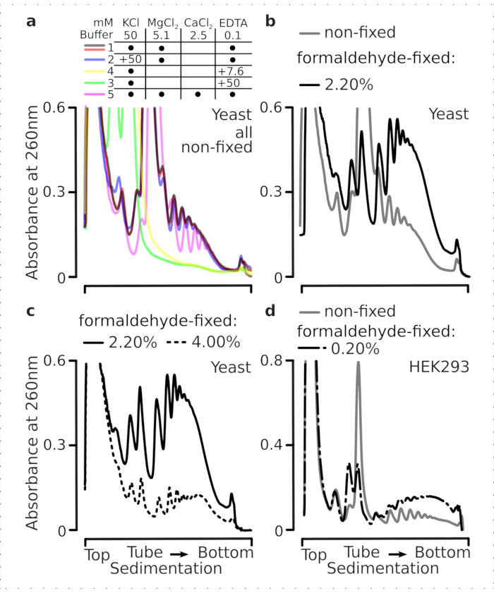
图2:翻译复合物提取的缓冲条件及固定对稳定性的评估。图中显示了通过10%–40% w/v蔗糖梯度分离的总酵母细胞裂解液在260 nm处测得的紫外吸光度谱图。(a) 单价和二价盐以及镁离子螯合对非固定酵母细胞提取物沉降行为的影响。红色和灰色线条代表典型的重复实验结果。(b,c) 来自非固定酵母细胞(灰色线条)、2.2%(黑色线条)和4.4% w/v甲醛固定的酵母细胞(黑色虚线)所获得裂解液的比较。(d) 与相同非固定细胞的样品(灰色线条)相比,经优化的0.2% w/v甲醛固定处理对HEK 293T细胞多聚核糖体的稳定作用(黑色点划线)。请点击此处查看该图的放大版本。
接下来,我们检测了使用不同浓度甲醛固定对多聚核糖体稳定化的影响。在细胞材料、缓冲液、细胞处理及操作时间等其他条件完全相同的情况下,我们比较了未固定细胞以及用2.2%和4%(w/v)甲醛固定的细胞所提取的材料(图2b,c)。结果表明,2.2%(w/v)甲醛更适用于固定,因为它不仅能很好地保留多聚核糖体(可通过多聚核糖体与单核糖体的比值判断,图2b),而且与4%(w/v)甲醛相比,并未降低核糖体材料的总体得率;后者则表现出明显的过度固定迹象(图2c)。
对于来源于哺乳动物细胞的材料,由于基于去垢剂的提取需要较大的裂解缓冲液体积与细胞体积之比,因此使用了缓冲液2(图2a)。该条件在蔗糖梯度离心中可产生分辨良好的翻译复合物(图2d)。值得注意的是,此处使用的甲醛浓度较低,仅为0.2%(w/v),因为较高浓度会导致多核糖体和核糖体物质显著丢失(数据未显示)。与酵母细胞实验结果相似,交联稳定的样品显示出多核糖体更好的保存效果以及更高的多核糖体与单核糖体比率(图2d)。
接下来,我们检测了所选的甲醛固定条件是否足以通过交联作用有效稳定多聚核糖体组分中正在活跃翻译的 mRNA,从而确认多聚核糖体产量的提高并非仅仅由于抑制了酶活性和翻译延伸进程。我们使用 EDTA 和高单价盐(KCl)来破坏多聚核糖体和核糖体的稳定性。这些试剂被添加至澄清的酵母细胞裂解液中,并分别在后续所有缓冲液及蔗糖梯度中,叠加于缓冲液 1 的组分之上。
事实上,15 mM EDTA 对来自固定细胞的多聚核糖体组分的去稳定作用较弱(图 3a),表明交联复合物具有更高的稳定性。通过提高甲醛浓度可在一定程度上抵消 EDTA 的去稳定作用,因为经 4% w/v 甲醛固定的细胞所提取的材料更耐解聚(图 3a)。然而,将 EDTA 浓度提高至 50 mM 时,在固定与未固定条件下均导致大部分翻译复合物发生去稳定,这可从物质沉降速度减慢以及缺乏清晰峰形得到推断(图 3b)。这一现象可归因于结构的部分解折叠及整体致密性的降低,而非多聚核糖体组分从 mRNA 上的完全解离。即便在此情况下,交联样品仍表现出更快的沉降速率(图 3b)。
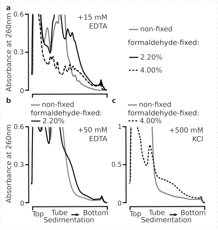
图3:影响 体内 酵母甲醛固定对多聚核糖体稳定性的影响 缓冲液1(参见正文和 图 2a) 在所有实验中均被使用。数据类型和绘图方法如文中所述 图2 图注。 (a) 比较在非固定细胞(灰色线)、2.2%(黑色线)和4%(黑色虚线,w/v)甲醛固定细胞所提取的多聚核糖体中,向细胞裂解液及后续缓冲液中添加15 mM EDTA对其稳定性的影响。b与(相同a),但需添加50 mM EDTA,并排除4% w/v的甲醛固定细胞。c与(相同a),但需添加 500 mM KCl,并排除 2.2% w/v 的甲醛固定细胞。 请点击此处以查看此图的放大版本。
与EDTA的作用类似,在500 mM KCl条件下,我们发现4% w/v的甲醛固定显著提高了结构的稳定性(图3c)。此种情况下观察到的致密性表观降低,也可归因于核糖体复合物组分的部分脱离,而非其完全从RNA上解离。总体而言,来自甲醛固定细胞的多聚核糖体表现出更强的抗解折叠和抗结构失稳能力,这与复合物内部形成了额外的共价键一致。
在促进生长的条件下,mRNA 可迅速启动翻译,导致多个核糖体结合于同一条 mRNA 分子上,形成称为多聚核糖体(polyribosomes)或简称多聚体(polysomes)的结构。多聚体可通过蔗糖密度梯度超速离心进行分离,其沉降行为取决于其所含核糖体的数量(即 mRNA 上同时结合的核糖体数目)。当翻译受到抑制时,核糖体无法及时进入下一轮翻译,导致多聚体发生(部分)“解聚”,表现为多聚体向低阶数形式迁移的模式性偏移以及单体核糖体(monosomes)的累积。4,26.
葡萄糖饥饿可提供一种能够在多核糖体分布水平上可视化的翻译应答模型。葡萄糖耗竭会引发酵母中最显著且最快速的翻译抑制效应之一1,3,40。先前的研究表明,在葡萄糖耗竭后1分钟内,即可发生多核糖体的丧失、单核糖体的积累以及翻译起始的抑制4。在重新补充葡萄糖5分钟内,翻译迅速恢复,并伴随多核糖体的明显增加3,4。此外还观察到,当细胞暴露于葡萄糖浓度为0.5%(w/v)或更低的培养基中时,翻译受到抑制;而在葡萄糖浓度为0.6%(w/v)或更高时,则未观察到抑制效应。
因此,我们希望确定所采用的固定条件是否适用于保存葡萄糖应激反应动态过程中存在的翻译水平差异,这种差异可通过多聚核糖体与单核糖体的比值进行评估。我们将高葡萄糖(添加2.00% w/v)培养至对数中期的细胞材料,与转移至无添加或低添加(分别为0.00%或0.25% w/v)葡萄糖培养基中10分钟后的细胞材料进行比较。固定处理采用2.2% w/v甲醛进行,同时对对照组(非饥饿组;快速更换为含2% w/v添加葡萄糖的标准培养基,孵育10分钟后固定)和饥饿10分钟组(快速更换为相同培养基但含低浓度0.25% w/v或无添加葡萄糖,孵育10分钟后固定)的细胞进行平行处理。
与先前的研究结果一致,我们观察到酵母细胞在葡萄糖饥饿应激条件下显著抑制翻译(图4a)。在未添加葡萄糖和低葡萄糖条件下,均诱导了多聚核糖体的解聚,其中低葡萄糖添加条件下保留的多聚核糖体略多,但差异明显。因此,酵母对葡萄糖去除的响应可能并非“全有或全无”型,而是逐步调节的。正如预期,甲醛交联具有稳定作用,固定细胞中的多聚核糖体物质在饥饿与非饥饿细胞之间表现出更明显的区分度,可能更好地保留了响应过程的动态范围(图4b)。有趣的是,对于固定后的样品,低葡萄糖添加浓度所导致的多聚核糖体丰度与未添加葡萄糖条件之间的差异,明显优于未固定细胞中的表现(图4a)。这强烈表明,甲醛固定法在保留和捕获高度动态过程(如翻译响应)中相对微小且短暂的平衡变化方面具有良好的适用性。
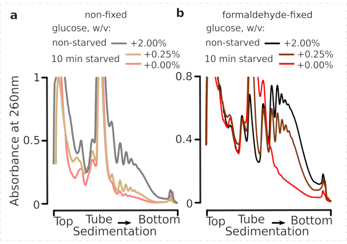
图 4:在葡萄糖饥饿条件下捕获酵母翻译过程的快速变化。 所有实验均使用缓冲液 1(见正文及图 2a)。数据类型与绘图方式参见图 2图注。(a) 来自未饥饿处理(灰色线)、限制性葡萄糖饥饿处理(添加 0.25% w/v 葡萄糖,处理 10 分钟;棕色线)和葡萄糖耗尽处理(不添加葡萄糖,处理 10 分钟;红色线)的未固定酵母细胞裂解液。(b) 与 (a) 相同,但使用 2.2% w/v 甲醛固定的细胞。请点击此处查看该图的放大版本。
利用蔗糖密度梯度离心法(“多聚核糖体分析”)检测与活跃翻译的mRNA结合的核糖体,以监测翻译状态,是一种广泛应用的技术26,27,28。结合定量微阵列分析,以及近年来的高通量测序技术28,44,多聚核糖体分析可在全转录组范围内提供关于核糖体结合mRNA的信息。在蛋白质生物合成研究领域,通常基于若干假设认为:mRNA存在于多聚核糖体中,表明其正活跃参与翻译过程。进一步地,通常(但并非总是)可以合理推断:在给定长度的mRNA上结合的核糖体越多(即多聚核糖体的阶数越高),该mRNA参与翻译的活跃程度也越高。因此,将多聚核糖体组分与其他组分分离,有助于富集正在活跃翻译的RNA。在核糖体印迹分析方法中,特别是TCP-seq10,38,39,该方法可产生来源于扫描、起始和终止密码子复合物的游离小亚基(SSU)的独立群体,进一步去除那些不与完整单体核糖体或多聚核糖体共沉降的核糖体亚基,可能提供额外的信息。
因此,我们将“非翻译”mRNP(例如游离SSU,即与单个SSU结合的mRNA或未结合mRNA的SSU)与“正在活跃翻译”的mRNA群体分离开来。为实现这一目的,我们假设与单个(单体)或多个核糖体(多聚核糖体)相互作用的mRNA可能正处于活跃翻译状态。这类复合物因其具有较高的沉降系数,可与其他成分分离。我们还建议将“正在活跃翻译”的mRNA群体分离至蔗糖垫层(50% w/v 蔗糖),而非直接在离心管壁上沉淀样品。通过将快速沉降的复合物离心进入垫层,可利用吸光度谱图监测分离效果,并获得比沉淀后复溶更高的可溶性、非聚集且未变性材料得率10,38。
总体而言,为了纯化单个小亚基(SSUs)、核糖体、双体(disomes)以及可能存在的高级有序紧密排列的多聚核糖体,将固定的澄清裂解液进行两步超速离心处理(图5)。在第一次蔗糖梯度离心中,超速离心导致游离的小亚基(SSUs)和大亚基(LSUs)分离并位于梯度上部(10%–20% w/v 蔗糖),而交联翻译产物组分(包括多聚核糖体及与完整核糖体结合的 mRNA)则富集于梯度底部(50% w/v 蔗糖)图 5a)。含有翻译后 mRNA 复合物的蔗糖溶液下层 50%(w/v)部分随后被浓缩,并用 RNase I 消化其 RNA,接着进行第二次蔗糖梯度超速离心,以分离出小亚基(SSU)、大亚基(LSU)、核糖体单体(RS)、RNase 抗性二聚体(DS)以及少量高阶核酸酶抗性多聚核糖体。 (图 5b)。经醋酸铀负染色及透射电子显微镜成像,证实了各沉降阶段所分离复合物的身份(图5).

图 5:将全部翻译后的 RNA 组分与未翻译的 RNA 分离。(a,c)示意图(左)及相应的代表性结果(右;数据类型和绘图方式如图 2图注所述),其中(a)为首次不连续蔗糖梯度分离,分离出非翻译型胞质组分,包括游离的小亚基(SSU)以及通过与核糖体和多聚核糖体共沉降而鉴定出的已翻译 mRNA 池;(c)为经受控的 RNase I 消化并穿过第二次线性蔗糖梯度超速离心后,从已翻译 mRNA 池中释放出的各个核糖体复合物的分离结果,分离为小亚基(SSU)、大亚基(LSU)、核糖体(RS)以及核酸酶抗性双体(DS)组分。实验中分别使用了高(15 AU260)和低(8 AU260)量的非饥饿状态消化材料,以展示在关注微量组分时增加超速离心上样量的可能性。也可识别出更高阶的核酸酶抗性多聚核糖体(例如,示例中所示的三聚体)。(b,d)分别为来自(a,c)的醋酸铀酰染色组分的代表性透射电子显微镜(TEM)图像,如图中标注。 请点击此处查看该图的放大版本。
为了检验固定方案对保留瞬时核糖体结合蛋白(特别是真核起始因子,eIFs)的适用性,我们检测了在核糖体各组分中eIF4A是否发生共沉淀。eIF4A是一种与核糖体动态结合且不稳定的真核起始因子。我们利用带有TAP标签的eIF4A酵母菌株(TIF1-TAP),检测了固定处理后所得样品中eIF4A的存在情况 与 使用抗TAP抗体对非固定细胞进行检测,并以Pab1p作为额外的RNA结合对照,通过SDS-PAGE电泳及随后的蛋白质印迹(western blotting)分析其丰度图6).
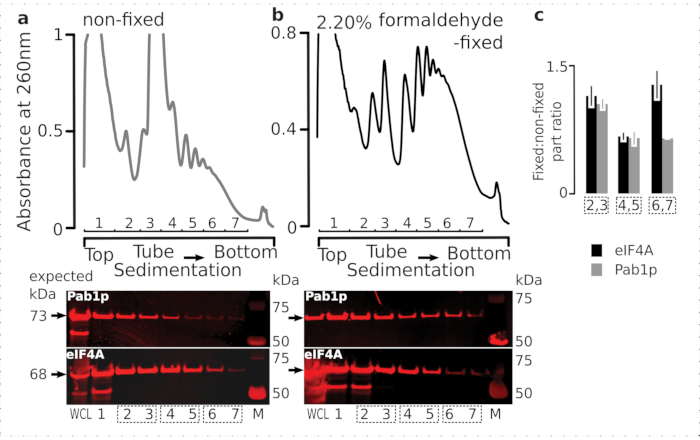
图6:翻译复合物中瞬时蛋白的稳定化 体内 甲醛固定 (a,b) (上方图示) 全细胞裂解液(WCL)的(a)未固定的和(b2.2% 甲醛固定的eIF4A-TAP酵母细胞经超速离心分离,并按所述方法进行观察 图2 图注。 (底部图示) 对相应蔗糖梯度分离样品(上图)及全细胞裂解液(WCL)对照进行 Western blot 成像。c固定材料与非固定材料组分中eIF4A或Pab1p丰度的平均比值。根据(a,b)的数据,计算了eIF4A(黑色柱)和Pab1p(灰色柱)在第2、3组分(SSU,LSU)、第4、5组分(RS,轻度多聚核糖体)以及第6、7组分(重度多聚核糖体)中的相对比例(归一化至第2–7组分总信号),并取其固定与非固定样品的比值(底部图)。误差线表示以合并组分(虚线框)作为重复样本时,比值相对于平均值的标准偏差。 请点击此处查看此图的放大版本。
与这两种蛋白在细胞中含量较高相一致,我们在未固定细胞的全细胞裂解液(WCL)以及沉降较慢的组分中均观察到较强的信号(图6a,底图)。我们还在固定细胞的全细胞裂解液中检测到大量这些蛋白,表明交联材料的提取效率较高,且未出现意外损失(图6b,底图)。然而,与未固定细胞不同的是,来自固定细胞的材料显示,在沉降较快的核糖体组分中,eIF4A相对于Pab1p的相对丰度升高(图6c)。该结果提示,在甲醛交联的样品中,eIF4A与多聚核糖体的结合更为牢固。
在确认交联对eIF4A在核糖体组分中存在具有阳性且特异的稳定作用后,我们利用带有eIF4A标签(TIF1-TAP)的酵母菌株的固定材料,通过磁性IgG磁珠进行亲和纯化,以捕获并富集含eIF4A的复合物。我们在蔗糖梯度离心第一次沉降后,对WCL、游离SSU以及多聚核糖体(翻译中的mRNA池)组分进行了亲和富集(例如,酵母实验方案第1.3节);此外,在第二次沉降中,将翻译产物池经RNase I处理解离为单个复合物后,进一步对SSU、LSU和RS组分进行了富集(例如,酵母实验方案第1.4节)(图7)。除LSU组分外,在所有情况下,与起始材料(上样量,I)中的β-actin相比,我们均能在纯化后的组分(洗脱组分,E)中观察到eIF4A的选择性富集(图7)。
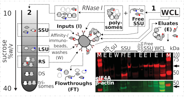
图 7选择性免疫纯化 体内 由瞬时结合的eIF4A稳定化的甲醛交联翻译复合物 示意图展示了不同翻译复合物的来源及eIF4A表位,包括未分级的eIF4A-TAP酵母细胞澄清WCL;通过第一次超速离心分离出的游离SSU和翻译RNA池(多聚核糖体);经RNase I消化释放出SSU、LSU和RS组分,并通过第二次超速离心进行分离(见正文)。Western印迹图像显示了各组分中eIF4A的丰度,与同时染色的β-actin对照的丰度进行比较。 请点击此处以查看此图的放大版本。
补充表 1. 请点击此处下载该表格。
讨论
甲醛固定是一种方便且常用的方法,可实现生物分子的快速体内交联10,36,45,46,47,48。与其他可能的生物分子靶标相比,成功捕获翻译复合物需要在细胞或其他材料快速冷冻的同时立即进行固定。若不及时稳定,多种与翻译相关的过程可能继续进行,导致复合物分布偏离未受干扰的体内状态49。与其他翻译抑制和核糖体复合物稳定方法相比,甲醛能够迅速穿透细胞膜,并以非特异性方式形成交联,因而有望最大限度地保留翻译复合物中间体的多样性,使其更接近天然分布状态50。
本文所述方法已在酵母和哺乳动物细胞中建立并优化,其他研究团队也已在此基础上开发出适用于更多样化生物材料的方法,例如在完整脊椎动物(如斑马鱼胚胎)中的应用10,38,39,49,51,52。尽管这些研究共同证实了该方法的通用性和广泛适用性,但由于需要进行优化和调整,翻译复合物的快速甲醛交联在转移至新型生物材料时仍被认为具有一定难度。
该方法成功的关键要求之一是对甲醛浓度以及细胞收集和破碎技术进行重新优化。通透性较差、体积小且呈圆形的酵母细胞需要更高浓度的甲醛(至少高10倍)并对固定后的细胞进行物理破碎。相比之下,培养中的大型扁平贴壁哺乳动物细胞容易发生过度固定,因此在固定过程中需轻柔操作,而固定复合物的提取则可通过使用去垢剂进行化学裂解膜结构来完成。交联不足可能导致较不稳定或寿命较短的中间产物解离或渗漏至后续状态;而交联过度则可能对核糖体组分的分离与研究产生不利影响,并引发选择性偏差,例如导致较重复合物更显著的损耗。据我们观察,即使是微小的变化,例如所用贴壁人源细胞的类型不同,也可能影响交联复合物的回收产量,因而需要重新优化交联方案。我们还可以预见,具有显著不同通透性特征的细胞(如植物细胞)将需要对固定条件进行更广泛的额外优化52。然而,很难设想有任何类型的生物材料会完全不适用于该方法。
与哺乳动物细胞固定方案相关的一个考虑因素是用作输入的细胞材料的密度和数量。建议细胞持续生长至少2天,期间不进行传代培养或其他干扰,以避免对外部细胞翻译动态产生影响。该建议适用于大多数细胞类型,但对于大多数贴壁细胞,保持汇合度不超过70%可确保避免显著的接触抑制效应,此类效应可能对翻译速率产生负面且不可预测的影响。
甲醛固定的另一个有趣且可能具有独特便利性的特征源于其无选择性的反应性,即对混合分类系统中翻译复合物的稳定作用。细菌,尤其是线粒体、叶绿体以及各种细胞内寄生生物的翻译复合物,历来难以通过特异性的翻译抑制剂进行靶向研究。相比之下,在TCP-seq数据中,映射到线粒体转录组的足迹序列可被清晰观测到38,39,50。一个值得关注的后续发展方向是利用该方法研究整个微生物群落(如土壤、水体或肠道样本)中的翻译过程,因为在这些复杂环境中,若采用其他方法实现可靠且快速的翻译终止及复合物稳定,将面临较大困难。
还应指出,对于最复杂的材料(如坚硬和/或体积较大的组织),在细胞破裂及材料均质化后可立即使用甲醛进行固定,且并无任何限制。该方法已被广泛用于在利用特定小分子抑制剂稳定翻译复合物时,消除细胞通透性带来的延迟。33,53,54,55鉴于甲醛固定在传统上一直被使用,并取得了极好的效果 离体/体外 样品稳定化在电子显微镜等应用中的作用45,56,57,58在这种情况下,我们可以预期负面效应会更少,特别是与从充分固定的细胞中提取翻译复合物效果不佳相关的问题。
我们的研究结果证实,快速甲醛固定法可用于稳定高度瞬时的复合物,例如包含 eIF4A 的复合物。值得注意的是,与哺乳动物不同,酵母中的 eIF4A 与帽结合复合物 eIF4F 的结合要弱得多,因此总体上与翻译复合物的结合也较弱。在酵母中,任何对核糖体材料进行的较彻底纯化过程通常都会导致 eIF4A 的丢失29,59,60,61,62,63。然而,在经过 in vivo 固定的酵母样品中,可以在所有预期存在 eIF4A 的翻译复合物组分中实现其可靠富集。先前发表的 Sel-TCP-seq 数据已展示了与核糖体结合更强的 eIF2 和 eIF3 的富集情况(同时也揭示了瞬时发生的共翻译蛋白复合物组装)39。因此,该方法适用于检测与翻译复合物结合较强和较弱的各类组分。
综上所述,我们介绍了一种可用于深入研究翻译起始阶段变化的方法,尤其适用于需要最小程度干扰mRNA上核糖体分布的情况。重要的是,该方法适用于稳定翻译复合物中相对不稳定且动态的组分(如eIF4A),并在经过必要优化后可广泛使用。我们还提供了证据,表明甲醛固定在研究翻译过程快速动态变化的情境中具有实用价值,从而为研究细胞对环境变化或应激条件的快速反应等课题开辟了新的方向。
披露
作者声明无利益冲突。
致谢
本工作得到了澳大利亚研究理事会发现项目基金(DP180100111,授予T.P.和N.E.S.)、国家健康与医学研究委员会研究者基金(GNT1175388,授予N.E.S.)以及研究奖学金(APP1135928,授予T.P.)的支持。作者们感谢澳大利亚国立大学先进显微镜中心的显微镜澳大利亚设施所提供的技术支持,该设施由大学及联邦政府共同资助。
材料
| 姓名 | 公司 | 目录编号 | 评论 |
|---|---|---|---|
| 酵母提取物 | Merck, Sigma-Aldrich | 70161 | |
| 蛋白胨 | Merck, Sigma-Aldrich | 70178 | |
| D-葡萄糖(右旋糖) | Merck, Sigma-Aldrich | 49139 | |
| 硫酸腺嘌呤 | Amresco | 0607-50G | |
| 甲醛溶液 | Merck Sigma-Aldrich | F11635-500ML | ACS级试剂,37 wt. % 溶于 H2O,含10-15%甲醇作为稳定剂(防止聚合) |
| RNaseOUT™ 重组核糖核酸酶抑制剂 | Invitrogen™ byThermo Fischer Scientific | 10777019 | |
| cOmplete™,无EDTA蛋白酶抑制剂混合物 | COEDTAF-RO Roche by Merck | 11873580001 | |
| 氯化镁溶液 | (Merck/Sigma-Aldrich) | M1028 | |
| 乙二胺四乙酸溶液 | (Merck/Sigma-Aldrich) | E7889 | |
| Ambion™ RNase I,克隆型,100 U/µL | Ambion | AM2294 | |
| SUPERase•In™ RNase抑制剂(20 U/μL) | Invitrogen™ by Thermo Fisher Scientific | AM2694 | |
| 酸性苯酚:氯仿:异戊醇 125:24:1(pH 4.0-5.0) | (Merck/Sigma-Aldrich) | P1944-100ML | |
| Dynabeads™ 羊抗小鼠IgG磁珠 | Invitrogen™ by Thermo Fisher Scientific) | 11033 | |
| 醋酸钠(3 M),pH 5.5 | Invitrogen™ by Thermo Fisher Scientific) | AM9740 | |
| 糖原(5 mg/ml) | Invitrogen™ by Thermo Fisher Scientific) | AM9510 | |
| 乙醇,纯级 | Merck; Sigma Aldrich | E7023 | |
| Amersham™ Hybond® P 蛋白印迹膜,PVDF | Merck | GE10600023 | PVDF膜用于蛋白印迹 |
| Bolt™ 4 至 12%,Bis-Tris,1.0 mm,Mini Protein Gel | Invitrogen™ by ThermoFischer Sientific | NW04120BOX | 蛋白凝胶 |
| 4X Bolt™ LDS 上样缓冲液 | Invitrogen™ by ThermoFischer Sientific | B0007 | LDS上样缓冲液 |
| Precision Plus Protein™ Kaleidoscope™ 预染蛋白标准品 | BioRad | 1610375 | 蛋白分子量标准 |
| 20X Bolt™ MES SDS 电泳缓冲液 | ThermoFischer Scientific | B0002 | PAGE电泳缓冲液 |
| Intercept® (PBS) 封闭缓冲液 | LI-COR | 927-70001 | Odyssey封闭缓冲液(PBS) |
| IRDye® 800CW 羊抗小鼠IgG二抗 | LI-COR | 92632210 | |
| IRDye® 800CW 羊抗兔IgG二抗 | LI-COR | 92632211 | |
| TAP标签多克隆抗体 | Invitrogen™ by ThermoFischer Sientific | CAB1001 | |
| 抗β-肌动蛋白抗体 | Abcam | ab8227 | |
| 蔗糖 | (Merck/Sigma-Aldrich) | 84097 | BioUltra,分子生物学级,≥99.5%(HPLC) |
| DL-二硫苏糖醇溶液 | (Merck/Sigma-Aldrich) | 43816 | BioUltra,分子生物学级,~1 M 溶于 H2O |
| Terumo 1CC/mL 注射器 | Terumo Syringe | 878499 | |
| 氯化钾 | (Merck/Sigma-Aldrich) | 60128 | |
| HEPES | (Merck/Sigma-Aldrich) | H3375 | |
| DMEM高糖培养基(Dulbecco改良Eagle培养基) | Sigma Aldrich | D5796 | |
| 胎牛血清 | Sigma Aldrich | 12003C | |
| 胰蛋白酶-EDTA(0.05%),含酚红 | Gibco | 25300062 | |
| 含钙镁的磷酸盐缓冲液(Dulbecco's Phosphate Buffered Saline) | Sigma-Aldrich | D8662 | |
| 甘氨酸 | Sigma-Aldrich | G7126 | |
| Tris盐酸盐 | Merck/Sigma-Aldrich | 10812846001 | |
| 十二烷基硫酸钠 | Merck/Sigma-Aldrich | 436143 | |
| IGEPAL CA-630 | Merck/Sigma-Aldrich | I3021 | |
| Rnasin 核糖核酸酶抑制剂 | Promega | N2111 | |
| 不锈钢研磨罐 | Retsch | 02.462.0059 | |
| MM400 混合研磨仪 | Retsch | 20.745.0001 | |
| 梯度分馏仪 | Brandel | BRN-BR-188 | |
| Thermomixer R | Eppendorf | Z605271 | |
| Nanodrop 分光光度计 | Thermo Fisher Scientific | ND-2000 | |
| 0.5 ml 带锁装置的微量离心管 | Eppendorf Safe-Lock | 30121023 | |
| Mini Gel 电泳槽 | (Thermo Fisher Scientific) | A25977 | PAGE电泳槽 |
| 5 mL 开口薄壁超透明离心管,13 x 51 mm | Beckman-Coulter | 344057 | |
| 13.2 mL 认证无菌开口薄壁聚丙烯离心管,14 x 89 mm - 50支装 | Beckman-Coulter | 331372 | |
| Amicon Ultra-0.5 超滤装置 | Merck | UFC5030 | Ultracel-30 再生纤维素膜,0.5 mL 样品体积 |
| Thermo Sorvall Evolution RC 地面超速离心机 | Cambridge Scientific | 15566 | |
| Beckman Coulter Optima L-90K | GMI | 8043-30-1191 | |
| Nunc EasYFlask 175 cm² 培养瓶 | Thermofisher Scientific | 159910 | |
| Falcon 50 mL 锥形离心管 | Thermofisher Scientific | 14-432-22 | |
| 25 mL 移液管 | Sigma-Aldrich | SIAL1250 | |
| 10 mL 移液管 | Sigma-Aldrich | SIAL1100 | |
| DNA低吸附管 | Eppendorf | 30108051 | |
| 5810 R 冷冻离心机 | Eppendorf | EP022628188 | 适用于50 mL离心管 |
| 轨道摇床培养箱 | Ratek | OM11 | |
| Frezco 17 微型离心机 | Thermofisher Scientific | 75002402 | |
| Eppendorf DNA低吸附管 | Merck/Sigma-Aldrich | EP0030108051 | |
| Eppendorf® Protein LoBind 管 | Merck/Sigma-Aldrich | EP0030108116 | |
| SW 41 Ti 摆动桶转子 | Beckman-Coulter | 331362 | |
| Heracell™ 150i CO2 培养箱,150 L | Thermofisher Scientific | 51026282 | |
| 0.3 mL 超细II型短胰岛素注射器 | BD Medical | 328822 | |
| 3 mL 带鲁尔锁扣头的注射器 | BD Medical | 302113 | |
| 25 G x 16 mm 皮下注射针头 | Terumo | TUAN2516R1 |
参考文献
- Janapala, Y., Preiss, T., Shirokikh, N. E. Control of translation at the initiation phase during glucose starvation in yeast. International Journal of Molecular Sciences. 20 (16), 4043(2019).
- Masvidal, L., Hulea, L., Furic, L., Topisirovic, I., Larsson, O. mTOR-sensitive translation: Cleared fog reveals more trees. RNA Biology. 14 (10), 1299-1305 (2017).
- Ashe, M. P., De Long, S. K., Sachs, A. B. Glucose depletion rapidly inhibits translation initiation in yeast. Molecular Biology of the Cell. 11 (3), 833-848 (2000).
- Crawford, R. A., Pavitt, G. D. Translational regulation in response to stress in Saccharomyces cerevisiae. Yeast. 36 (1), 5-21 (2019).
- Melamed, D., Pnueli, L., Arava, Y. Yeast translational response to high salinity: global analysis reveals regulation at multiple levels. RNA. 14 (7), 1337-1351 (2008).
- Hershey, J. W., Sonenberg, N., Mathews, M. B. Principles of translational control: An overview. Cold Spring Harbor Perspectives in Biology. 4 (12), 011528(2012).
- Mata, J., Marguerat, S., Bähler, J. Post-transcriptional control of gene expression: a genome-wide perspective. Trends in Biochemical Sciences. 30 (9), 506-514 (2005).
- Spriggs, K. A., Bushell, M., Willis, A. E. Translational regulation of gene expression during conditions of cell stress. Molecular Cell. 40 (2), 228-237 (2010).
- Liu, B., Qian, S. B. Translational reprogramming in cellular stress response. Wiley Interdisciplinary Reviews RNA. 5 (3), 301-315 (2014).
- Archer, S. K., Shirokikh, N. E., Beilharz, T. H., Preiss, T. Dynamics of ribosome scanning and recycling revealed by translation complex profiling. Nature. 535 (7613), 570-574 (2016).
- Hinnebusch, A. G., Ivanov, I. P., Sonenberg, N. Translational control by 5'-untranslated regions of eukaryotic mRNAs. Science. 352 (6292), 1413-1416 (2016).
- Dever, T. E., Green, R. The elongation, termination, and recycling phases of translation in eukaryotes. Cold Spring Harbor Perspectives in Biology. 4 (7), 013706(2012).
- Shirokikh, N. E., Preiss, T. Translation initiation by cap-dependent ribosome recruitment: Recent insights and open questions. Wiley Interdisciplinary Reviews RNA. 9 (4), 1473(2018).
- Jiménez-Díaz, A., Remacha, M., Ballesta, J. P., Berlanga, J. J. Phosphorylation of initiation factor eIF2 in response to stress conditions is mediated by acidic ribosomal P1/P2 proteins in Saccharomyces cerevisiae. PLoS One. 8 (12), 84219(2013).
- Sonenberg, N., Hinnebusch, A. G. Regulation of translation initiation in eukaryotes: mechanisms and biological targets. Cell. 136 (4), 731-745 (2009).
- Majmundar, A. J., Wong, W. J., Simon, M. C. Hypoxia-inducible factors and the response to hypoxic stress. Molecular Cell. 40 (2), 294-309 (2010).
- Barraza, C. E., et al. The role of PKA in the translational response to heat stress in Saccharomyces cerevisiae. PLoS One. 12 (10), 0185416(2017).
- Richter, K., Haslbeck, M., Buchner, J. The heat shock response: Life on the verge of death. Molecular Cell. 40 (2), 253-266 (2010).
- Jamar, N. H., Kritsiligkou, P., Grant, C. M. The non-stop decay mRNA surveillance pathway is required for oxidative stress tolerance. Nucleic Acids Research. 45 (11), 6881-6893 (2017).
- Chen, Z., et al. The complete pathway for thiosulfate utilization in Saccharomyces cerevisiae. Applied and Environmental Microbiology. 84 (22), (2018).
- Marzluf, G. A. Molecular genetics of sulfur assimilation in filamentous fungi and yeast. Annual Review of Microbiology. 51, 73-96 (1997).
- Miller, D., Brandt, N., Gresham, D. Systematic identification of factors mediating accelerated mRNA degradation in response to changes in environmental nitrogen. PLoS Genetics. 14 (5), 1007406(2018).
- Zhang, W., Du, G., Zhou, J., Chen, J. Regulation of sensing, transportation, and catabolism of nitrogen sources in Saccharomyces cerevisiae. Microbiology and Molecular Biology Reviews. 82 (1), (2018).
- Tokpohozin, S. E., Fischer, S., Becker, T. Selection of a new Saccharomyces yeast to enhance relevant sorghum beer aroma components, higher alcohols, and esters. Food Microbiology. 83, 181-186 (2019).
- Walker, G. M., Stewart, G. G. Saccharomyces cerevisiae in the production of fermented beverages. Beverages. 2 (4), 30(2016).
- Chassé, H., Boulben, S., Costache, V., Cormier, P., Morales, J. Analysis of translation using polysome profiling. Nucleic Acids Research. 45 (3), 15(2017).
- Jin, H. Y., Xiao, C. An integrated polysome profiling and ribosome profiling method to investigate in vivo translatome. Methods in Molecular Biology. 1712, 1-18 (2018).
- Arava, Y., et al. Genome-wide analysis of mRNA translation profiles in Saccharomyces cerevisiae. Proceedings of the National Academy of Sciences of the United States of America. 100 (7), 3889-3894 (2003).
- Lackner, D. H., et al. A network of multiple regulatory layers shapes gene expression in fission yeast. Molecular Cell. 26 (1), 145-155 (2007).
- Ingolia, N. T., Ghaemmaghami, S., Newman, J. R. S., Weissman, J. S. Genome-Wide Analysis in Vivo of Translation with Nucleotide Resolution Using Ribosome Profiling. Science. 324 (5924), 218-223 (2009).
- Ingolia, N. T., Hussmann, J. A., Weissman, J. S. Ribosome Profiling: Global Views of Translation. Cold Spring Harbor Perspectives in Biology. 11 (5), (2019).
- Gerashchenko, M. V., Lobanov, A. V., Gladyshev, V. N. Genome-wide ribosome profiling reveals complex translational regulation in response to oxidative stress. Proceedings of the National Academy of Sciences of the United States of America. 109 (43), 17394-17399 (2012).
- Hussmann, J. A., Patchett, S., Johnson, A., Sawyer, S., Press, W. H. Understanding biases in ribosome profiling experiments reveals signatures of translation dynamics in yeast. Proceedings of the National Academy of Sciences Genetics. 11 (12), 1005732(2015).
- Santos, D. A., Shi, L., Tu, B. P., Weissman, J. S. Cycloheximide can distort measurements of mRNA levels and translation efficiency. Nucleic Acids Research. 47 (10), 4974-4985 (2019).
- Schneider-Poetsch, T., et al. Inhibition of eukaryotic translation elongation by cycloheximide and lactimidomycin. Nature Chemical Biology. 6 (3), 209-217 (2010).
- Hoffman, E. A., Frey, B. L., Smith, L. M., Auble, D. T. Formaldehyde crosslinking: A tool for the study of chromatin complexes. Journal of Biological Chemistry. 290 (44), 26404-26411 (2015).
- Kage, U., Powell, J. J., Gardiner, D. M., Kazan, K. Ribosome profiling in plants: What is not lost in translation. Journal of Experimental Botany. 71 (18), 5323-5332 (2020).
- Shirokikh, N. E., Archer, S. K., Beilharz, T. H., Powell, D., Preiss, T. Translation complex profile sequencing to study the in vivo dynamics of mRNA-ribosome interactions during translation initiation, elongation and termination. Nature Protocols. 12 (4), 697-731 (2017).
- Wagner, S., et al. Selective translation complex profiling reveals staged initiation and co-translational assembly of initiation factor complexes. Molecular Cell. 79 (4), 546-560 (2020).
- Zlotorynski, E. Profiling ribosome dynamics. Nature Reviews Molecular Cell Biology. 17 (9), 535-535 (2016).
- Sen, N. D., Gupta, N., S, K. A., Preiss, T., Lorsch, J. R., Hinnebusch, A. G. Functional interplay between DEAD-box RNA helicases Ded1 and Dbp1 in preinitiation complex attachment and scanning on structured mRNAs in vivo. Nucleic Acids Research. 47 (16), 8785-8806 (2019).
- Zhao, J., Qin, B., Nikolay, R., Spahn, C. M. T., Zhang, G. Translatomics: The global view of translation. International Journal of Molecular Sciences. 20 (1), 20010212(2019).
- Luthe, D. S. A simple technique for the preparation and storage of sucrose gradients. Analytical Biochemistry. 135 (1), 230-232 (1983).
- Wang, Z., Gerstein, M., Snyder, M. RNA-Seq: A revolutionary tool for transcriptomics. Nature Review Genetics. 10 (1), 57-63 (2009).
- Orlando, V. Mapping chromosomal proteins in vivo by formaldehyde-crosslinked-chromatin immunoprecipitation. Trends in Biochemical Sciences. 25 (3), 99-104 (2000).
- Schmiedeberg, L., Skene, P., Deaton, A., Bird, A. A Temporal Threshold for Formaldehyde Crosslinking and Fixation. PLoS One. 4 (2), 4636(2009).
- Solomon, M. J., Varshavsky, A. Formaldehyde-mediated DNA-protein crosslinking: A probe for in vivo chromatin structures. Proceedings of the National Academy of Sciences. 82 (19), 6470-6474 (1985).
- Solomon, M. J., Larsen, P. L., Varshavsky, A. Mapping proteinDNA interactions in vivo with formaldehyde: Evidence that histone H4 is retained on a highly transcribed gene. Cell. 53 (6), 937-947 (1988).
- Bohlen, J., Fenzl, K., Kramer, G., Bukau, B., Teleman, A. A. Selective 40S footprinting reveals cap-tethered ribosome scanning in human cells. Molecular Cell. 79 (4), 561-574 (2020).
- Shirokikh, N. E. Translation complex stabilization on messenger RNA and footprint profiling to study the RNA responses and dynamics of protein biosynthesis in the cells. Critical Reviews in Biochemistry and Molecular Biology. , (2021).
- Giess, A., et al. Profiling of small ribosomal subunits reveals modes and regulation of translation initiation. Cell Reports. 31 (3), 107534(2020).
- Firmino, A. A. P., et al. Separation and paired proteome profiling of plant chloroplast and cytoplasmic ribosomes. Plants (Basel). 9 (7), (2020).
- Gerashchenko, M. V., Gladyshev, V. N. Translation inhibitors cause abnormalities in ribosome profiling experiments. Nucleic Acids Research. 42 (17), 134(2014).
- Santos, D. A., Shi, L., Tu, B. P., Weissman, J. S. Cycloheximide can distort measurements of mRNA levels and translation efficiency. Nucleic Acids Research. 47 (10), 4974-4985 (2019).
- Schneider-Poetsch, T., et al. Inhibition of eukaryotic translation elongation by cycloheximide and lactimidomycin. Nature Chemical Biology. 6 (3), 209-217 (2010).
- Plénat, F., et al. Formaldehyde fixation in the third millennium. Annales De Pathologie. 21 (1), 29-47 (2001).
- Salic, A., Mitchison, T. J. A chemical method for fast and sensitive detection of DNA synthesis in vivo. Proceedings of the National Academy of Sciences. 105 (7), 2415-2420 (2008).
- Wang, N. S., Minassian, H. The formaldehyde-fixed and paraffin-embedded tissues for diagnostic transmission electron microscopy: A retrospective and prospective study. Human Pathology. 18 (7), 715-727 (1987).
- Grifo, J. A., et al. Characterization of eukaryotic initiation factor 4A, a protein involved in ATP-dependent binding of globin mRNA. Journal of Biological Chemistry. 257 (9), 5246-5252 (1982).
- Li, Y. Commonly used tag combinations for tandem affinity purification. Biotechnology and Applied Biochemistry. 55 (2), 73-83 (2010).
- Blum, S., et al. ATP hydrolysis by initiation factor 4A is required for translation initiation in Saccharomyces cerevisiae. Proceedings of the National Academy of Sciences. 89 (16), 7664-7668 (1992).
- Merrick, W. C. eIF4F: A Retrospective. Journal of Biological Chemistry. 290 (40), 24091-24099 (2015).
- Rogers, G. W., Komar, A. A., Merrick, W. C. eIF4A: The godfather of the DEAD box helicases. Progress in Nucleic Acid Research and Molecular Biology. 72, 307-331 (2002).
重印与许可
