本方案旨在描述如何研究金黄色葡萄球菌(Staphylococcus aureus)在人宿主细胞内的内化程度及其在细胞内存活的能力,以及抗菌化合物在细胞内的有效性。
方法文章
改良的酶保护实验用于研究Staphylococcus aureus的内化作用及抗菌化合物的胞内活性
* These authors contributed equally
本文内容
勘误通知
Important: There has been an erratum issued for this article. View Erratum Notice
摘要
摘要
Staphylococcus aureus 表达毒力因子以触发其被真核细胞内吞,并在不同的亚细胞区室中存活。本文介绍一种酶保护实验,用于研究 S. aureus 在贴壁的非专职吞噬细胞(NPPCs)中的内化程度及其胞内存活情况,以及抗菌化合物在细胞内的效力。将 NPPCs 在多孔板中培养至完全汇合。S. aureus 菌种在细胞培养基中过夜培养。根据每孔细胞数量对细菌悬液进行稀释,以控制感染复数对细胞进行接种。接种后的细胞孵育 2 小时,使细菌被 NPPCs 内化,随后向培养基中加入溶葡萄球菌素,以选择性杀灭胞外细菌。在整个实验的其余阶段,培养基中均保留溶葡萄球菌素。
此时,可将感染的细胞与抗菌化合物共同孵育,以评估这些化合物对金黄色葡萄球菌(S. aureus)的胞内活性。随后,细胞需洗涤三次以去除药物,再通过在琼脂平板上培养来定量胞内金黄色葡萄球菌(S. aureus)的载量。或者,在研究参与胞内存活及细胞毒性的葡萄球菌毒力因子时,可使用蛋白酶K使溶葡萄球菌酶(lysostaphin)失活,从而省去洗涤步骤。该方法可提高胞内细菌载量定量的可靠性,尤其适用于因胞内金黄色葡萄球菌(S. aureus)大量增殖而导致细胞容易从培养板上脱落的情况。这些实验方案几乎适用于所有类型的贴壁原代人多能细胞(NPPCs),以及类器官等三维细胞培养模型。
引言
Staphylococcus aureus 是一种可危及生命的病原体,同时也是皮肤和黏膜的共生细菌,全球约有二十亿人携带该菌1。在人类中,鼻腔携带 S. aureus 者更易感染自身携带的菌株;然而,S. aureus 黏膜定植的多因素决定机制仍不明确1,2。除急性感染外,患者还可能发展为慢性 S. aureus 感染,此类感染通常难以治愈3。深入理解定植和感染过程中宿主与病原体之间的相互作用,对于开发新型治疗策略和改善患者管理至关重要。
体外, S. aureus 可触发其内化进入表达 α5β1 整合素的宿主细胞4金黄色葡萄球菌细胞壁锚定的纤连蛋白结合蛋白与宿主细胞及细胞外基质之间的三元相互作用 S. aureus,纤连蛋白以及宿主细胞表面表达的β1整合素被公认为主要通路 S. aureus 在角质形成细胞、成骨细胞、成纤维细胞以及上皮和内皮细胞等NPPC中的内化作用4近期研究表明 S. aureus 可在人类细胞内发现于鼻腔定植期间5,6 和感染7然而,细胞内储存库在发病机制中的作用 S. aureus 感染机制仍不明确。宿主细胞可能为 S. aureus 可免受免疫系统的攻击8 以及大多数抗菌化合物6,9.
溶葡萄球菌素保护实验,由Proctor描述10 20世纪80年代初期,可实现对细菌和宿主因子在细菌内化过程中作用的研究 S. aureus 分离株。溶葡萄球菌素是由 Staphylococcus simulans,这 对几乎所有病原体均表现出强效活性 S. aureus 分离株,包括耐药菌株11溶葡萄球菌素已用于特异性破坏胞外菌 S. aureus 以实现仅对存活的胞内细菌进行计数12该技术已被广泛应用,并有助于发现多种病原体的毒力因子 S. aureus庆大霉素单独使用或与溶葡萄球菌素联合使用,也被广泛用于研究细胞内细菌。
然而,最近一项研究表明,庆大霉素以时间和浓度依赖的方式进入真核细胞,并到达细胞内化的细菌13本研究还表明,溶葡萄球菌素不会进入真核细胞,证实基于溶葡萄球菌素的酶保护实验(EPA)是定量胞内细菌的最准确方法 S. aureus 通过培养接种13无论使用何种化合物(如溶葡球菌素或庆大霉素)来杀灭细胞外细菌,在进行细胞内细菌铺板前,均应通过洗涤细胞的方式去除该化合物。 S. aureus 在琼脂平板上。连续洗涤可能导致细胞脱落,尤其是贴壁性较差的细胞(如重度感染的细胞),从而导致对胞内菌数量的低估 S. aureus 负载。本文详细描述了如何利用EPA来定量细胞内 S. aureus 加载 并利用细胞内抗菌化合物功效测定方法 体外 模型。值得注意的是,已有研究提出一种简单方法,通过避免剧烈洗涤来提高细胞内载荷定量的可靠性。
方案
1. 人上皮细胞的培养
- 使用含酚红的高糖 Dulbecco 改良 Eagle 培养基(DMEM),添加 10% 胎牛血清(FBS),不加抗生素,配制完全培养基。
- 在完全培养基中培养 A549 上皮细胞,培养条件为 36 ± 1 °C in 5% CO₂2确保使用合适尺寸的培养容器,以获得足够数量的细胞用于后续步骤(参见步骤 1.10)。
注意:一个 75 cm² 培养瓶2 (T-75)培养瓶足以用于接种两个24孔板并进行细胞传代。 - 感染前两天,准备一块24孔板。
- 弃去 T-75 培养瓶中的用过的培养基,用 10 mL 的 Dulbecco 洗涤细胞一次′s 磷酸盐缓冲液(DPBS)。
- 加入5 mL胰蛋白酶-EDTA,于36℃孵育细胞5分钟 ± 1 °C in 5% CO₂2.
- 加入5 mL完全培养基,将细胞转移至离心管中。
- 离心细胞5分钟,300 × g × g.
- 弃去上清液,将细胞重悬于10 mL新鲜完全培养基中。
- 使用自动细胞计数器(或计数板)对细胞进行计数。
- 用完全培养基稀释细胞,制备30 mL浓度为2.0的细胞悬液 × 105 细胞/毫升
- 向24孔板的每孔中加入1 mL细胞悬液,相当于细胞密度约为1.0 × 10⁵ 细胞/厘米² 对于2 cm²的孔面积².
- 在36℃下孵育细胞48小时 ± 1 °C in 5% CO₂2 直至细胞达到100%汇合度。
注意:除待测条件外,应预留三个孔用于感染当天的细胞计数(见步骤 3.1.4)。根据待测条件的数量,可同时准备最多两块 24 孔板。方案中 указ的体积应相应增加。
2. 培养 S. aureus 菌株
- 感染前两天,配制完全感染培养基:含高糖DMEM(不含酚红),并添加10%不含抗生素的胎牛血清(FBS)。
- 将待测的S. aureus菌株在琼脂平板上解冻。
- 在36 ± 1 °C条件下孵育琼脂平板18–24小时。
- 接种前一天,挑取一个待测S. aureus菌株的单菌落,接种至10 mL完全感染培养基中。
- 在36 ± 1 °C、160 rpm振荡条件下培养细菌18–24小时。使用50 mL试管并倾斜45°放置,以防止细菌沉降。
注意:在开始使用新菌株前,建议在与后续实验相同的培养条件下(培养基、细菌载量、溶葡萄球菌素浓度及孵育时间)验证其对溶葡萄球菌素的敏感性。同时,应确定OD600nm为0.5时对应的细菌载量,因为不同菌株之间可能存在轻微差异。可根据实验目的调整菌株的培养条件。
3. 感染实验 S. aureus
- 细胞密度和活力的测定
- 移除并弃去用于计数A549细胞的三个孔中的用过的培养基。
- 向每个孔中加入1 mL含5 µg/mL Hoechst 33342和1 µg/mL碘化丙啶的完全感染培养基。
注意:Hoechst 33342为已知致突变剂,应小心操作。碘化丙啶为潜在致突变剂,必须小心操作,并根据相关法规安全处置。 - 在36 ± 1 °C、5% CO2条件下孵育细胞30分钟。
- 使用宽场荧光显微镜对细胞数量进行计数,并计算细胞活力。
注意:若无荧光显微镜,可使用台盼蓝染色结合细胞计数板来计算细胞密度和活力。
- 细菌悬液的制备
- 在试管中加入25 mL完全感染培养基,并在36 ± 1 °C预热。
- 使用细胞密度计,在完全感染培养基中将S. aureus悬液调整至OD600nm为0.5。
- 根据每孔细胞数量,用完全感染培养基稀释OD600nm为0.5的菌液,制备20 mL用于细胞接种的细菌悬液,使感染复数(MOI)为1。
注意:MOI表示每孔中每个细胞所对应的细菌数量。例如,若每孔有1.0 × 10⁶个细胞,为达到MOI为1,应制备浓度为2.0 × 10⁶ CFU/mL的细菌悬液,以便在500 µL体积中加入106 CFU(见步骤3.3.3)。可根据待测细胞类型和菌株调整MOI。 - 使用自动螺旋接种仪测定用于细胞接种的稀释后细菌悬液中的S. aureus载量。
- 将琼脂平板在36 ± 1 °C孵育18–24小时。
- 次日,使用菌落计数器计数菌落数量,以计算每种测试菌株的准确MOI。
注意:若无自动螺旋接种仪,可通过在琼脂平板上进行系列稀释来测定细菌载量。详见细菌学分析手册14。
- 细胞接种
- 使用低倍显微镜观察24孔板的每一孔,确保细胞状态健康且生长正常。
- 移除并弃去24孔板中的用过的细胞培养基。
- 向每孔含100%汇合度细胞的孔中加入500 µL细菌悬液进行接种。
- 在36 ± 1 °C、5% CO2条件下孵育细胞2小时。
注意:建议每种实验条件使用三个孔(三复孔),并至少进行三次独立实验。孵育时间可根据实验目的进行调整。
- 采用改进的酶保护法(iEPA)定量细胞内细菌
- 将3.5 mL 2% Triton X-100溶于无菌水中,与3.5 mL胰蛋白酶-EDTA混合,制备7 mL 4x裂解缓冲液。
- 在乙酸盐缓冲液中配制10 mg/mL的溶葡萄球菌素原液,并分装25 µL至冻存管中。于-80 °C保存,最长6个月。
- 取25 µL溶葡萄球菌素原液(10 mg/mL)与225 µL 0.1 M Tris-HCl混合,新鲜配制250 µL浓度为1 mg/mL的工作液。4 °C保存,最长48小时。
- 将250 µL溶葡萄球菌素工作液加入6 mL完全感染培养基中,制备6.25 mL含溶葡萄球菌素的完全感染培养基。
- 向每孔中加入250 µL含溶葡萄球菌素的完全感染培养基,并用手轻轻旋转摇动培养板,使液体均匀分布。
- 在36 ± 1 °C、5% CO2条件下孵育1小时,使溶葡萄球菌素杀灭细胞外细菌。
- 孵育结束后,向每孔中加入10 µL浓度为20 mg/mL的蛋白酶K,以灭活溶葡萄球菌素。
- 室温孵育2分钟。
- 向每孔中加入250 µL 4x裂解缓冲液,通过渗透压冲击裂解细胞。
- 在36 ± 1 °C孵育10分钟。
- 用移液器在孔底反复吹打10次,充分混匀,确保细胞完全裂解并均质化。
- 使用自动螺旋接种仪测定每孔中S. aureus的载量。
- 将琼脂平板在36 ± 1 °C孵育18–24小时。
- 次日,使用菌落计数器计数菌落数量,计算每孔的细胞内S. aureus载量。
- 采用酶保护法(EPA)测定抗菌化合物的细胞内效力
- 将3.125 mL 2% Triton X-100溶于无菌水中,加入6.25 mL胰蛋白酶-EDTA和15.625 mL无菌水,配制25 mL 1x裂解缓冲液。
- 取25 µL溶葡萄球菌素原液(10 mg/mL)与225 µL 0.1 M Tris-HCl混合,新鲜配制250 µL浓度为1 mg/mL的工作液。
- 将250 µL溶葡萄球菌素工作液加入24.75 mL完全感染培养基中,制备25 mL含溶葡萄球菌素的完全感染培养基。
- 针对每种待测抗菌化合物,准备3.1 mL含溶葡萄球菌素及目标浓度抗菌化合物的完全感染培养基。
- 移除并弃去24孔板中的用过的细胞培养基。
- 向每孔中加入1 mL含溶葡萄球菌素的完全感染培养基。
- 在36 ± 1 °C、5% CO2条件下孵育1小时,使溶葡萄球菌素杀灭细胞外细菌。
- 移除并弃去24孔板中含溶葡萄球菌素的培养基。
- 向三个孔中各加入1 mL含溶葡萄球菌素及待测抗菌化合物的培养基。
- 对每种待测抗菌化合物重复步骤3.5.9。
- 对照条件下,向三个孔中各加入1 mL含溶葡萄球菌素但不含任何抗菌化合物的培养基。
- 在36 ± 1 °C、5% CO2条件下孵育24小时。
- 孵育结束后,移除并弃去用过的培养基,并用含CaCl2和MgCl2的无菌DPBS轻轻洗涤每孔三次。
- 向每孔中加入1 mL 1x裂解缓冲液,通过渗透压冲击使细胞脱落并裂解。
- 在36 ± 1 °C孵育10分钟。
- 用移液器在孔内反复吹打10次,充分混匀,确保细胞完全裂解并均质化。
- 使用自动螺旋接种仪测定每孔中S. aureus的载量。
- 将琼脂平板在36 ± 1 °C孵育18–24小时。
- 次日,使用菌落计数器计数菌落数量,计算每孔的细胞内S. aureus载量。
注意:每种抗菌化合物的细胞内活性应根据对照组的细菌载量进行计算。同时,必须检测所有抗菌化合物的细胞毒性,以确认对照组与处理组之间的差异并非由细胞死亡引起。
结果
S. aureus 被 A549 上皮细胞内化的实验结果如图1A所示。将 A549 细胞以感染复数(MOI)为1的条件分别接种 S. aureus SF8300 WT 和缺乏纤连蛋白结合蛋白A与B的 SF8300 ΔfnbA/B,孵育2小时。为清除胞外 S. aureus,向培养基中加入溶葡萄球菌素(lysostaphin),并继续孵育1小时。随后,通过洗涤去除溶葡萄球菌素以进行EPA检测,或使用蛋白酶K灭活溶葡萄球菌素以进行iEPA检测。接着,用裂解缓冲液裂解细胞,并通过培养法对细菌载量进行定量。采用EPA方法时,SF8300 WT 和 SF8300 ΔfnbA/B 的平均胞内载量分别为 4.46 和 0.49 Log CFU/mL(图1A,绿色柱状图)。采用iEPA方法时,SF8300 WT 和 SF8300 ΔfnbA/B 的平均胞内载量分别为 4.53 和 0.56 Log CFU/mL(图1A,红色柱状图)。值得注意的是,EPA 与 iEPA 两种方法所得结果相似,这可归因于当细胞状态良好时洗涤操作易于完成,且在本实验条件下 S. aureus 诱导的细胞毒性极低(数据未显示)。
万古霉素、利福平和左氧氟沙星对S. aureus的胞内活性结果如图1B所示。为测定这些抗生素的胞内活性,使用MOI为1的S. aureus ATCC 29213感染HaCaT细胞,感染2小时。随后,将细胞与溶葡萄球菌素(含或不含待测抗菌化合物)共同孵育24小时。通过洗涤去除溶葡萄球菌素和抗菌化合物后,用裂解缓冲液裂解细胞,并通过培养法定量细菌载量。对照组、万古霉素(50 µg/mL)、利福平(7 µg/mL)和左氧氟沙星(10 µg/mL)处理组的平均胞内载量分别为4.57、4.51、3.03和2.91 log CFU/mL(图1B)。
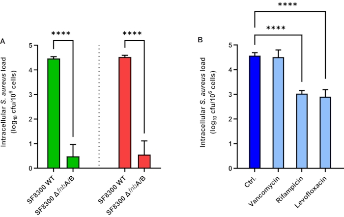
图1细胞内 金黄色葡萄球菌 上皮细胞中的负载。 (A酶保护实验(绿色柱状图)与改良酶保护实验(红色柱状图)在感染的 A549 细胞中的结果 S. aureus SF8300 WT 和 Δ纤维蛋白原A/B。B) 抗菌化合物在感染金黄色葡萄球菌的 HaCaT 细胞内的活性 S. aureus ATCC 29213。柱状图表示三次独立实验(每组设三个重复)的平均值。误差线表示标准差。**** p < 0.0001。缩写:Ctrl = 对照;cfu = 菌落形成单位。 请点击此处以查看此图的放大版本。
讨论
本文所述的检测方法对于研究金黄色葡萄球菌(S. aureus)在NPPCs中的内化程度、胞内存活情况以及抗菌化合物的胞内抗菌效果具有重要价值6,15,16。两种检测方案中的某些步骤尤为关键。细胞的健康状态和密度必须在各次独立实验之间得到精确控制并保持一致。细菌接种物必须经过仔细标准化,以获得接近目标理论感染复数(MOI)的实际MOI。通常情况下,加样时需格外小心,避免细胞脱落。在EPA中,去除溶葡萄球菌素和抗生素的洗涤步骤至关重要。研究发现,在未使用抗生素的情况下,使用蛋白酶K可改善该步骤(见下文)。最后但同样重要的是,在裂解缓冲液孵育后,应确保每个孔中的细胞完全脱离并充分匀浆,以准确量化金黄色葡萄球菌(S. aureus)的胞内载量。
在某些情况下可能会遇到问题,需要首先检查几个要点。如果实验结果缺乏可重复性,必须注意S. aureus可能形成菌团,导致通过吸光度进行定量不准确。当更换培养基(例如为了去除分泌蛋白)而进行离心和洗涤步骤时,细菌的聚集现象可能加剧。细菌悬液应尽快使用,因为细菌在室温下会持续生长。溶葡球菌酶(lysostaphin)的效力可能因储存条件不当、培养基中酶活性的pH非最适、不同批次或供应商之间的酶活性差异,以及某些菌株在特定生长条件下对溶葡球菌酶不敏感而降低。酚红可能具有轻微的抑菌作用,尤其是在培养基营养成分相对于常规细菌培养肉汤较为贫乏的情况下。因此,建议使用不含酚红的细胞培养基,这也有助于减少背景噪声,从而改善荧光显微镜观察效果。
尽管该方法是研究不同菌株细胞内命运的有力工具,但仍需考虑其某些局限性。使用过高的感染复数(MOI)可能会超出NPPCs的内化能力,从而抹平不同测试菌株之间的差异。由于溶葡球菌酶(或抗生素)会迅速破坏受损细胞所释放的S. aureus,细胞毒性最强的菌株其内化程度可能被低估。因此,对于细胞毒性较低的菌株,更易于开展长时间孵育的实验(例如研究细胞内存活或抗生素的细胞内活性)。因此,孵育时间与MOI应根据菌株毒力、细胞类型及实验目的进行精确调整。
本文所述使用溶葡萄球菌酶的方法比基于庆大霉素的方法更可靠,因为与溶葡萄球菌酶不同,庆大霉素容易被宿主细胞内化13。另一个优势在于溶葡萄球菌酶可被灭活。Kim 等人13曾报道使用 EDTA 螯合锌离子或使用 1,10-菲啰啉抑制溶葡萄球菌酶活性;然而,在细菌涂板前仍需进行多次洗涤以彻底去除酶。本方法中,蛋白酶 K 可快速灭活溶葡萄球菌酶。我们观察到,由于胞内金黄色葡萄球菌(S. aureus)的增殖,细胞在严重感染时容易从培养板上脱落。通过省略最后的洗涤步骤,iEPA 方法显著简化了操作流程,并能够回收那些松散贴壁或已脱落细胞中的内化细菌。
iEPA 中使用的更高浓度的试剂和缓冲液还有助于减少移液操作并最大限度降低细胞损失。此外,iEPA 可用于悬浮细胞,也可用于难以洗涤的类器官。综上所述,酶保护实验可用于研究内化程度及内化后分子的细胞内命运 S. aureus,以及不同抗菌药物的细胞内活性 体外 模型。应进一步改进以更准确地表征内化作用与细胞毒性的关系,从而更充分地认识开发能够到达靶点的药物的重要性 S. aureus 细胞内
披露
作者声明无任何利益冲突。
致谢
S. aureus 菌株 SF8300 WT 和 SF8300 ΔfnbA/B 由美国加州大学旧金山分校的 Binh Diep 教授慷慨惠赠。本研究由里昂大学基金会支持下的 FINOVI 协会资助(项目编号:AO13 FINOVI)。
材料
| 姓名 | 公司 | 目录编号 | 评论 |
|---|---|---|---|
| 24孔板 | CORNING-FALCON | 353047 | |
| A549细胞系 | ATCC | CCL-185 | |
| 乙酸盐缓冲液,pH 4.6 | Fluka | 31048 | 用于在20 mM醋酸钠溶液中配制10 mg/mL的溶葡萄球菌素储备液。 |
| AMBICIN(重组溶葡萄球菌素) | AMBI | LSPN-50 | 冻干重组溶葡萄球菌素。长期保存需在-80 °C下冷冻。 |
| COS - 哥伦比亚琼脂 + 5% 羊血 | Biomerieux | 43049 | 可使用任何适合培养葡萄球菌的琼脂平板替代。 |
| 密度计 WPA CO8000 | Biochrom Ltd. | 80-3000-45 | 细胞密度测定仪 |
| Dulbecco’s 改良Eagle’s 培养基,高糖含酚红 | Sigma-Aldrich | D6429 | |
| Dulbecco’s 改良Eagle’s 培养基,高糖不含酚红 | Sigma-Aldrich | D1145 | |
| Dulbecco’s 磷酸盐缓冲液 | Sigma-Aldrich | D8537 | |
| Dulbecco′s 含MgCl2和CaCl2的磷酸盐缓冲液,经无菌过滤 | Sigma-Aldrich | D8662 | |
| Dulbecco′s 磷酸盐缓冲液,经无菌过滤 | Sigma-Aldrich | D8537 | |
| Easyspiral 稀释仪 | Interscience | 414000 | 自动稀释仪和螺旋接种仪 |
| 胎牛血清 | Gibco | 10270-106 | |
| HaCaT细胞系 | 细胞系服务(CLS) | 300493 | |
| Hoechst 33342,三盐酸盐,三水合物,水中10 mg/mL溶液 | Fisher scientific | 11534886 | |
| 碘化丙啶,水中1.0 mg/mL溶液 | Invitrogen | P3566 | |
| 蛋白酶K,重组型,20 mg/mL | Eurobio | GEXPRK01-B5 | > 30 U/mg,批号901727 |
| Scan 4000 | Interscience | 438000 | 自动菌落计数器 |
| 无菌水 | OTEC | 600500 | |
| T-75培养瓶 | CORNING-FALCON | 353136 | |
| TC20自动细胞计数仪 | Biorad | 1450102 | 自动细胞计数仪 |
| Tris 1 M,pH 8.0 | Invitrogen | AM9855G | 用于在0.1 M Tris-HCl中配制1 mg/mL的溶葡萄球菌素工作液。 |
| Triton X-100 | Sigma-Aldrich | T8787 | |
| 胰蛋白酶-EDTA溶液 | Sigma-Aldrich | T3924 | 含0.05%猪胰蛋白酶和0.02% EDTA的Hanks′平衡盐溶液,含酚红 |
| 宽场荧光显微镜 | Nikon | Ti2 |
参考文献
- Verhoeven, P. O., et al. Detection and clinical relevance of Staphylococcus aureus nasal carriage: an update. Expert Review of Anti-Infective Therapy. 12 (1), 75-89 (2014).
- Gagnaire, J., et al. Epidemiology and clinical relevance of Staphylococcus aureus intestinal carriage: a systematic review and meta-analysis. Expert Review of Anti-Infective Therapy. 15 (8), 767-785 (2017).
- Tong, S. Y. C., Davis, J. S., Eichenberger, E., Holland, T. L., Fowler, V. G. Staphylococcus aureus infections: epidemiology, pathophysiology, clinical manifestations, and management. Clinical Microbiology Reviews. 28 (3), 603-661 (2015).
- Josse, J., Laurent, F., Diot, A. Staphylococcal adhesion and host cell invasion: fibronectin-binding and other mechanisms. Frontiers in Microbiology. 8, 2433(2017).
- Hanssen, A. -M., et al. Localization of Staphylococcus aureus in tissue from the nasal vestibule in healthy carriers. BMC Microbiology. 17 (1), 89(2017).
- Rigaill, J., et al. Evaluation of the intracellular efficacy of antimicrobial agents used for Staphylococcus aureus decolonization in a cell model mimicking nasal colonization. Journal of Antimicrobial Chemotherapy. 73 (11), 3044-3048 (2018).
- Yang, D., et al. Novel insights into Staphylococcus aureus deep bone infections: the Involvement of osteocytes. mBio. 9 (2), 00415-00418 (2018).
- Tuchscherr, L., et al. Staphylococcus aureus phenotype switching: an effective bacterial strategy to escape host immune response and establish a chronic infection. EMBO Molecular Medicine. 3 (3), 129-141 (2011).
- Valour, F., et al. Antimicrobial activity against intraosteoblastic Staphylococcus aureus. Antimicrobial Agents and Chemotherapy. 59 (4), 2029-2036 (2015).
- Proctor, R. A., Prendergast, E., Mosher, D. F. Fibronectin mediates attachment of Staphylococcus aureus to human neutrophils. Blood. 59 (4), 681-687 (1982).
- Climo, M. W., Ehlert, K., Archer, G. L. Mechanism and suppression of lysostaphin resistance in oxacillin-resistant Staphylococcus aureus. Antimicrobial Agents and Chemotherapy. 45 (5), 1431-1437 (2001).
- Bur, S., Preissner, K. T., Herrmann, M., Bischoff, M. The Staphylococcus aureus extracellular adherence protein promotes bacterial internalisation by keratinocytes independent of fibronectin-binding proteins. Journal of Investigative Dermatology. 133 (8), 2004-2012 (2013).
- Kim, J. -H., Chaurasia, A. K., Batool, N., Ko, K. S., Kim, K. K. Alternative enzyme protection assay to overcome the drawbacks of the gentamicin protection assay for measuring entry and intracellular survival of Staphylococci. Infection and Immunity. 87 (5), 00119(2019).
- Maturin, L., Peeler, J. T. Aerobic plate count. Bacteriological Analytical Manual., Edition 8, Revision A. 3, Available from: https://www.fda.gov/food/laboratory-methods-food/bam-chapter-3-aerobic-plate-count 3(2021).
- Kolenda, C., et al. Evaluation of the activity of a combination of three bacteriophages alone or in association with antibiotics on Staphylococcus aureus embedded in biofilm or internalized in Osteoblasts. Antimicrobial Agents and Chemotherapy. 64 (3), 02231(2020).
- Abad, L., et al. Antibiofilm and intraosteoblastic activities of rifamycins against Staphylococcus aureus: promising in vitro profile of rifabutin. Journal of Antimicrobial Chemotherapy. 75 (6), 1466-1473 (2020).
重印与许可
勘误
Formal Correction: Erratum: Improved Enzyme Protection Assay to Study Staphylococcus aureus Internalization and Intracellular Efficacy of Antimicrobial Compounds
Posted by JoVE Editors on 1/19/2022. Citeable Link.
An erratum was issued for: Improved Enzyme Protection Assay to Study Staphylococcus aureus Internalization and Intracellular Efficacy of Antimicrobial Compounds. One of the affiliations was updated.
The fifth affiliation was updated from:
Département de Bactériologie, Institut des Agents Infectieux, Hospices Civils de Lyon, Lyon, France
to:
Department of Bacteriology, Institute for Infectious Agents, Hospices Civils de Lyon, Lyon, France
