坏死性小肠结肠炎(NEC)是最严重的胃肠道(GI)疾病,常发生于早产儿,尤其是极低出生体重儿,具有高死亡率且发病机制尚不明确。NEC的病因可能与炎症性免疫调节系统异常有关。建立NEC动物模型是研究NEC疾病免疫机制不可或缺的工具。目前NEC动物模型通常采用C57BL/6J新生小鼠;而BALB/c新生小鼠则较少被使用。相关研究表明,小鼠感染时,与C57BL/6J小鼠相比,BALB/c小鼠更倾向于Th2细胞分化。已有研究提示,NEC的发生与发展与辅助性T细胞2型(Th2)细胞的增加相关,并常伴随感染。因此,本研究采用新生BALB/c小鼠建立NEC模型,该模型具有与NEC患儿相似的临床特征和肠道病理改变。尚需进一步研究以明确该动物模型是否可用于NEC中Th2细胞应答的研究。
需要JoVE订阅才能观看此内容。 请登录或开始免费试用
本文内容
勘误通知
Important: There has been an erratum issued for this article. View Erratum Notice
摘要
摘要
坏死性小肠结肠炎(NEC)是最严重的胃肠道(GI)疾病,常发生于早产儿,尤其是极低出生体重儿,具有高死亡率且发病机制尚不明确。NEC的病因可能与炎症性免疫调节系统异常有关。建立NEC动物模型是研究NEC疾病免疫机制不可或缺的工具。目前NEC动物模型通常采用C57BL/6J新生小鼠;而BALB/c新生小鼠则较少被使用。相关研究表明,小鼠感染时,与C57BL/6J小鼠相比,BALB/c小鼠更倾向于Th2细胞分化。已有研究提示,NEC的发生与发展与辅助性T细胞2型(Th2)细胞的增加相关,并常伴随感染。因此,本研究采用新生BALB/c小鼠建立NEC模型,该模型具有与NEC患儿相似的临床特征和肠道病理改变。尚需进一步研究以明确该动物模型是否可用于NEC中Th2细胞应答的研究。
引言
坏死性小肠结肠炎(necrotizing enterocolitis, NEC)是最严重的胃肠道(GI)疾病,主要发生于早产儿(>90%),尤其是极低出生体重(VLBW)的婴儿1。在极低出生体重婴儿中,该病的发病率为10%至12%,被诊断为NEC的患儿死亡率介于20%至30%之间2,3。NEC的病因可能与黏膜损伤、致病菌侵入以及肠道喂养有关,这些因素可导致易感宿主产生炎症反应并诱发肠道损伤3。NEC的发病机制尚不明确。相关研究表明,患儿的免疫反应异常,遗传易感性、微血管张力以及肠道菌群变化可能在疾病发生中起重要作用3。
坏死性小肠结肠炎(NEC)动物模型是研究NEC发病机制不可或缺的工具。用于建立NEC模型的动物种类包括猪、大鼠和小鼠。然而,由于猪的妊娠期较长、生长周期久且饲养成本高,近年来猪已不再是建立NEC模型的首选动物,逐渐被大鼠或小鼠所取代4。由于不同品系小鼠的免疫背景存在差异5,不同研究需要选用不同的小鼠品系来建立NEC动物模型。BALB/c小鼠具有一个重要特征:当其受到感染或应对外界损伤时,体内TH2细胞在感染过程中的极化程度显著强于其他品系小鼠6,7,8。辅助性T细胞在NEC的发生与发展过程中起着关键作用,尤其是TH2细胞的发育与分化3,9,10,11。因此,本研究采用BALB/c小鼠建立NEC模型,可能有助于开展针对T细胞的NEC疾病相关研究。
访问受限。请登录或开始试用以查看此内容。
方案
本研究经广州妇女儿童医疗中心医学伦理委员会(编号:174A01)和广州赛业生物实验室动物中心动物伦理委员会(IACUC-G160100)批准。所有动物均在同一房间内的SPF(特定病原体-free)环境中饲养,实验在常规环境中进行。用于繁殖的小鼠年龄为7–8周;用于诱导坏死性小肠结肠炎(NEC)的小鼠(n = 72)于出生第4天与母鼠分离, 母鼠(n=14)被保留在原始笼具中并进行哺乳 对照(Cont.)组小鼠(n=24)。
1. 试剂与器械的准备
- 按相应比例配制BALB/c小鼠的代乳液(早产儿奶粉:羊奶粉 = 2:1)。
注意:配方奶的最终营养成分12见表1。 - LPS溶液(2.5 mg/mL)
- 将总共10 mg的LPS粉末溶解于4 mL灭菌的双蒸水中,充分混匀,分装后于-20 °C冰箱中保存。
注意:LPS溶液可于2-8 °C避光保存,用于短期使用;或于-20 °C长期保存。
- 将总共10 mg的LPS粉末溶解于4 mL灭菌的双蒸水中,充分混匀,分装后于-20 °C冰箱中保存。
2. 在新生 BALB/c 小鼠中诱导坏死性小肠结肠炎
- 喂养新生小鼠。
- 将新生小鼠与母鼠同笼饲养,在出生后第0至4天由母鼠哺乳。
- 在第4天晚上(当新生小鼠体重为2.5–3 g时),将坏死性小肠结肠炎(NEC)组的新生小鼠与母鼠分离以诱导NEC,将其置于动物培养箱中,并用配方奶喂养。而对照组(Cont.组)小鼠则继续与母鼠同笼并由母鼠哺乳。
注意:由于新生小鼠体温调节能力较弱,与母鼠分离后必须在培养箱中饲养。
- 将灌胃管浸入75%酒精容器中1–2分钟进行消毒,随后用洁净的双蒸水冲洗两次。
注意:为避免小鼠之间的交叉污染,每次喂养后均需重复上述处理步骤。 - 建立NEC模型。
- 在第4天将新生小鼠从母鼠身边取出,并禁食一晚。
- 在第5天通过灌胃给予脂多糖(LPS)(每次20–30 µL),并用配方奶喂养(每次40–50 µL)。
- 从第5天开始,每天进行两次缺氧-复氧-冷刺激循环,持续5天。将小鼠置于含5% O2的缺氧装置中90秒,随后复氧3分钟;重复此过程五次。接着将小鼠置于4 °C环境中15分钟,然后转移回培养箱。诱导过程详见图1A、B。
注意:缺氧-复氧-冷刺激循环每天上午和下午各进行一次。容器中预先配制5% O2与95% N2的混合气体,并使用氧浓度检测仪测定气体浓度。
- 密切观察所有小鼠,每日称重,记录诱导期间小鼠的存活情况,并记录粪便性状(是否出现黏液便或血便)。
注意:建立的NEC模型持续5天。 - 在第10天或更早时间,当小鼠出现NEC症状(肠梗阻、便血、腹泻)13时,采用异氟烷吸入麻醉实施安乐死,随后立即 采集肠道组织。不得采集自然死亡小鼠的组织。
注意:本研究中,小鼠安乐死的终点标准为出现便血及全身发绀。
3. 对小鼠进行灌胃
- 固定小鼠头部,右手持胃管,从小鼠口腔左侧口角处插入胃管。
注意:用食指固定小鼠头部,并轻轻向后下方按压,以防止小鼠在操作过程中向前弯曲,影响胃管插入。 - 缓慢将胃管移至口腔中央。插入约 2-3 cm 后,向消化道内推注 40-50 µL 配方液或 20-30 µL LPS。胃灌注操作参见 图 2A、B。
注意:正常情况下,胃管可顺利插入消化道。若小鼠出现强烈呕吐反射,提示胃管误入气管,此时应轻柔拔出胃管,让小鼠休息片刻后再尝试灌胃。此外,胃灌注操作用于在小鼠安乐死后建立 NEC 模型。
4. 采集新鲜肠道组织标本用于苏木精和伊红(H&E)染色
- 将小鼠新鲜回肠组织浸入10%福尔马林中固定24小时。
- 将组织包埋于石蜡中,并切成4 µm厚的切片。
- 用二甲苯脱蜡,并依次在无水乙醇、95%乙醇、80%乙醇、70%乙醇和蒸馏水中进行梯度水化,每步浸泡5分钟。用苏木精染液染色5分钟,然后在75%乙醇中的1%盐酸中分化5秒。最后用伊红染液染色1分钟。
注意:苏木精染色后,必须用乙醇中的1%盐酸进行分化,以去除过度结合的苏木精以及细胞质中的苏木精染料。1%盐酸的浓度适用于肠道组织。 - 在40倍放大倍数下观察肠组织的组织病理学变化。
访问受限。请登录或开始试用以查看此内容。
结果
通过配方奶喂养、LPS喂养、缺氧和冷刺激诱导BALB/c小鼠坏死性小肠结肠炎(NEC)模型。在造模期间,观察小鼠的肠道病理变化、粪便特征、体重变化及每日存活情况。NEC造模过程中小肠的代表性图像;图中数字表示肠道病理评分,范围从0(正常上皮)到4(最严重)(图3A)。NEC组的肠道病理评分显著高于对照组(Cont.组)(图3B)。图中数字表示粪便评分,范围从0(成形粪粒)到3(液态粪便)(图3C)。第10天时,NEC组的粪便评分显著高于对照组,表明NEC组小鼠的肠道功能障碍更为严重(图3D)。在第5天(即NEC模型造模的第一天),两组小鼠的体型无显著差异;然而,到第10天时,NEC组小鼠从头部至尾部均明显比对照组小鼠更瘦小(图4A)。
在建立模型的5天期间,NEC组小鼠的体重增长缓慢,甚...
访问受限。请登录或开始试用以查看此内容。
讨论
坏死性小肠结肠炎(NEC)是新生儿最常见的胃肠道急症,具有较高的发病率和死亡率,尤其在早产儿中更为显著1,2,3。然而,其发病机制仍不明确。目前认为,黏膜损伤、病原体侵袭和经肠喂养是NEC的高危因素3。迄今为止,用于建立NEC模型的动物主要包括猪、大鼠和小鼠。大多数研究采用新生C57BL/6小鼠来诱导NEC13,14,15,16,极少有研究使用BALB/c新生小鼠来诱导NEC。然而,BALB/c小鼠在Th细胞极化方面具有优势6,7
访问受限。请登录或开始试用以查看此内容。
披露
作者声明无利益冲突。
致谢
作者感谢广州妇女儿童医疗中心临床生物资源库提供临床样本,以及广州南方基因科技有限公司实验动物中心提供小鼠。本研究由国家自然科学基金 中国国家自然科学基金资助项目 81770510(R.Z.)
访问受限。请登录或开始试用以查看此内容。
材料
| 姓名 | 公司 | 目录编号 | 评论 |
|---|---|---|---|
| 无水乙醇 | Sinopharm Chemical Reagent Co., LTD. | 100092683 | |
| 山羊奶粉 | Petag | 71795558417 | |
| HE染色液 | Sinopharm Chemical Reagent Co., LTD. | G1003 | |
| 异氟烷 | RWD, Shenzhen Reward Life Technology Co., LTD. | R510 | |
| LPS | Sigma-Adrich | L2880 | |
| 医用氧气 | 多种 | 多种 | |
| 显微镜 | NIKON | NIKON成像系统 (DS-Ri2) | |
| 中性树脂 | Sinopharm Chemical Reagent Co., LTD. | 10004160 | |
| 石蜡 | 多种 | 多种 | |
| 早产儿奶粉 | 雅培 | 57430 | |
| 二甲苯 | Sinopharm Chemical Reagent Co., LTD. | 10023418 | |
| 1% 盐酸 | 多种 | 多种 | |
| 10% 甲醛 | LEAGENE | DF0110 |
参考文献
- Horbar, J. D., et al. Mortality and neonatal morbidity among infants 501 to 1500 grams from 2000 to 2009. Pediatrics. 129 (6), 1019-1026 (2012).
- Stoll, B. J., et al. Neonatal outcomes of extremely preterm infants from the NICHD Neonatal Research Network. Pediatrics. 126 (3), 443-456 (2010).
- Neu, J., Walker, W. A. Necrotizing enterocolitis. New England Journal of Medicine. 364 (3), 255-264 (2011).
- Sangild, P. T., et al. Invited Review: The preterm pig as a model in pediatric gastroenterology. Journal of Animal Science. 91 (10), 4713-4729 (2013).
- Cancro, M. P., Sigal, N. H., Klinman, N. R. Differential expression of an equivalent clonotype among BALB/c and C57BL/6 mice. Journal of Experimental Medicine. 147 (1), 1-12 (1978).
- Kuroda, E., Yamashita, U. Mechanisms of enhanced macrophage-mediated prostaglandin E2 production and its suppressive role in Th1 activation in Th2-dominant BALB/c mice. Journal of Immunology. 170 (2), 757-764 (2003).
- Fornefett, J., et al. Comparative analysis of clinics, pathologies and immune responses in BALB/c and C57BL/6 mice infected with Streptobacillus moniliformis. Microbes and Infection. 20 (2), 101-110 (2018).
- Rosas, L. E., et al. Genetic background influences immune responses and disease outcome of cutaneous L. mexicana infection in mice. International Immunology. 17 (10), 1347-1357 (2005).
- Sproat, T., Payne, R. P., Embleton, N. D., Berrington, J., Hambleton, S. T cells in preterm infants and the influence of milk diet. Frontiers in Immunology. 11, 1035(2020).
- Nanthakumar, N., et al. The mechanism of excessive intestinal inflammation in necrotizing enterocolitis: an immature innate immune response. PLoS One. 6 (3), 17776(2011).
- Afrazi, A., et al. New insights into the pathogenesis and treatment of necrotizing enterocolitis: Toll-like receptors and beyond. Pediatric Research. 69 (3), 183-188 (2011).
- Auestad, N., Korsak, R. A., Bergstrom, J. D., Edmond, J. Milk-substitutes comparable to rat's milk; their preparation, composition and impact on development and metabolism in the artificially reared rat. British Journal of Nutrition. 61 (3), 495-518 (1989).
- Liu, Y., et al. Lactoferrin-induced myeloid-derived suppressor cell therapy attenuates pathologic inflammatory conditions in newborn mice. Journal of Clinical Investigation. 129 (10), 4261-4275 (2019).
- MohanKumar, K., et al. A murine neonatal model of necrotizing enterocolitis caused by anemia and red blood cell transfusions. Nature Communications. 10 (1), 3494(2019).
- He, Y. M., et al. Transitory presence of myeloid-derived suppressor cells in neonates is critical for control of inflammation. Nature Medicine. 24 (2), 224-231 (2018).
- Cho, S. X., et al. Characterization of the pathoimmunology of necrotizing enterocolitis reveals novel therapeutic opportunities. Nature Communications. 11 (1), 5794(2020).
- Halpern, M. D., et al. Decreased development of necrotizing enterocolitis in IL-18-deficient mice. American Journal of Physiology. Gastrointestinal and Liver Physiology. 294 (1), 20-26 (2007).
- Wu, N., et al. MAP3K2-regulated intestinal stromal cells define a distinct stem cell niche. Nature. 592 (7855), 606-610 (2021).
- Nino, D. F., Sodhi, C. P., Hackam, D. J. Necrotizing enterocolitis: new insights into pathogenesis and mechanisms. Nature Reviews. Gastroenterology & Hepatology. 13 (10), 590-600 (2016).
- Chuang, S. L., et al. Cow's milk protein-specific T-helper type I/II cytokine responses in infants with necrotizing enterocolitis. Pediatric Allergy & Immunology. 20 (1), 45-52 (2009).
访问受限。请登录或开始试用以查看此内容。
重印与许可
勘误
Formal Correction: Erratum: A Neonatal BALB/c Mouse Model of Necrotizing Enterocolitis
Posted by JoVE Editors on 3/07/2022. Citeable Link.
An erratum was issued for: A Neonatal BALB/c Mouse Model of Necrotizing Enterocolitis. The Representative Results section was updated.
Figure 1 was updated from:
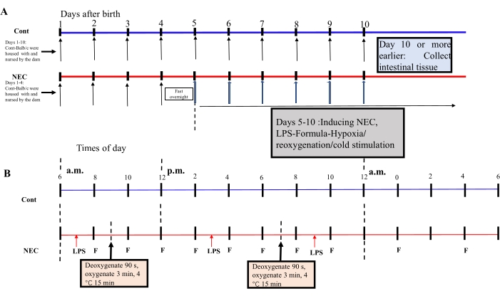
Figure 1: Induction of the BALB/c NEC model process. (A) The mice in the NEC group were separated from the dam at birth until they were 4 days old (on Day 4) and fasted that night. The NEC model was induced from Day 5 onwards after birth and lasted for 5 days. Intestinal tissue specimens were collected on Day 10 or earlier. The mice in the Cont. group were housed with and nursed by the dam. (B) The sequence of operations for each day after inducing the NEC model. Abbreviations: Cont. = control; NEC = necrotizing enterocolitis; LPS = lipopolysaccharide. Please click here to view a larger version of this figure.
to:
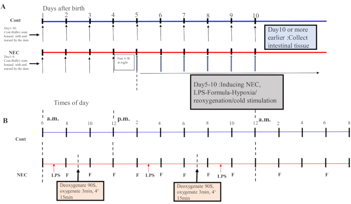
Figure 1: Induction of the BALB/c NEC model process. (A) The mice in the NEC group were separated from the dam at birth until they were 4 days old (on Day 4) and fasted that night. The NEC model was induced from Day 5 onwards after birth and lasted for 5 days. Intestinal tissue specimens were collected on Day 10 or earlier. The mice in the Cont. group were housed with and nursed by the dam. (B) The sequence of operations for each day after inducing the NEC model. Abbreviations: Cont. = control; NEC = necrotizing enterocolitis; LPS = lipopolysaccharide. Please click here to view a larger version of this figure.
