本文介绍了一种设计和测试二肽抑制剂的方案,该二肽可利用无细胞和细胞实验检测其对Src激酶酶活性的抑制作用,以用于抗癌应用。本文所配制的肽(W-RCH3、WCH3-RCH3 和 W-R(CH3)2) 对Src激酶的抑制IC50值分别为510 nM、916 nM和1 µM。
需要JoVE订阅才能观看此内容。 请登录或开始免费试用
本文内容
Retraction Notice
The article Developing and Testing Methylated Nano-Structured Dipeptides that Inhibit Src Kinase Activity In Vitro for Anti-Cancer Applications has been retracted by the journal due to authorship and reproducibility concerns.
摘要
摘要
本研究旨在开发一种新型抗癌治疗方法,设计并采用Fmoc固相肽合成法合成了七种含有甲基化色氨酸和/或甲基化精氨酸的二肽。Src酪氨酸激酶的过表达已被证实与多种癌症的发生发展相关。先前研究表明,含有非天然氨基酸(如甲基化精氨酸(RCH3)、二甲基化精氨酸(R(CH3)2)和/或甲基化色氨酸(WCH3)残基)的二肽能够抑制Src激酶活性。在本研究中,通过无细胞实验测试了其中三种二肽W-RCH3、WCH3-RCH3和W-R(CH3)2,其IC50值(抑制50%活性时的浓度)分别为510 nM、916 nM和1 µM。这些数值与先前研究中合成的环状五至九聚W-R肽([W-R]5-[W-R]9)所获得的结果相当。然而,未甲基化的二肽则未表现出对Src激酶的任何抑制活性。所有这些二肽(50 µM)在与三种不同癌细胞系——白血病(CCRF-CEM)、乳腺腺癌(MDA-MB-231)和卵巢腺癌(SK-OV-3)细胞——共孵育长达72小时后,均未表现出细胞毒性,表明这些肽穿过细胞膜的通透性有限。因此,为进一步将其应用于抗癌治疗,尚需开展研究以提高这些肽的细胞通透性,例如通过使用肽载体或对肽进行额外的功能化修饰。总之,本研究提供了一套用于合成和测试可抑制Src激酶活性、因而具有潜在抗癌能力的肽的实验方案,并通过无细胞和细胞实验验证了其效果。
引言
癌症是由正常细胞异常增殖引起的疾病,是全球最致命的疾病之一。这些异常细胞通过一种称为转移的过程扩散到身体的不同器官。最常见的癌症类型是乳腺癌,2020年全球有226万人罹患该病。此外,2020年约有180万人死于肺癌1。根据世界卫生组织的数据,2020年全球约有1000万人死于癌症2。癌细胞与正常细胞的不同之处在于其会过度表达某些酶,例如蛋白酪氨酸激酶(PTKs)。美国国家癌症研究所将激酶定义为能够磷酸化其他蛋白质或糖类的酶3。了解激酶的调控功能有助于设计有效的抗癌药物。例如,PTKs催化其他蛋白质或糖类的磷酸化反应,导致ATP因失去一个磷酸基团而转化为ADP。所有癌基因和原癌基因中,约有80%编码PTKs4。Src激酶是一类非受体酪氨酸激酶家族,包括Lck、Fyn、Hck、Blk、Yes和Yrk,在癌细胞中尤其是乳腺癌细胞中呈高表达5,6。Src酪氨酸激酶与有丝分裂原作用、细胞分化、T细胞活化以及细胞转化相关。由于Src能够降低癌细胞的黏附能力,因而促进癌细胞的侵袭和转移。Src激酶包含五个不同的结构域,从N端到C端依次为:脂肪酸结构域、Src同源3结构域(SH3)、Src同源2结构域(SH2)、酪氨酸激酶结构域(SH1)以及C端调控结构域7。

图1:Src激酶酶中的靶向结构域,包括SH3结构域、SH2结构域、激酶结构域(SH1)以及一段短的C端调控区段。 请点击此处查看该图的放大版本。
激酶结构域SH1在设计Src激酶抑制剂时最常作为靶点,因为它包含两个保守的ATP和底物结合位点(图1)。如果已知激酶结构域的氨基酸序列,也可利用该底物作为靶标来设计一种化合物,以模拟其与Src激酶的结合。8此外,其他位点如 SH3 和 SH2 结构域也可作为靶点。与其他化疗药物相比,激酶抑制剂具有更低的毒性和更高的疗效9截至2021年9月,已有73种小分子激酶抑制剂获得美国食品药品监督管理局(FDA)批准10伊马替尼是一种选择性抑制酪氨酸激酶活性的抗癌药物;然而,由于激酶结构域出现点突变,部分患者对该药物产生耐药性。11阿斯利康发布了saracatinib,这是一种抑制Src家族酪氨酸激酶的药物,其IC50 50%抑制浓度(IC50)为2.7 nM,但在二期临床试验中被弃用12截至2020年初,美国FDA批准的52种蛋白酪氨酸激酶(PTK)抑制剂13,仅28种靶向受体酪氨酸蛋白激酶,11种阻断非受体酪氨酸蛋白激酶,11种抑制蛋白丝氨酸/苏氨酸蛋白激酶,2种阻断MEK1/213在肿瘤学领域日益增长的研究兴趣将继续推动激酶抑制剂作为潜在抗癌药物的发现。然而,迄今为止,仅针对500种蛋白激酶中的50种开展了治疗研究;因此,预计在不久的将来将有更多激酶被纳入药物开发的研究范围。14此外,还需要发现激酶抑制剂,以探索尚未明确的、可导致癌症的激酶突变。
因此,本研究旨在开发可用于抑制Src家族激酶的多肽,并靶向ATP结合位点,因其在不同激酶之间具有保守性。为此,合成了一系列含有甲基化色氨酸和/或甲基化精氨酸的二肽,并检测其协同抑制Src激酶的能力。色氨酸的吲哚环可模拟ATP中的腺嘌呤,从而与ATP竞争结合ATP结合位点。此外,配体中的甲基化精氨酸可与Src的SH3结构域发生竞争性结合。研究人员发现,含有去甲基化精氨酸的多肽能够抑制SH3结构域,这可能是由于SH3结合基序上存在一个特定的保守序列(即PXXP),该序列对N端含有两至三个精氨酸残基或C端含有一至两个精氨酸残基的配体具有结合亲和力。15,16,17Arg的胍基与SH3结构域中保守的Asp-99残基结合18,19,而精氨酸的其余部分与酶中保守的Trp-118结合,这一点已通过NMR分析及多个SH3结构域的晶体结构得到证实19. 本文介绍了一种合成七种甲基化二肽的实验方案,并检测了这些二肽对Src激酶的抑制能力。此外,还进一步评估了这些肽对多种癌细胞系的杀伤能力 体外 进行了检测。
访问受限。请登录或开始试用以查看此内容。
方案
1. 肽的合成
注意:W-R 的合成被描述为一个代表性示例(图 2)。
- 称取 566 mg(0.3 mmol)H-Arg(Pbf)-2-氯三苯甲基树脂,加入肽合成反应瓶中(参见 材料清单),按照 Mandal 等人所采用的实验步骤20采用成熟的固相多肽合成(SPPS)策略进行多肽合成。21.
注意:含有非天然氨基酸的甲基化或二甲基化二肽在 Rink 酰胺树脂(载量为 0.3 mmol/g)上合成,而含有天然氨基酸的二肽则在已连接第一个氨基酸的树脂上合成,例如 H-Arg(Pbf)-2-氯三苯甲基树脂(载量为 0.3 mmol/g)。重要的是,Rink 酰胺树脂合成的肽段其 C 末端以 NH 封端2. - 在连接肽反应容器的情况下,用5 mL N,N-二甲基甲酰胺(DMF)在氮气压力下溶胀干树脂1小时。
注意:整个合成过程需在通风橱中进行。实验全程必须佩戴护目镜、手套和实验服。计时器对于准确控制各化学试剂添加步骤之间的时间间隔也至关重要。 - 在整个合成过程中,使用与肽合成反应器相连的氮气压力去除多余的 DMF。
- 称取 473.9 mg Fmoc-Trp(Boc)-OH(0.9 mmol)和 341 mg 2-(1H-苯并三唑-1-基)-1,1,3,3-四甲基脲六氟磷酸盐(HBTU)(0.9 mmol)作为偶联试剂。将粉末加入试管中,加入 5 mL 无水 DMF 溶解。
- 添加 313.4 µL (1.8 mmol) 的 N,N将二异丙基乙胺(DIPEA)作为活化剂加入试管(步骤 1.4),并通过振荡试管混合。
- 将试管中的内容物加入树脂中,在室温下通入氮气,使反应进行1小时。随后,用氮气压力排干多余的DMF,并用DMF洗涤树脂3次。
- 脱保护 N用 5 mL 含 20% 哌啶的 DMF 溶液(v/v)在氮气保护下脱除 Fmoc 保护基,反应 20 分钟。随后用 DMF 洗涤树脂 3 次(每次 5 mL)。
- 在氮气氛围下加入5 mL二氯甲烷(DCM),干燥树脂,静置5 min,然后通过氮气压力排出DCM。再在氮气氛围下加入5 mL甲醇,处理5 min,以进一步提高树脂的干燥程度。
- 加入 10 mL 新鲜配制的裂解混合液(TFA/硫代苯醚/二硫苏糖醇/苯甲醚,90:3:5:2 v/v/w/v),反应 2.5 小时,以脱除所有侧链保护基并将二肽从树脂上裂解下来。
注意:裂解缓冲液必须加入玻璃量筒中,因为三氟乙酸(TFA)具有酸性且危险;使用时需格外小心。 - 反应2.5小时后,用氮气压力将反应液从肽反应瓶转移至圆底烧瓶中。加入250 mL冷的乙醚(Et₂O)。2将 O)加入含有粗肽的圆底烧瓶中,以沉淀该肽。
- 使用带有侧管(侧管连接水源,利用水压进行过滤)的锥形漏斗和滤纸过滤析出的肽,收集沉淀物(肽),以彻底去除乙醚。粗肽沉淀在10分钟内完成。
- 使用反相高效液相色谱(HPLC)柱纯化粗肽(参见 材料表) 采用梯度系统,使用含0.1% TFA的水溶液与含0.1% TFA的乙腈进行梯度洗脱,乙腈比例从0%升至100%,时间30–60分钟,流速为1 mL/min。
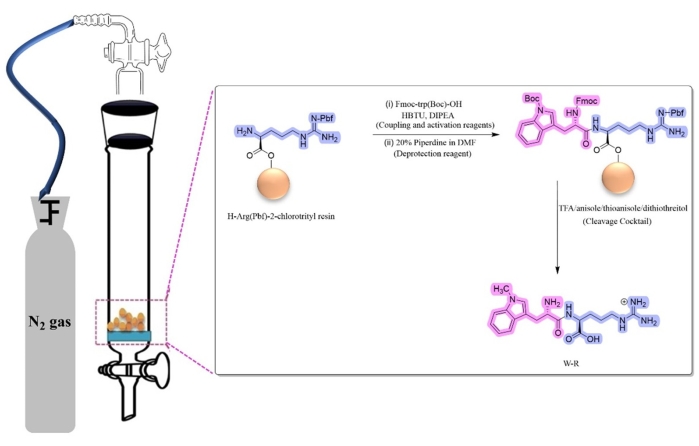
图 2:W-R 的固相多肽合成。缩写:HBTU = 2-(1H-苯并三唑-1-基)-1,1,3,3-四甲基脲六氟磷酸盐;DIPEA = N,N-二异丙基乙胺;TFA = 三氟乙酸;DMF = 二甲基甲酰胺。请点击此处查看该图的放大版本。
2. 合成肽的细胞毒性测定
- 将 SK-OV-3 细胞以 2,000 个细胞/孔、MDA-MB-231 细胞以 5,000 个细胞/孔、或 CCRF-CEM 细胞以 5 × 10⁶ 个细胞/孔(总体积为 100 µL/孔)接种至 96 孔微孔板中。将细胞置于 37 °C、含 5% CO2 和 95% 空气的湿润环境中培养,直至细胞融合度达到 75%–80%。
注意:CCRF-CEM 细胞使用 RPMI-16 培养基,MDA-MB-231 和 SK-OV-3 细胞系均使用 Eagle 最低必需培养基(EMEM)。上述培养基均添加 10% 胎牛血清(FBS)和 1% 青霉素/链霉素(青霉素 10,000 单位/mL,链霉素 10 mg/mL,溶于 0.9% NaCl 中)。实验开始前,将所有培养基和添加剂置于 37 °C 水浴中预热 30 分钟。实验操作应在生物安全柜中进行,并佩戴手套和实验服。 - 用移液器吸除培养基,向 96 孔板中已接种的三种不同类型癌细胞孔中,每孔加入 100 µL 的 50 µM 多肽溶液或作为阳性对照的 10 µM 阿霉素(Dox),每个处理设三个复孔。将微孔板放回培养箱,在与步骤 2.1 相同的条件下继续培养 72 小时。
- 培养 72 小时后,向每孔加入 20 µL 的 3-(4,5-二甲基噻唑-2-基)-5-(3-羧甲氧基苯基)-2-(4-磺基苯基)-2H-四氮唑(MTS)试剂。用铝箔纸包裹微孔板,于 37 °C 湿润环境中孵育 1–4 小时。
注意:MTS 是一种用于检测活细胞(未经处理或经多肽处理)的染料,因为只有活细胞才能将 MTS 四氮唑还原为有色的甲臜染料,其吸光度可在 490 nm 处测定。 - 使用微孔板读数仪(见材料表)测定甲臜产物在 490 nm 处的吸光度(A490)。以仅含培养基与 MTS 的混合液作为空白对照。以纯水(因多肽可溶于水)和 0.1 N HCl(因 WCH3 需用 HCl 溶解)作为空白对照。
- 使用电子表格软件(见材料表)根据以下公式(公式 1)计算细胞存活率。该方法与 Mandal 等人20 使用的方法相同。
 公式 (1)
3. c-Src 激酶活性检测
注意:Src 激酶活性检测采用商用检测试剂盒(参见材料表),按照 Chhikara 等人22的方法进行三复孔实验。使用单独的 WCH3 和 RCH3 作为对照,以比较甲基化氨基酸单独存在时对激酶的效应,以及与其他甲基化或非甲基化氨基酸共同存在时的效应。
- 在384孔低体积黑色非结合表面圆底微孔板中,加入2.5 µL反应混合液,其中包含检测试剂盒提供的激酶缓冲液中的0.7 nM His6-Src激酶结构域,再加入2.5 µL溶于10% DMSO中的预稀释肽段(4倍目标浓度)。按照生产商说明书,使用微孔板摇床(5 rpm)在室温下孵育10分钟。
注:反应混合液由激酶缓冲液(200 mM HEPES,pH 7.5)、MgCl2 (16 mM)、DMSO(4%)、EGTA(8 mM)、2-巯基乙醇(43 mM)和Brij-35(0.04%)组成。本实验激酶反应的最佳底物为(AEEEIYGEFEAKKKK)。 - 向微孔板中加入5 µL ATP/底物混合液(40 µM/600 µM),并在室温下于微孔板摇床上孵育30分钟。
- 同时,将试剂盒中的终止与检测缓冲液B(1x)与含8 nM ADP示踪剂和10 µg/mL染料偶联ADP2抗体的溶液(见材料表)混合,配制成1x ADP检测混合液。加入10 µL该1x ADP检测混合液以终止激酶反应(步骤3.2),并在室温下孵育1小时。
- 使用微孔板读板仪测定荧光强度,激发波长为580 nm,发射波长为630 nm,带宽为10 nm。从仪器获取相对荧光单位(RFUs),并根据公式(2)计算酶抑制率。
 公式(2)
公式(2) - 根据该公司网站上列出的方法计算IC50值23。
访问受限。请登录或开始试用以查看此内容。
结果
采用Fmoc固相肽合成法合成了W-RCH3、WCH3-RCH3、W-R(CH3)2、WCH3-R、WCH3-R(CH3)2以及对照肽W-R(图3),纯度分别为95%、98.7%、99%、100%、100%和99.5%。通过ESI-MS确认了这些二肽的化学结构。这些二肽的质荷比(m/z)值分别为374.1624、388.1949、388.1794、374.1815、402.2022和361.1457。
访问受限。请登录或开始试用以查看此内容。
讨论
本文中用于抑制 Src 激酶并由此杀伤癌细胞的多肽含有甲基化色氨酸和/或甲基化精氨酸,这些属于非天然氨基酸。在合成这些多肽的过程中,加入乙醚后形成白色沉淀是一个关键步骤。然而,并非所有合成的多肽都能形成沉淀;因此,即使未观察到沉淀生成,仍可通过液相色谱-质谱联用技术(LC-MS)检测目标分子量来确认多肽合成是否成功。所获得的多肽分子量还可用于预测其1H-NMR 谱图。已知非天然氨基酸相较于天然氨基酸对蛋白酶的蛋白水解降解具有更高的稳定性与抗性25,因而可能更有效地抑制 Src 激酶活性。此外,诸如甲基化丙氨酸(即 α-氨基异丁酸)等非天然甲基化氨基酸已被报道具有抗癌和抗菌活性26。精氨酸氨基酸的胍基含有五个氢键供体,通过添加甲基可消除其中的部分氢键供体。这种修饰将改变氨基酸的结构,进而影响其结合相互作用及生物学功能。
含色氨酸的二肽,例如 H-L-色氨酸-L-精氨酸-OH(W-R),具有多种应用。例如,WR 二肽作为过氧化物酶体增殖物激活受体 γ(PPARγ)的拮抗...
访问受限。请登录或开始试用以查看此内容。
披露
作者无任何利益冲突需要披露。
致谢
我们感谢沙特阿拉伯吉达阿卜杜勒阿齐兹国王大学(KAU)科研事务副院长办公室(DSR)对本项目提供的资助,资助编号为(G: 031-130-1443)。
访问受限。请登录或开始试用以查看此内容。
材料
| 姓名 | 公司 | 目录编号 | 评论 |
|---|---|---|---|
| 1,4-二硫苏糖醇 | Sigma-Aldrich | 10708984001 | 多肽合成 |
| 用于固相合成的 Aldrich 砂芯过滤漏斗 | Sigma-Aldrich | Z283304 | 多肽合成反应器 |
| Alexa594 Tracer | Bell Brook Labs, Madison, WI | 3013 | |
| 茴香醚 | Sigma-Aldrich | 8014520500 | |
| CellTiter 96 AQueous One 溶液 细胞增殖检测试剂 | Promega | G3582 | 细胞增殖检测(MTS 试剂) |
| 二氯甲烷,99.9%,超干燥 | Fishersci | AC326850025 | |
| Fmoc-ADMA(Pbf)-OH | Sigma-Aldrich | 8521070001 | |
| Fmoc-Arg(Me,Pbf)-OH | Sigma-Aldrich | 8521050001 | |
| Fmoc-Arg(Pbf)-OH | Sigma-Aldrich | 8520670025 | |
| Fmoc-Trp(Boc)-OH | Sigma-Aldrich | 47561-25G-F | |
| HPLC C18 色谱柱 | Shimadzu (RP-HPLC 系统) | 水/乙腈梯度洗脱 | |
| IRDye QC-1 淬灭剂 | Bell Brook Labs, Madison, WI | 3013 | |
| 微孔板读板仪 SpectraMax M2e | Molecular devices | ||
| Microsoft Excel | Microsoft | 电子表格软件 | |
| N,N-二异丙基乙胺(DIPEA) | Sigma-Aldrich | 496219 | |
| N,N-二甲基甲酰胺,无水,99.8% | Fishersci | AA43997M1 | |
| 20% 哌啶 | Sigma-Aldrich | 80645 | |
| Rink 酰胺树脂(100–200 目) | Sigma-Aldrich | 8550010025 | |
| 硫代茴香醚 | Sigma-Aldrich | 92358 | |
| Transcreener ADP2 FI 检测试剂盒 | Bell Brook Labs, Madison, WI | 3013 | c-Src 激酶活性检测试剂盒 |
参考文献
- Sung, H., et al. Global Cancer Statistics 2020: GLOBOCAN estimates of incidence and mortality worldwide for 36 cancers in 185 countries. CA: A Cancer Journal for Clinicians. 71 (3), 209-249 (2021).
- Cancer. World Health Organization. , Available from: https://www.who.int/news-room/fact-sheets/detail/cancer#:~:text=Cancer%20a%20leading%20cause,and%20rectum%20and%20prostate%20cancers (2022).
- Negi, P., Cheke, R. S., Patil, V. M. Recent advances in pharmacological diversification of Src family kinase inhibitors. Egyptian Journal of Medical Human Genetics. 22 (1), 52(2021).
- Huang, X. L., et al. Role of receptor tyrosine kinases mediated signal transduction pathways in tumor growth and angiogenesis-New insight and futuristic vision. International Journal of Biological Macromolecules. 180, 739-752 (2021).
- Mohammed, S., et al. Sublethal doxorubicin promotes migration and invasion of breast cancer cells: role of Src Family non-receptor tyrosine kinases. Breast Cancer Research. 23 (1), 76(2021).
- Biscardi, J. S., Ishizawar, R. C., Silva, C. M., Parsons, S. J. Tyrosine kinase signalling in breast cancer: epidermal growth factor receptor and c-Src interactions in breast cancer. Breast Cancer Research. 2 (3), 1-8 (2000).
- Ortiz, M. A., et al. Src family kinases, adaptor proteins and the actin cytoskeleton in epithelial-to-mesenchymal transition. Cell Communication and Signaling. 19 (1), 67(2021).
- Hill, Z. B., Perera, B. G., Andrews, S. S., Maly, D. J. Targeting diverse signaling interaction sites allows the rapid generation of bivalent kinase inhibitors. ACS Chemical Biology. 7 (3), 487-495 (2012).
- Wu, S., Fu, L. Tyrosine kinase inhibitors enhanced the efficacy of conventional chemotherapeutic agent in multidrug resistant cancer cells. Molecular Cancer. 17 (1), 25(2018).
- Ayala-Aguilera, C. C., et al. Small molecule kinase inhibitor drugs (1995-2021): Medical indication, pharmacology, and synthesis. Journal of Medicinal Chemistry. 65 (2), 1047-1131 (2022).
- Lyczek, A., et al. Mutation in Abl kinase with altered drug-binding kinetics indicates a novel mechanism of imatinib resistance. Proceedings of the National Academy of Sciences. 118 (46), 2111451118(2021).
- Musumeci, F., Schenone, S., Brullo, C., Botta, M. An update on dual Src/Abl inhibitors. Future Medicinal Chemistry. 4 (6), 799-822 (2012).
- Roskoski, R. Properties of FDA-approved small molecule protein kinase inhibitors: A 2020 update. Pharmacological Research. 152, 104609(2020).
- Cohen, P., Cross, D., Jänne, P. A. Kinase drug discovery 20 years after imatinib: Progress and future directions. Nature Reviews Drug Discovery. 20 (7), 551-569 (2021).
- Feng, S., Chen, J. K., Yu, H., Simon, J. A., Schreiber, S. L. Two binding orientations for peptides to the Src SH3 domain: development of a general model for SH3-ligand interactions. Science. 266 (5188), 1241-1247 (1994).
- Alexandropoulos, K., Cheng, G., Baltimore, D. Proline-rich sequences that bind to Src homology 3 domains with individual specificities. Proceedings of the National Academy of Sciences. 92 (8), 3110-3114 (1995).
- Feng, S., Kasahara, C., Rickles, R. J., Schreiber, S. L. Specific interactions outside the proline-rich core of two classes of Src homology 3 ligands. Proceedings of the National Academy of Sciences. 92 (26), 12408-12415 (1995).
- Polverini, E., Rangaraj, G., Libich, D. S., Boggs, J. M., Harauz, G. Binding of the proline-rich segment of myelin basic protein to SH3 domains: Spectroscopic, microarray, and modeling studies of ligand conformation and effects of posttranslational modifications. Biochemistry. 47 (1), 267-282 (2008).
- Weng, Z., et al. Structure-function analysis of SH3 domains: SH3 binding specificity altered by single amino acid substitutions. Molecular and Cellular Biology. 15 (10), 5627-5634 (1995).
- Mandal, D., Nasrolahi Shirazi, A., Parang, K. Cell-penetrating homochiral cyclic peptides as nuclear-targeting molecular transporters. Angewandte Chemie. 50 (41), 9633-9637 (2011).
- Hussein, W. M., Skwarczynski, M., Toth, I. Peptide Synthesis: Methods and Protocols. , Humana Press. (2020).
- Chhikara, B. S., et al. Phenylpyrazalopyrimidines as tyrosine kinase inhibitors: Synthesis, antiproliferative activity, and molecular simulations. Molecules. 25 (9), 2135(2020).
- TRANSCREENER ADP2 Fl Assay. BellBrook Labs. , Available from: https://www.bellbrooklabs.com/wp-content/uploads/2021/03/Tech-Manual-ADP2-Fl-v031621.pdf (2022).
- Sanner, M. F., et al. Cyclic peptides as protein kinase inhibitors: Structure-activity relationship and molecular modeling. Journal of Chemical Information and Modeling. 61 (6), 3015-3026 (2021).
- Nahhas, A. F., Nahhas, A. F., Webster, T. J. The physical properties of tripeptide stereocomplex nano-formations. Journal of Biomedical Nanotechnology. 16 (10), 1495-1503 (2020).
- Nahhas, A. F., Chang, R., Webster, T. J. Introducing unnatural amino acids-containing tripeptides as antimicrobial and anticancer agents. Journal of Biomedical Nanotechnology. 14 (5), 987-993 (2018).
- Deng, G., et al. Tryptophan-containing dipeptide derivatives as potent PPARγ antagonists: design, synthesis, biological evaluation, and molecular modeling. European Journal of Medicinal Chemistry. 43 (12), 2699-2716 (2008).
- Chetty, V. T., Sharma, A. M. Can PPARγ agonists have a role in the management of obesity-related hypertension. Vascular Pharmacology. 45 (1), 46-53 (2006).
- Skeggs, L. T., Kahn, J. R., Shumway, N. P. The preparation and function of the hypertensin-converting enzyme. Journal of Experimental Medicine. 103 (3), 295-299 (1956).
- Lunow, D., Kaiser, S., Brückner, S., Gotsch, A., Henle, T. Selective release of ACE-inhibiting tryptophan-containing dipeptides from food proteins by enzymatic hydrolysis. European Food Research and Technology. 237 (1), 27-37 (2013).
- Weber, J., Wilke-Mounts, S., Grell, E., Senior, A. E. Tryptophan fluorescence provides a direct probe of nucleotide binding in the noncatalytic sites of Escherichia coli F1-ATPase. The Journal of Biological Chemistry. 269 (15), 11261-11268 (1994).
- Iavarone, A. T., Patriksson, A., vander Spoel, D., Parks, J. H. Fluorescence probe of Trp-cage protein conformation in solution and in gas phase. Journal of the American Chemical Society. 129 (21), 6726-6735 (2007).
- Nongonierma, A. B., Fitzgerald, R. J. Inhibition of dipeptidyl peptidase IV (DPP-IV) by tryptophan containing dipeptides. Food & Function. 4 (12), 1843-1849 (2013).
- Bjelke, J. R., et al. Dipeptidyl peptidases 8 and 9: specificity and molecular characterization compared with dipeptidyl peptidase IV. The Biochemical Journal. 396 (2), 391-399 (2006).
访问受限。请登录或开始试用以查看此内容。