本研究描述了一种从人粪便中富集、分离和纯化细菌胞外囊泡(BEVs)的方法 通过 密度梯度离心(DGC)可识别BEV的形态、粒径和浓度等物理特性,并探讨DGC方法在临床与科学研究中的潜在应用。
方法文章
利用密度梯度离心法从人粪便中分离和纯化细菌胞外囊泡
* These authors contributed equally
本文内容
摘要
摘要
细菌胞外囊泡(BEVs)是由细菌产生的纳米级囊泡,在细菌与细菌之间以及细菌与宿主之间的通讯中发挥重要作用,可传递来自亲代细菌的蛋白质、脂质和核酸等生物活性分子。源自肠道微生物群的BEVs不仅在胃肠道内发挥作用,还可到达远端器官,对生理和病理过程产生重要影响。理论上深入探究来源于人类粪便的BEVs的类型、数量及其功能,对于理解肠道微生物群分泌BEVs的机制及其生物学作用至关重要。这些研究同时也要求进一步优化当前BEVs的分离与纯化策略。
本研究通过建立两种密度梯度离心(DGC)模式——自上而下法(Top-down)和自下而上法(Bottom-up),优化了BEV的分离与纯化工艺。BEV在第6至第8组分(F6-F8)中富集分布。根据颗粒形态、大小、浓度及蛋白质含量评估该方法的有效性。计算了颗粒和蛋白质的回收率,并通过检测特异性标志物分析两种DGC模式的回收率与纯度。结果表明,自上而下离心模式的污染水平较低,且回收率和纯度与自下而上模式相当。7小时的离心时间足以实现每毫克粪便样本中108个BEV的浓度。
除了粪便外,根据成分和黏度的差异进行适当修改后,该方法也可应用于其他类型的体液。总之,这一详细且可靠的方案将有助于标准化地分离和纯化BEVs,从而为后续的多组学分析和功能实验奠定基础。
引言
肠道被广泛认为是人体内定植微生物群落最丰富的器官,其中参与定植和增殖的细菌超过90%1,2。大量证据表明,肠道微生物群通过受损的肠屏障调节肠道微环境,并与远端器官的功能障碍发生相互作用3,4。越来越多的研究表明,肠道微生物群失衡与炎症性肠病(IBD)的进展5,6以及通过肠-脑轴介导的认知障碍密切相关5,6,7,8。细菌产生的细菌胞外囊泡(BEVs)在这些病理过程中发挥着重要作用。
细菌外膜囊泡(BEVs)是包裹细菌衍生物的纳米级颗粒,直径范围为20至400 nm。研究表明,这些颗粒能够促进细菌与其宿主生物之间的相互作用9,10。尽管无法直接观察到,但由于其在诊断生物标志物、治疗靶点和药物递送载体方面的广阔应用前景,这些颗粒已引起研究人员越来越多的关注11。人类粪便常被用作研究BEVs的生物样本,主要来源于肠道细菌,其中含有水、细菌、脂质、蛋白质、未消化的食物残渣以及脱落的上皮细胞等复杂混合物。粪便成分的复杂性给BEVs的分离和纯度带来了挑战,从而阻碍了对BEVs进行全面、客观和真实地分析。因此,如何有效减少污染物成分的干扰并提高BEVs的得率,已成为亟待解决的关键问题。
现有的分离策略主要依赖于超高速离心(UC)、密度梯度离心(DGC)和尺寸排阻色谱(SEC)等技术12,13,14,15,16,17。目前,DGC 是细菌胞外囊泡(BEV)分离领域应用最广泛的方法之一,包含两种沉降-漂浮模式:"Top-down" 和 "Bottom-up",其模式由样品的初始加样位置决定。这些方法基于大小和密度差异将细胞外囊泡(EVs)与其他成分区分开来,但所得纯度和回收率存在差异。先前的研究表明,单一方法难以充分将体液样本中的EVs与可溶性蛋白质(如血液中的脂蛋白18和尿液中的Tamm-Horsfall蛋白19)有效分离。此外,真核细胞来源的细胞外囊泡(EEVs)的尺寸分布常与BEVs重叠,因此需要进一步优化方法以提高BEV的得率。因此,推进BEV研究的关键在于发展高效分离与纯化技术。值得注意的是,Tulkens 等人15采用正交生物物理策略实现了粪便BEVs与EEVs的分离,其中Bottom-up DGC模式的离心时间长达18小时。相比之下,本研究将其缩短至7小时,显著节省了梯度超速离心时间并简化了操作流程。
在本研究中,我们通过一系列从低速到超高速的差速离心富集细菌外膜囊泡(BEVs)后,在优化的缓冲条件下采用两种密度梯度离心(DGC)模式对粪便来源的BEVs进行了分离与纯化。基于形态学、粒径及浓度的评估结果表明,该改进方法具有良好的分离效果。本研究可为未来的研究奠定基础,将其应用拓展至更广泛的领域,并有助于揭示人体内BEVs的异质性特征,同时也促进了BEV分离与分析技术的标准化进程。
方案
本研究经南方医科大学南方医院伦理委员会批准,并在取得参与者知情同意后进行。本研究所采用的所有方法均遵循国际人类微生物组标准(IHMS: http://www.microbiome-standards.org/)提供的标准操作指南。所有后续液体操作步骤均须在生物安全柜或超净工作台内进行。
1. 粪便样本的采集与分装
- 向参与者分发粪便采样器、密封袋和冰盒,并提供详细的指导,说明如何采集和保存样本。
- 指导每位参与者使用提供的采样器收集粪便样本,并在24小时内将样本运送至实验室,运输过程中保持温度为4 °C。
- 样本送达实验室后,使用无菌勺将少于3.5 g的粪便分装至已预先称重的50 mL离心管中。在管壁上标注粪便样本的重量,以供后续预处理步骤使用。
暂停点:若无法立即处理样本,应在做好相应记录后,将样本置于-80 °C长期保存。
2. 粪便样本制备
- 将500 mL磷酸盐缓冲液(PBS)置于4 °C预冷。使用50 mL注射器将预冷的PBS通过0.22 µm聚醚砜(PES)滤膜过滤,分装至十个50 mL离心管中。
- 将两个50 mL离心管置于冰上,每管加入3.5 g粪便样本。向每管中加入35 mL PBS。
注意:根据粪便溶解所需计算PBS用量,确保样本浓度不超过10%(w/v)。若处理更多样本,可根据需要增加离心管数量。 - 在4 °C、300 rpm条件下振荡样本2小时,或在4 °C静置至少8小时,直至粪便完全可见地悬浮于溶液中。
3. 差速离心
- 将高速冷冻离心机预冷至 4 °C。
- 用 PBS 调节步骤 2.3 中制备的两个样品管的重量,使其总重量达到 0.1 g。
- 在 4 °C 条件下,以 3,000 × g 离心 20 分钟。
- 小心用移液器将上清液转移至两个洁净的 50 mL 离心管中,保留约 1 mL 液体在沉淀上方。此过程使用一次性塑料巴斯德吸管。
- 用 PBS 调节两个管的重量,使其总重量在 ± 0.1 g 范围内。
- 将转移的上清液在 4 °C 条件下,以 12,000 × g 离心 30 分钟。
- 使用 20 mL 注射器吸取上清液,取下注射器针头,通过 0.22 µm 滤膜过滤至 50 mL 管中。此时,上清液体积应约为 30 mL。
注意:若在 12,000 × g 离心后仍可见大量杂质,则需在 4 °C 条件下重复以 12,000 × g 离心 30 分钟的步骤。若不进行此操作,可能导致过滤过程中因孔隙堵塞而造成 BEV 的显著损失。
4. 超高速离心
- 用75%(v/v)酒精擦拭转子和吊桶,以去除任何残留污染物。
- 将一支38.5 mL超速离心管放入试管架中。
注意:根据样品体积选择合适的超速离心管。避免使用产品说明书中标明不适用的紫外线或化学试剂对离心管进行灭菌。 - 从步骤3.7中转移约30 mL过滤后的粪便上清液至超速离心管中。
- 向超速离心管中加入约8 mL PBS,使液面距离管口留有3 mm的间隙。
- 将装有样品的两支超速离心管分别置于相对位置的离心吊桶中,例如,1号对应4号(1-4)、2-5、3-6。
- 使用PBS调节两个吊桶的重量,使其总重量差异在±0.005 g以内。
- 无论是否装载离心管,均将所有吊桶安装到转子上。
- 启动真空,于4 °C条件下以160,000 × g 离心70分钟。
- 释放腔体真空,待仪器 Home 页面显示 Ready 后,打开舱门。
- 从超速离心机中取出转子。
- 将吊桶从转子转移至支架上,并使用镊子取出离心管。
- 弃去上清液,管底可见明显的棕褐色沉淀。
- 使用1,000 µL移液器吸取1 mL预冷(4 °C)PBS,反复吹打沉淀使其充分重悬。随后用PBS补至约37 mL,使液面距离管口留有3 mm的间隙。
- 按照步骤4.5–4.11再次进行超速离心,于4 °C条件下以160,000 × g 离心70分钟,以去除附着在管壁上的部分杂质成分。
- 弃去上清液,将两支超速离心管倒置5分钟,并用擦拭纸清除内壁残留液体。
注意:清洁内壁可最大限度减少残余污染物对BEV分析的干扰,尤其在颗粒尺寸方面。应选择不会影响BEV分析结果的擦拭纸。 - 使用1,000 µL移液器吸取1.2 mL预冷(4 °C)PBS,反复吹打每支管中的沉淀,使其重悬。
- 将一支超速离心管中的1.2 mL PBS/BEV溶液转移至一支洁净的1.5 mL微量离心管中。
暂停点:从一支超速离心管中收集的PBS/BEV溶液可继续进行步骤6。另一管溶液应储存于-80 °C。
5. 密度梯度离心溶液的制备
- 0.02 M HEPES 缓冲液的制备
- 将 0.477 g HEPES 粉末和 0.8 g NaCl 加入 90 mL 去离子灭菌水中混合。
- 通过加入 1 M 氢氧化钠(NaOH)调节 pH 至 7.2。用去离子水定容至 100 mL,并通过 0.22 µm PES 膜过滤溶液。
注意:氢氧化钠为强腐蚀性碱类物质。操作人员必须在通风橱中小心操作和配制。
- 密度梯度缓冲液的制备
- 根据用于密度梯度离心的超速离心管数量(步骤 6)计算每种密度梯度缓冲液所需的体积(在本方案中,60%、50%、40%、20% 和 10% 梯度溶液的体积分别为 2.5 mL、3 mL、6 mL、6 mL 和 6 mL)。
- 配制 50%(w/v)碘克沙醇工作液时,按体积比 1:5(0.5 mL:2.5 mL)混合 0.02 M HEPES 缓冲液与 60%(w/v)碘克沙醇储备液。
注意:使用一次性 20 mL 注射器吸取碘克沙醇储备液,避免引入空气。 - 为配制不同浓度的碘克沙醇缓冲液,使用 1,000 µL 移液器按照 表 1 所示比例,将步骤 5.2.2 中获得的 50% 碘克沙醇工作液与步骤 5.1 中获得的 HEPES 缓冲液混合。
注意:步骤 5 应在关闭光源的超净工作台中进行。打开的碘克沙醇储备液应于 4 °C 冰箱中保存,以防细菌生长。碘克沙醇溶液和 HEPES 缓冲液均需避光保存。
6. 建立密度梯度离心系统
- 自上而下密度梯度离心模式
- 将第4步分离得到的500 µL PBS/BEV溶液与3 mL PBS混合。使用1,000 µL移液器轻轻混匀,得到3.5 mL PBS/BEV溶液。
- 将一个31 mL超速离心管放置在带孔的泡沫板上,并标记为“↓”。
- 使用1,000 µL移液器垂直将3 mL 50%碘克沙醇溶液加入离心管底部。
- 将离心管倾斜至70°角,并将试管架或其他支撑物置于泡沫板水平略上方,位于管口下方。
- 使用1,000 µL移液器将3 mL 40%碘克沙醇溶液加至50%碘克沙醇溶液上方。
- 使用1,000 µL移液器将3 mL 20%碘克沙醇溶液加至40%碘克沙醇溶液上方。
- 使用1,000 µL移液器将3 mL 10%碘克沙醇溶液加至20%碘克沙醇溶液上方。
- 使用1,000 µL移液器将6.1.1步得到的3.5 mL PBS/BEV溶液加至10%碘克沙醇溶液上方。
- 轻轻将离心管恢复至直立位置。
注:移液完成后应可见分层现象。
- 自下而上密度梯度离心模式
- 将一个31 mL超速离心管放置在带孔的泡沫板上,并标记为“↑”。
- 使用1,000 µL移液器垂直将2.5 mL 60%碘克沙醇储备液加入离心管底部。再加入第4步分离得到的500 µL PBS/BEV溶液,轻轻混匀,得到3 mL 50%碘克沙醇/BEV溶液。
- 将离心管倾斜至70°角,并将试管架或其他支撑物置于泡沫板水平略上方,位于管口下方。
- 使用1,000 µL移液器将3 mL 40%碘克沙醇溶液加至50%碘克沙醇/BEV溶液上方。
- 使用1,000 µL移液器将3 mL 20%碘克沙醇溶液加至40%碘克沙醇溶液上方。
- 使用1,000 µL移液器将3 mL 10%碘克沙醇溶液加至20%碘克沙醇溶液上方。
- 使用1,000 µL移液器将3.5 mL PBS加至10%碘克沙醇溶液上方。
- 轻轻将离心管恢复至直立位置。
- 此时,第4.17步获得的悬浮液剩余200 µL,可作为名为 "UC" 的组别进行后续分析。
注:在第6步转移溶液时,移液器吸头应始终紧贴超速离心管管壁,并与管轴垂直。
7. 密度梯度离心与组分收集
- 用 PBS 调节两个离心管的重量,使总重量在 ± 0.005 g 范围内。
- 按照步骤 4.5 将离心管放入转子吊桶中,在 4 °C 条件下以 160,000 × g 离心 7 小时。
- 使用移液器(1000 µL)从管壁一侧由上至下依次收集组分,顺序为:3 mL、2 mL、1 mL、1 mL、1 mL、1 mL、1 mL、1 mL、1.5 mL 和 3 mL,收集至一支 38.5 mL 超速离心管中。
注意:保持视线与液面处于同一水平。 - 对步骤 7.3 中获得的每一份组分,按照步骤 4 的条件在 4 °C 下以 160,000 × g 进行超速离心 70 分钟。
- 弃去上清液,并将超速离心管倒置 5 分钟。
- 用 200 µL 移液器将沉淀重悬于 200 µL 预冷(4 °C)的 PBS 中。
- 将 PBS/BEV 溶液转移至一支洁净的 1.5 mL 微量离心管中。
- 将组分标记为 F1 至 F10,并根据步骤 8 进行分析。
暂停点:若步骤 7.8 中获得的样品无法立即处理,可于 -80 °C 保存。
8. 收集组分的表征与定量分析
- 使用微孔板检测仪,以空白对照测定各组分的吸光度值(OD 340 nm),并计算其相应的密度。
注意:将 PBS/BEV 溶液(步骤 6.1.1 和步骤 6.2.2)替换为 PBS,以建立空白对照。- 根据 50% 储备液(步骤 5.2.2),用 0.02 M HEPES 将碘克沙醇溶液稀释为 0%、5%、10%、12.5% 和 20% 的溶液,每种各取 100 µL 作为标准品,其对应的密度分别为 1.0058 g/mL、1.0318 g/mL、1.058 g/mL、1.0708 g/mL 和 1.111 g/mL。
- 将空白对照(步骤 7.3 制备)的每种组分各取 50 µL,以及步骤 8.1.1 制备的每种标准溶液各取 50 µL,分别加入 96 孔板的复孔中。
- 将波长设为 340 nm,测定终点光密度,并计算各组分的密度。
- 根据密度确定 F4–F9 为 BEV 组分的范围。
- 使用二辛可宁酸法(BCA)测定 BEV 组分的蛋白浓度。
- 按照生产商提供的 PBS 将物质稀释十倍,制备成 0.5 µg/µL 的标准蛋白溶液。
- 根据试剂说明书,向 96 孔板各孔中加入不同体积的步骤 8.2.1 制备的标准蛋白溶液,并用 PBS 补足至每孔总体积为 20 µL。
- 向各孔中分别加入步骤 7.8 制备的样品以及步骤 6.2.9 定义的超速离心(UC)后剩余的样品各 20 µL。
- 将试剂 A 和试剂 B 按 50:1 的比例混合,每孔转移 200 µL。
- 将 96 孔板置于 37 °C 水浴中孵育 30 分钟。
- 使用微孔板读数仪测定吸光度值,根据步骤 8.2.2 中稀释的标准溶液所生成的标准曲线(x:光密度;y:蛋白浓度)计算样品的蛋白浓度(µg/µL),并确定样品的蛋白含量(µg)。
- 使用透射电子显微镜(TEM)对步骤 8.1.4 定义的 BEV 组分进行表征,以验证 BEVs 的存在。以下所有操作均应在冰上进行。
- 将步骤 7.8 分离得到的 F4–F9 各组分以及步骤 6.2.9 定义的超速离心后剩余的样品各取 10 µL,滴加至 Formvar/碳支持铜网上,孵育 20 分钟,用滤纸吸去多余液体。
- 用 100 µL PBS 洗涤样品三次,每次 1 分钟,用滤纸吸去液体。
- 用 100 µL 1%(w/v)戊二醛固定样品 5 分钟,用滤纸吸去液体。
- 用 100 µL PBS 洗涤铜网十次,每次 2 分钟,用滤纸吸去液体。
- 用 50 µL 1.5%(w/v)醋酸铀染色 10 分钟,用滤纸吸去液体。
注意:醋酸铀具有放射性,接触皮肤或吸入时具有极高毒性。操作含醋酸铀的溶液时应在通风橱中进行,并采取适当的安全防护措施。 - 将铜网转移至 1%(w/v)甲基纤维素液滴中 5 分钟,用滤纸吸去液体。
- 将风干后的铜网置于避光、无尘环境中保存,直至观察。
- 使用透射电子显微镜拍摄图像并进行分析。
- 使用纳米颗粒追踪分析(NTA)对步骤 8.1.4 定义的 BEV 组分进行表征,以计数颗粒数量。
- 使用 110 nm 聚苯乙烯颗粒校准系统。
- 用 PBS 清洗样品池。
- 将步骤 7.8 获得的不同组分样品以及步骤 6.2.9 定义的超速离心后剩余的样品稀释至浓度范围为 105–109 颗粒/mL,最佳浓度为 107 颗粒/mL。
- 在 23–30 °C 温度范围内,记录并分析 11 个位置的数据,每个位置重复三次。
- 通过考马斯亮蓝染色(CBBS)和蛋白质印迹(WB)分析样品的蛋白组成。
- 若需定量,使用 PBS 和 5 × 上样缓冲液将步骤 7.8 获得的不同组分 BEV 样品以及步骤 6.2.9 定义的超速离心后剩余的样品稀释至终浓度为 0.5 或 1 µg/µL,确保步骤 8.5.2 中每孔上样量足够(20 µL,即 10 µg 或 20 µg)。将样品在 95 °C 下煮沸 10 分钟。
- 将预制聚丙烯酰胺凝胶组装至电泳槽中,并将样品加入加样孔。
- 进行垂直电泳,参数设置为:160 V,50 分钟。
- 将凝胶置于考马斯亮蓝染色液中染色 1 小时,用去离子水冲洗至蓝色背景变浅,用相机拍摄图像。
- 对其他凝胶进行蛋白转膜,参数设置为:400 mA,20 分钟。
- 用 5%(w/v)BSA 溶液在室温下封闭转印膜 1 小时。
- 用 TBST 缓冲液洗涤膜三次,每次 5 分钟。
- 在 4 °C 下,用一抗(BEV 标记物:LPS、OmpA、LTA;EEV 标记物:CD63、CD9、TSG-101、Syntenin、Integrin β1;其他污染标记物:Flagellin、Calnexin)孵育膜 8–12 小时。
- 用 TBST 缓冲液洗涤膜三次,每次 10 分钟。
- 在室温下,用二抗孵育膜 1 小时。
- 重复步骤 8.5.9。
- 使用化学发光装置显影转印膜。
- 将 PBS/BEV 溶液(步骤 6.1.1 和步骤 6.2.2)替换为大肠杆菌(Escherichia coli)来源的 BEVs,建立阳性对照(参见材料表中粗提 E. coli-BEVs 的操作流程),并完成上述所有实验以表征 BEVs。
暂停点:根据上述表征结果,确定 F6–F8 为富含 BEV 的组分分布范围,并在后续分析中使用此缩小后的范围。
- 计算颗粒和蛋白的回收率,以评估两种密度梯度离心(DGC)模式的效率。以下结果以百分比形式表示。
- 计算自上而下(Top-down)模式的颗粒回收率:F6–F8 中的总颗粒数 / 步骤 6.2.9 定义的超速离心后颗粒数。
- 计算自下而上(Bottom-up)模式的颗粒回收率:F6–F8 中的总颗粒数 / 步骤 6.2.9 定义的超速离心后颗粒数。
- 计算自上而下(Top-down)模式的蛋白回收率:F6–F8 中的总蛋白含量(µg)/ 步骤 6.2.9 定义的超速离心后蛋白含量(µg)。
- 计算自下而上(Bottom-up)模式的蛋白回收率:F6–F8 中的总蛋白含量(µg)/ 步骤 6.2.9 定义的超速离心后蛋白含量(µg)。
- 计算颗粒/蛋白比值,以评估超速离心(UC)及两种 DGC 模式传统定义的纯度,以下结果均以百分比形式表示。
- 步骤 6.2.9 定义的超速离心后的颗粒/蛋白比值:超速离心后颗粒数 / 超速离心后蛋白含量(µg)。
- 自上而下(Top-down)模式的颗粒/蛋白比值:F6–F8 中的总颗粒数 / F6–F8 中的蛋白含量(µg)。
- 自下而上(Bottom-up)模式的颗粒/蛋白比值:F6–F8 中的总颗粒数 / F6–F8 中的蛋白含量(µg)。
- 选择最适合粪便 BEV 纯化及后续分析的离心模式(本方案中选择自上而下模式)。
结果
确定富含细菌胞外囊泡(BEV)的组分分布
为确定富含细菌胞外囊泡(BEVs)的组分分布,设立空白对照以测定在OD 340 nm处的吸光度值,并根据测定结果及碘克沙醇说明书(步骤8.1)计算各组分的密度。表2展示了密度测定结果,显示F4至F9组分的密度处于通常与胞外囊泡相关的范围内。该结果表明,大部分BEVs被分离于这些组分中,因此将F4–F9定义为BEVs的粗略范围。采用透射电子显微镜(TEM)对从F4–F9组分中分离得到的粪便BEVs的形态特征进行表征,如图1所示。TEM图像(图2)显示存在典型的杯状结构,这是BEVs的特征性形态。通过纳米颗粒追踪分析(NTA)和双辛可宁酸蛋白定量法(BCA)分别测定颗粒数量和蛋白质浓度,以进一步评估回收率和纯度。图3展示了各组分的蛋白质和颗粒浓度,结果显示F6和F7组分的浓度最高,其次是F5和F8组分。随后进行考马斯亮蓝染色(CBBS)和蛋白质印迹(WB)分析,以获得总体蛋白质信息。CBBS结果与蛋白质浓度测定结果一致,F6和F7组分显示出最明显的条带(图4)。为确认BEVs的分布,采用来源于Escherichia coli的胞外囊泡作为阳性对照(见步骤8.5.13)。其分离与纯化步骤依据材料表及前述实验方案部分(步骤3、4)进行。WB分析进一步证实,在F6–F8组分中存在外膜蛋白A(OmpA),该蛋白是细菌外膜的主要组分,也是BEVs的特异性标志物。基于上述结果,将F6–F8组分定义为适用于后续实验与分析的BEV富集组分。
干扰组分分布范围的验证
真核细胞外囊泡(EEVs)与细菌外膜囊泡(BEVs)具有相似的大小和密度,被确认为BEV分析中的主要干扰因素。为解决这一问题,在自上而下(Top-down)和自下而上(Bottom-up)两种模式中,分别对7小时和15小时离心时间所得的第5至第8组分检测了三种EEV相关蛋白。EEV相关的跨膜蛋白CD63主要在F5中被检测到,而BEV标志物LPS则在第6–7组分中富集。该分布模式在两种模式中均被观察到,尽管在Bottom-up模式中F7组分出现CD63-EEVs的微弱条带。然而,其他EEV标志物CD9和TSG-101无论在何种模式或离心时间下,在这些组分中均未检测到明显信号。在另一项独立实验中,使用靶向Syntenin和Integrin β1的抗体,以确定EEV特有蛋白标志物在第5–8组分中的富集情况。Syntenin在F5中显著检出,尤其在Top-down模式下更为明显,而Integrin β1则在F6中被检测到(图5)。为进一步评估干扰颗粒的存在情况,将Dil标记的低密度脂蛋白(Dil-LDL)引入密度梯度系统,并测定各组分的荧光强度。在Top-down模式中,第1–4组分表现出相对较高的荧光值;而在Bottom-up模式中,F1–F6均显示较高荧光强度(图6)。
评估两种密度梯度离心模式在浓度、粒径及细菌外膜囊泡(BEV)标志物方面的回收率与纯度
在确定F6-F8组分为富含细菌外膜囊泡(BEV)的组分后,进一步评估了两种密度梯度离心(DGC)模式的回收率与纯度。比较了自“自上而下”(Top-down)模式、“自下而上”(Bottom-up)模式以及仅采用超速离心(UC)所获得的BEV富集组分的粒径分布。两种离心模式下观测到的颗粒粒径范围均为90至300 nm,符合BEV定义的直径范围(图7)。随后,基于DGC前后颗粒与蛋白浓度计算了两种模式的颗粒与蛋白回收率(表3 和 图8)。值得注意的是,“自下而上”模式的回收率高于另一种模式。在另一独立实验组中,分别在7 h和15 h两种不同离心时间下评估了两种模式的蛋白回收情况。重要的是,无论采用何种梯度模式,在不同离心时间后F6-F8组分中的蛋白含量均无统计学显著差异(图9)。通过计算颗粒数与蛋白量的比值,对两种模式的纯度进行了粗略评估。数据显示,两种模式间的纯度无显著差异。此外,采用考马斯亮蓝染色(CBBS)和蛋白质印迹(WB)进一步比较纯度,重点检测了三种BEV标志物(LPS、OmpA和LTA)、一种真核细胞外囊泡(EEV)标志物(CD63)以及两种污染标志物(Calnexin、Flagellin)。结果表明,DGC后干扰物质有所部分减少,且在两种DGC模式中,LPS与OmpA的信号较单独使用超速离心更为明显(图10)。
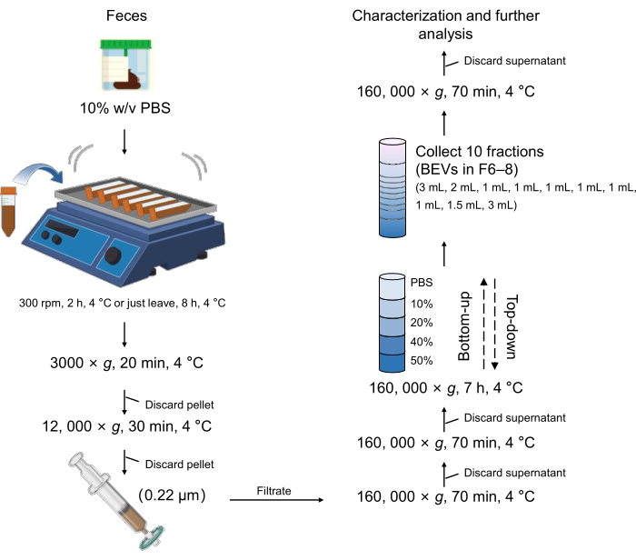
图1:细菌胞外囊泡(BEVs)分离与纯化的示意图流程。 缩写:BEVs = 细菌胞外囊泡。 请点击此处查看该图的放大版本。
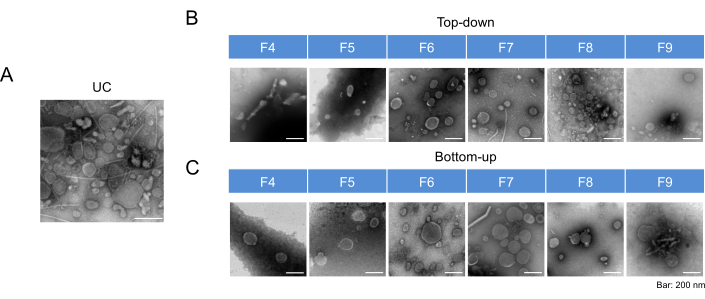
图2:BEV组分的代表性透射电镜图像。(A)经超速离心(UC)分离获得的BEVs的透射电镜图像。(B)采用自上而下模式经密度梯度离心(DGC)分离获得的BEVs的透射电镜图像。(C)采用自下而上模式经密度梯度离心(DGC)分离获得的BEVs的透射电镜图像。所有图像均以200 nm的统一比例尺呈现。缩写:TEM = 透射电子显微镜;BEVs = 细菌胞外囊泡;UC = 超速离心;DGC = 密度梯度离心。请点击此处查看该图的放大版本。
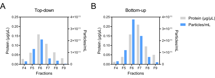
图 3:自上而下和自下而上密度梯度离心模式后BEV组分的蛋白质浓度和颗粒浓度。采用BCA法测定BEV组分的蛋白质浓度,以灰色柱状图表示。颗粒浓度通过NTA测定,以蓝色柱状图表示。缩写:BEV = 细菌胞外囊泡;DGC = 密度梯度离心;BCA = 双辛可宁酸法;NTA = 纳米颗粒追踪分析。请点击此处查看该图的放大版本。
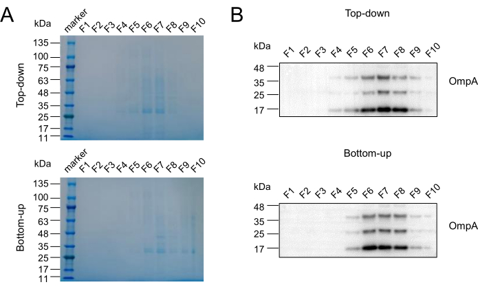
图4:BEV富集组分的确定。(A)采用考马斯亮蓝染色法(CBBS)分析在自上而下和自下而上两种模式中富含粪便细菌胞外囊泡(BEVs)的组分。(B)从Escherichia coli中分离、纯化胞外囊泡(EVs),并作为阳性对照用于Western blot(WB)检测,以确认BEVs的存在与分布。OmpA:外膜蛋白A,一种BEV标志物。缩写:BEV = 细菌胞外囊泡;CBBS = 考马斯亮蓝染色;EVs = 胞外囊泡;WB = Western blotting。请点击此处查看该图的放大版本。
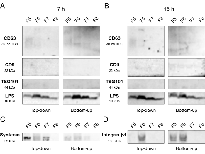
图5:EEV分布的验证。 对通过自上而下和自下而上密度梯度离心(DGC)方法获得的组分5–8进行Western印迹分析,密度梯度超速离心时间分别为7 h(A)和15 h(B)。另进行一次独立实验,重点分析7小时离心后得到的F5至F8组分,结果如(C)和(D)所示。所选标志物包括与BEV(LPS)和EEV(CD63、CD9、TSG-101、Syntenin、Integrin β1)相关的蛋白。缩写:DGC = density gradient centrifugation;BEV = bacterial extracellular vesicle;EEV = eukaryotic extracellular vesicle。 请点击此处查看该图的放大版本。
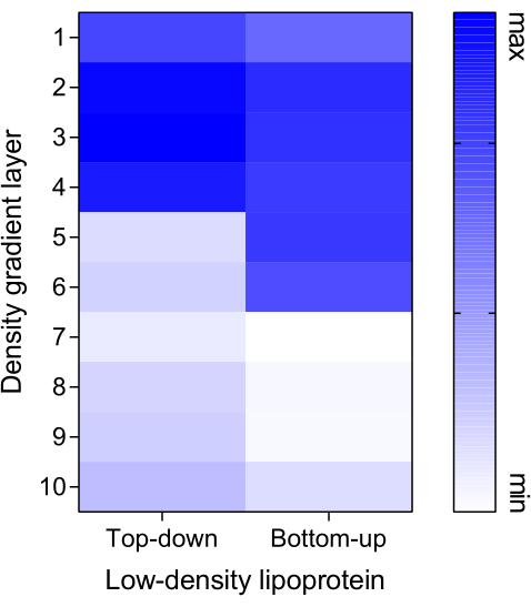
图6:低密度脂蛋白的分布情况。将 Dil 标记的低密度脂蛋白(Dil-LDL,作为污染标志物)以 10 µg 的浓度分别加入自上而下(Top-down)和自下而上(Bottom-up)两种模式的密度梯度系统中。Dil-LDL 替代了 PBS/BEV 溶液(步骤 6.1.1 和 步骤 6.2.2),以建立检测模型。使用微孔板检测仪在激发/发射波长 549/565 nm 下测定各组分的荧光强度。缩写:BEV = 细菌胞外囊泡。请点击此处查看该图的放大版本。
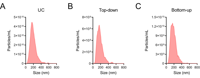
图 7:BEV 富集组分的粒径分布。(A)超速离心(UC)后获得的粗提 BEV 的粒径分布。(B)采用自上而下密度梯度离心(DGC)模式获得的 BEV 富集组分(F6–F8)的粒径分布。(C)采用自下而上 DGC 模式获得的 BEV 富集组分(F6–F8)的粒径分布。采用纳米颗粒追踪分析(NTA)对颗粒尺寸进行定量分析。图(A–C)所用的稀释倍数分别为 10,000、2,000 和 5,000,并对结果进行了相应校准。缩写:BEV = 细菌胞外囊泡;UC = 超速离心;DGC = 密度梯度离心;NTA = 纳米颗粒追踪分析。请点击此处查看该图的放大版本。
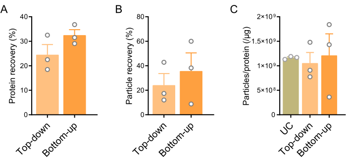
图8:自上而下和自下而上DGC模式的蛋白质与颗粒回收率。(A)两种模式的蛋白质回收率,以DGC(UC)前后蛋白质浓度的比值计算。(B)两种模式的颗粒回收率,以DGC(UC)前后颗粒浓度的比值计算。(C)UC、自上而下和自下而上模式的颗粒/蛋白质比值。数据以均值±标准误表示。统计分析采用双尾非配对t-检验对图A和B进行分析,图C采用单因素方差分析(ANOVA)结合Tukey事后检验。未观察到显著差异。缩写:DGC = 密度梯度离心;UC = 超速离心。请点击此处查看该图的放大版本。
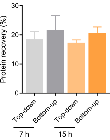
图9:基于自上而下法和自下而上法密度梯度离心(DGC)模式,采用不同密度相关超速离心时间的蛋白质回收率比较。 在独立实验中,分别采用自上而下法和自下而上法,对经7小时和15小时梯度超速离心后获得的F6–F8组分中的蛋白质回收率进行了评估。数据以均值±标准误(SEM)表示。采用双因素方差分析(ANOVA)结合Fisher最小显著性差异检验进行统计学分析。未观察到显著差异。缩写:DGC = 密度梯度离心。 请点击此处查看该图的放大版本。
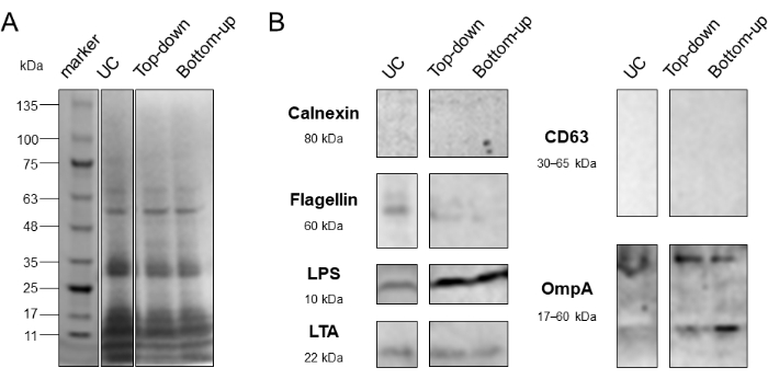
图 10:自上而下和自下而上密度梯度离心模式的纯度评估。(A)采用考马斯亮蓝染色(CBBS)比较超速离心法(UC)、自上而下法和自下而上法的蛋白质分布。(B)采用 Western 印迹法(WB)评估 UC、自上而下法和自下而上法的纯度。Calnexin:内质网相关蛋白标志物;Flagellin:细菌污染蛋白标志物。CD63:真核细胞外囊泡跨膜蛋白标志物;LTA:革兰氏阳性菌细菌外膜囊泡(BEVs)标志物;LPS、OmpA:革兰氏阴性菌 BEVs 标志物。缩写:DGC = 密度梯度离心;UC = 超速离心;CBBS = 考马斯亮蓝染色;WB = Western 印迹法。请点击此处查看该图的放大版本。
| 碘克沙醇溶液浓度 (%) | 0.02 M HEPES 缓冲液 | 50% 碘克沙醇工作液 |
| 40 | 1 | 4 |
| 20 | 3 | 2 |
| 10 | 4 | 1 |
表1:制备碘克沙醇密度梯度缓冲液的配比。 本表提供了用于获得特定浓度碘克沙醇溶液的0.02 M HEPES缓冲液与50%碘克沙醇工作液的配比。
| F1 | F2 | F3 | F4 | F5 | F6 | F7 | F8 | F9 | F10 | |
| 密度 (g/mL) | 1.023 | 1.038 | 1.049 | 1.062 | 1.074 | 1.098 | 1.144 | 1.185 | 1.239 | 1.293 |
表2:基于碘克沙醇的密度梯度离心系统的密度。 该表格显示了基于碘克沙醇的密度梯度离心系统中各组分的密度。空白对照:基于磷酸盐缓冲液(PBS)的梯度系统。
| 自上而下模式 | 自下而上模式 | |
| 颗粒回收率 (%) | 24.08 | 35.69 |
| 蛋白质回收率 (%) | 24.5 | 32.45 |
表3:两种模式的颗粒和蛋白质回收率。 该表格显示了自上而下模式和自下而上模式的颗粒和蛋白质回收率(图8)。
讨论
细菌胞外囊泡(BEVs)是由细菌分泌的脂质双层纳米颗粒,携带大量蛋白质、脂质、核酸及其他生物活性分子,有助于介导细菌的功能效应20。源自肠道的BEVs已被证实参与炎症性肠病、克罗恩病和结直肠癌等疾病的发生发展,同时还影响整体代谢并介导认知功能障碍4,16,17,20,21,22,23,24,25,26。目前,从人体样本——粪便中获取关于肠道细菌来源BEVs完整且客观的生物学信息仍受到一系列因素的限制,其中首要的关键问题是如何从复杂的宿主环境中分离出BEVs。
分离BEVs的主要方法,如超速离心法(UC)、密度梯度离心法(DGC)和尺寸排阻色谱法(SEC)13,14,15,16,17,均具有各自的优点和局限性。难以完全去除某些干扰物质,以及缺乏统一的操作流程,严重影响了对BEVs更真实、全面的分析,仍是当前分离过程中的挑战。为减轻干扰物带来的不利影响,许多研究15,27,28已结合使用上述两种或多种方法以实现BEVs的分离,尤其是从体液中分离BEVs,但复杂的操作流程往往会影响结果的稳定性。对于BEVs而言,其与其他污染物(如蛋白质聚集体、脂质组分、真核细胞外囊泡(EEVs)15)在密度上的差异,如今可能成为一种特异性的分离手段。
目前,碘克沙醇因其等渗特性已成为分离细胞外囊泡(EVs)的密度梯度离心首选介质,取代了蔗糖。这些特性有助于保持EV的形态完整性,并便于对各组分进行密度估算29。与Tulkens等15的研究一致,本研究采用相同的DGC系统浓度,使用50% (w/v)、40% (w/v)、20% (w/v)、10% (w/v) 和0%浓度梯度层的碘克沙醇。第一步是使用HEPES缓冲液配制不同浓度的碘克沙醇溶液。与Tris (-EDTA)-蔗糖缓冲液相比,适当浓度的HEPES缓冲液因其固有性质,可确保离心系统内长期的pH稳定性。同时,在离心系统的最上层使用无菌PBS缓冲液,不仅可在溶液配制过程中防止细菌及其代谢产物的潜在污染,还能维持DGC前后样本缓冲体系的一致性。具体操作包括:在超速离心(UC)后用PBS缓冲液重悬沉淀,并在DGC后用PBS替代碘克沙醇,从而便于结果的比较分析。此外,DGC系统中各梯度层的体积分别校准为:50%、40%、20%、10%和0%(PBS层)碘克沙醇溶液对应3 mL、3 mL、3 mL、3 mL和3.5 mL。该策略有助于建立相对较宽的密度范围(1.02–1.30 g/mL),可能增强细菌来源的囊泡(BEVs)与其他干扰成分的分离效果,从而使该梯度体系适用于其他含有复杂污染物的体液样本。
需要特别强调的是,该实验方案中的某些步骤至关重要。例如,第2.2步中所述的稀释倍数,即将1 g粪便样本与至少10 mL PBS混合,对于防止过饱和以及节约实验资源具有关键作用。在这些条件下,假设其他粪便特性保持不变,样本中颗粒和蛋白质的浓度通常会随粪便质量的增加而成比例升高。如有需要,可进一步分析粪便的一致性、脂蛋白或血液含量等因素。在第3.4步操作过程中,应避免直接倾倒溶液,以防扰动沉淀物,导致较大颗粒堵塞0.22 µm滤膜。若滤膜迅速饱和(例如,少于10 mL液体通过滤膜),建议增加一次12,000 × g的离心步骤。在密度梯度离心过程中,梯度溶液的精确配制是获得可靠结果的前提条件。此外,在叠加上层低密度溶液时,必须小心操作离心管,以防止分层结构被破坏,同时需通过仔细移液排除所有气泡。最后,在吸取各组分时,应沿管壁谨慎吸取,避免过度或不足吸取,以免造成BEV分布不准确。定期观察液面位置并保持规范操作有助于缓解这一问题。
本研究采用多种表征方法确定了富含BEVs的组分(F6-F8),并探讨了两种不同的梯度超速离心模式:自上而下法(Top-down)和自下而上法(Bottom-up)。通过对F4-F8组分中蛋白质浓度和颗粒浓度的测定(图3)并比较回收率和纯度(图8 和 图10),在从粪便样本中分离和纯化BEVs时,自下而上的离心模式在蛋白质和颗粒水平上检测到的数值均高于另一种方法。该观察结果暗示了不同的动态过程。研究提出了一种假设,即非囊泡性细胞外纳米颗粒(NVEPs)29,例如密度低于BEV的脂蛋白,若样品从底部加样,则可能无法达到其浮力密度图6),可能导致回收率偏高,即使在DGC 15小时后图9)。这一观察结果提示,有必要考虑基于颗粒与蛋白质比例的传统纯度定义是否仍然适用。此外,值得注意的是,EEV 主要富集于 F5 组分,而 BEV 则在 F6–F7 组分中显著检出。图4 和 图5值得注意的是,采用自上而下(Top-down)模式时,这种差异似乎更为显著,因此该模式可能更适用于本实验方案。然而,必须强调的是,在F6组分中检测到整合素β1(Integrin β1)的存在,可能提示EEV之间存在内在异质性。当将该方法应用于其他体液(如血液和尿液)时,研究人员应考虑样本的独特特性。本方案中设定的离心时间为7小时,明显短于其他研究中报道的时间,后者最长可达18小时。与本研究中以相同模式进行的15小时蛋白回收率相比(图9),此时已足以实现所需的分离纯度。在这些操作条件下,自上而下模式下的颗粒回收率可达108 (4.5 × 10⁷-1.5 × 10⁸)每毫克粪便,与先前的报道一致(1011 每克湿粪便中的BEV数量15,29,30最后,空白对照组各组分的密度测量结果证实,BEVs 的密度范围为 1.09–1.19 g/mL,与早期研究结果一致。
该方法尽管有效,但由于饮食、肠道功能及其他因素对粪便成分的影响而存在局限性。这些变量可能改变粪便的性状,进而影响细菌丰度及细菌外膜囊泡(BEV)的回收率。因此,至关重要的是根据粪便特征的评估结果,对BEV的分离与纯化过程进行标准化31,32。未来的研究应致力于确定一种用于归一化BEV产量的标准,类似于尿肌酐或尿流速的标准化方法。此类基准将有助于提升方法学评估的准确性,并揭示粪便BEV与机体生理及病理状态之间的关联。此外,粪便BEV与多种疾病之间存在显著关联,凸显其重要的临床潜力。然而,本方案中规定的离心时间尚不能满足临床医生对更便捷、更快速检测的需求。因此,还需进一步研究以确定更短的离心时间(例如少于3小时)是否能够获得相当的结果。同时,也有必要进一步探究两种离心模式下发生的生物学事件。尽管推测在自下而上(Bottom-up)模式中,非囊泡成分在F6-F8组分中更为富集,但优化梯度浓度可能有助于识别具有特定功能的更多亚型。
本研究成功证明了使用密度梯度离心法(DGC)从人类粪便中分离和纯化细菌外膜囊泡(BEVs)。该策略有望应用于其他生物样本,如血液、尿液和唾液,为研究人员提供一种有效的方法以揭示BEVs中隐藏的信息,从而推动其在临床中的应用。
披露
作者声明不存在利益冲突。
致谢
本工作由以下机构资助: 国家杰出青年科学基金(82025024); 国家自然科学基金重点项目(82230080); 国家重点研发计划&中国国家重点研发计划(2021YFA1300604);国家自然科学基金(81871735、82272438 和 82002245); 中国广东 自然科学基金 用于 杰出青年学者(2023B1515020058); 广东省自然科学基金(2021A1515011639); 中国山东省自然科学基金重大基础研究发展计划(ZR2020ZD11); 中国博士后科学基金(2022M720059);南方医科大学南方医院优秀青年发展计划(2022J001)。
材料
| 姓名 | 公司 | 目录编号 | 评论 |
|---|---|---|---|
| 1 %(w/v)戊二醛(用去离子水将2.5 %储备液稀释配制) | ACMEC | AP1126 | 在步骤8.3.3中使用透射电子显微镜对BEV进行形态学观察 |
| 1 %(w/v)甲基纤维素(以去离子水从原始粉末配制) | Sigma-Aldrich | M7027 | 在步骤 8.3.6 中使用透射电子显微镜对 BEV 进行形态学观察 |
| 1.5 % (w/v) 醋酸铀酰(用去离子水从原始粉末配制) | Polysciences | 21447-25 | 在步骤8.3.5中使用透射电子显微镜对BEV进行形态学观察 |
| 1000 μL,200 μL, 10 μL 移液管 | KIRGEN | KG1313, KG1212, KG1011 | 转移溶液 |
| 5 %(w/v)牛血清白蛋白溶液(用TBST缓冲液将原始粉末配制而成) | Fdbio 科学 | FD0030 | 用于第 8.5.6 步的 Western 印迹封闭 |
| 5 × 上样缓冲液 | Fdbio 科学 | FD006 | 在第8.5.1步中用于蛋白质印迹和考马斯亮蓝染色 |
| 75 % (v/v) 乙醇 | LIRCON | LIRCON-500 mL | 表面消毒 |
| 96孔板 | Rar | A8096 | 测定吸光度值 |
| 抗钙联蛋白抗体 | Abcam | ab92573 | 蛋白质印迹(一抗) |
| 抗CD63抗体 | Abcam | ab134045 | Western 印迹(一抗) |
| 抗CD9抗体 | Abcam | ab236630 | 蛋白质印迹(一抗) |
| 抗鞭毛蛋白抗体 | Sino Biological | 40067-MM06 | 蛋白质印迹(一抗) |
| 抗整合素β1抗体 | Abcam | ab30394 | 蛋白质印迹(一抗) |
| 抗LPS抗体 | 赛默飞世尔科技 | MA1-83152 | 蛋白质印迹(一抗) |
| 抗LTA抗体 | 赛默飞世尔科技 | MA1-7402 | 蛋白质印迹(一抗) |
| 抗OmpA抗体 | CUSABIO | CSB-PA359226ZA01EOD,https://www.cusabio.com/ | 蛋白质印迹(一抗) |
| 抗Syntenin抗体 | Abcam | ab133267 | 蛋白质印迹(一抗) |
| 抗TSG101抗体 | Abcam | ab125011 | 蛋白质印迹(一抗) |
| 高压灭菌 | ZEALWAY | GR110DP | 实验所用物品和培养基的灭菌 |
| 平衡 | Mettler Toledo | AL104 | 用PBS平衡已加样的离心管 |
| 二辛可宁酸法测定 | Fdbio 科学 | FD2001 | 在步骤 8.2 测定 BEVs 的蛋白质含量 |
| BioRender | BioRender | https://app.biorender.com | 制备图1所示的BEVs分离与纯化的示意图工作流程 |
| 生物安全柜 | Haier | HR1200-II B2 | 进行粪便样本处理的相关操作 |
| Centrifuge 5810 R;转子 F-34-6-38 | Eppendorf | 5805000092;5804727002,适配器:5804774000 | BEVs 预处理(步骤 3) |
| 化学发光装置 | BIO-OI | OI600SE-MF | 用于第 8.5.12 步 Western 印迹中的信号检测 |
| Cytation 5 | BioTek | F01 | 用于测量吸光度(步骤 8.1)和荧光值(图 6)的微孔板检测仪 |
| Dil标记的低密度脂蛋白 | ACMEC | AC12038 | 干扰组分分布的定义 |
| 电泳设备 | Bio-rad | 1658033 | 用于第8.5.2、8.5.3和8.5.5步的蛋白质印迹实验中的蛋白质分离与转膜 |
| 增强型化学发光HRP试剂盒 | Fdbio科学 | FD8020 | 用于第8.5.12步的Western印迹信号检测 |
| 大肠杆菌 | 美国典型培养物保藏中心 | ATCC8739 | 以BEVs作为阳性对照进行分离。方案:将25 g LB培养基粉末溶解于1 L去离子水中,高压灭菌。转移800 μ将保存的 L 型大肠杆菌接种至培养基中。37 ℃培养 °在培养摇床中孵育。然后在3,000离心 × 4℃下20分钟2000 × g离心 °C, 12, 000 × 4℃下30分钟3000 × g离心 °C,将上清液通过0.22 μm滤膜过滤 μm 膜,并在 160,000 下进行超速离心 × 4℃下70分钟离心,转速为g °C. 将定义为大肠杆菌粗提BEV的沉淀重悬于1.2 mL PBS中(步骤3、4)。 |
| 50 mL Falcon管 | KIRGEN | KG2811 | BEVs 预处理(步骤 3) |
| 甲胎蛋白染色缓冲液 | AbSci | ab.001.50 | 第8.5.4步考马斯亮蓝染色 |
| 滤纸 | Biosharp | BS-TFP-070B | 在步骤 8.3(吸取溶液)中使用透射电子显微镜对 BEV 进行形态学观察 |
| 铜网,Formvar/碳膜支持 | Sigma-Aldrich | TEM-FCF200CU50 | 在步骤8.3中使用透射电子显微镜对BEV进行形态学观察 |
| HEPES 粉末 | Meilunbio | MB6078 | 制备不同浓度的碘克沙醇缓冲液用于密度梯度离心 |
| HRP AffiniPure 山羊抗小鼠 IgG(H+L) | Fdbio 科学 | FDM007 | 蛋白质印迹(二抗) |
| HRP AffiniPure 山羊抗兔 IgG(H+L) | Fdbio 科学 | FDR007 | 蛋白质印迹(二抗) |
| 摇床培养箱 | Qiangwen | DHZ-L | 培养大肠杆菌 |
| Kimwipes™ 精细操作擦拭布 | Kimtech Science | 34155 | 在步骤 4.15 中擦拭超速离心管的内壁 |
| LB肉汤 | Hopebio | HB0128 | 培养大肠杆菌 |
| 低温冰箱(-80) °C) | Haier | DW-86L338J | 储存样品 |
| 甲醇 | Alalddin | M116118 | 用于第8.5.5步Western blot实验中PVDF膜的活化 |
| 1.5 mL 微量离心管 | KIRGEN | KG2211 | 密度梯度离心后回收组分 |
| 2 mL 微量离心管 | KIRGEN | KG2911 | 密度梯度离心后回收组分 |
| 5 mL 微量离心管 | BBI | F610888-0001 | 密度梯度离心后回收组分 |
| 酶标仪 | 赛默飞世尔科技 | Multiskan MK3 | 在步骤 8.2 测定 BEVs 的蛋白质含量 |
| Millipore 滤膜 0.22 μm | 默克密理博 | SLGP033RB | 过滤除菌;材质:聚醚砜,PES |
| NaCl | GHTECH | 1.01307.040 | 密度梯度离心液 |
| NaOH | GHTECH | 1.01394.068 | 密度梯度离心溶液(pH 调整) |
| Optima™ XPN-100 | 贝克曼库尔特 | A94469 | 第4步和第7步中用于BEVs分离的超速离心 |
| OptiPrep™ | Serumwerk Bernburg AG | 1893 | 密度梯度离心储备液 |
| 轨道摇床 | 尤宁 | CS-100 | 在步骤2中溶解粪便 |
| 磷酸盐缓冲液 | Procell | PB180327 | 在第2步溶解粪便 |
| 移液器 | Eppendorf | 3120000267, 3120000259 | 转移溶液 |
| 塑料巴斯德吸管 | ABCbio | ABC217003-4 | 在预处理步骤3.4中去除上清液 |
| 聚偏二氟乙烯(PVDF)膜 | Millipore | ISEQ00010, IPVH00010 | 用于第8.5.5步蛋白质印迹中的蛋白转膜 |
| 预制聚丙烯酰胺凝胶,4–20% 15孔板 | ACE | F15420Gel | 用于第8.5.2、8.5.3步的蛋白质印迹实验中的蛋白质分离 |
| 一抗稀释液 | Fdbio 科学 | FD0040 | 在第 8.5.8 步的蛋白质印迹实验中使用 |
| 蛋白标准品 | Fdbio 科学 | FD0672 | 在第8.5步中用于蛋白质印迹和考马斯亮蓝染色 |
| 快速蛋白质印迹溶液 | UBIO | UW0500 | 用于第8.5.5步蛋白质印迹中的蛋白转印 |
| SW 32 Ti 摆动桶转子 | 贝克曼库尔特 | 369650 | 第4步和第7步中用于BEVs分离的超速离心 |
| 20 mL、50 mL 注射器 | 捷运通道 | ZSQ-20ML,YCXWJZSQ-50 mL | 转移缓冲液并去除预处理中的上清液 |
| TBS 粉末 | Fdbio 科学 | FD1021 | 在第 8.5 步的蛋白质印迹实验中使用 |
| 透射电子显微镜(TEM) | 日立 | H-7650 | 第8.3步中对BEV的形态学观察 |
| Tween-20 | Fdbio 科学 | FD0020 | 在第8.5步的蛋白质印迹实验中使用 |
| 超速离心管 | 贝克曼 | 326823, 355642 | 第4步和第7步中用于BEVs分离的超速离心 |
| 超净工作台 | AIRTECH | SW-CJ-2FD | 执行液体处理的相关操作 |
| 水浴锅 | Bluepard | CU600 | 用于第8.2.5步中测量BEVs的蛋白质含量 |
| ZetaView | Particle Metrix | S/N 21-734,软件 ZetaView(版本 8.05.14 SP7) | 纳米颗粒跟踪分析(NTA)用于在步骤 8.4 中测量 BEV 的粒径和浓度 |
参考文献
- Costello, E. K., et al. Bacterial community variation in human body habitats across space and time. Science. 326 (5960), 1694-1697 (2009).
- Greenhalgh, K., Meyer, K. M., Aagaard, K. M., Wilmes, P. The human gut microbiome in health: establishment and resilience of microbiota over a lifetime. Environmental Microbiology. 18 (7), 2103-2116 (2016).
- de Vos, W. M., Tilg, H., Van Hul, M., Cani, P. D. Gut microbiome and health: mechanistic insights. Gut. 71 (5), 1020-1032 (2022).
- Zhou, P., Yang, D., Sun, D., Zhou, Y. Gut microbiome: New biomarkers in early screening of colorectal cancer. Journal of Clinical Laboratory Analysis. 36 (5), 24359(2022).
- Paik, D., et al. Human gut bacteria produce Τ(Η)17-modulating bile acid metabolites. Nature. 603 (7903), 907-912 (2022).
- Parada Venegas, D., et al. Short chain fatty acids (SCFAs)-mediated gut epithelial and immune regulation and its relevance for inflammatory bowel diseases. Frontiers in Immunology. 10, 277(2019).
- Jiang, C., Li, G., Huang, P., Liu, Z., Zhao, B. The Gut microbiota and Alzheimer's disease. Journal of Alzheimer's Disease. 58 (1), 1-15 (2017).
- Morais, L. H., Schreiber, H. L. t, Mazmanian, S. K. The gut microbiota-brain axis in behaviour and brain disorders. Nature Reviews Microbiology. 19 (4), 241-255 (2021).
- Kim, J. H., Lee, J., Park, J., Gho, Y. S. Gram-negative and Gram-positive bacterial extracellular vesicles. Seminars in Cell and Developmental Biology. 40, 97-104 (2015).
- Toyofuku, M., Nomura, N., Eberl, L. Types and origins of bacterial membrane vesicles. Nature Reviews Microbiology. 17 (1), 13-24 (2019).
- Xie, J., Li, Q., Haesebrouck, F., Van Hoecke, L., Vandenbroucke, R. E. The tremendous biomedical potential of bacterial extracellular vesicles. Trends in Biotechnology. 40 (10), 1173-1194 (2022).
- Coumans, F. A. W., et al. Methodological guidelines to study extracellular vesicles. Circulation Research. 120 (10), 1632-1648 (2017).
- Northrop-Albrecht, E. J., Taylor, W. R., Huang, B. Q., Kisiel, J. B., Lucien, F. Assessment of extracellular vesicle isolation methods from human stool supernatant. Journal of Extracellular Vesicles. 11 (4), 12208(2022).
- Park, Y. E., et al. Microbial changes in stool, saliva, serum, and urine before and after anti-TNF-α therapy in patients with inflammatory bowel diseases. Scientific Reports. 12 (1), 6359(2022).
- Tulkens, J., De Wever, O., Hendrix, A. Analyzing bacterial extracellular vesicles in human body fluids by orthogonal biophysical separation and biochemical characterization. Nature Protocols. 15 (1), 40-67 (2020).
- Tulkens, J., et al. Increased levels of systemic LPS-positive bacterial extracellular vesicles in patients with intestinal barrier dysfunction. Gut. 69 (1), 191-193 (2020).
- Kang, C. S., et al. Extracellular vesicles derived from gut microbiota, especially Akkermansia muciniphila, protect the progression of dextran sulfate sodium-induced colitis. PloS one. 8 (10), 76520(2013).
- Simonsen, J. B. What are we looking at? Extracellular vesicles, lipoproteins, or both. Circulation Research. 121 (8), 920-922 (2017).
- Correll, V. L., et al. Optimization of small extracellular vesicle isolation from expressed prostatic secretions in urine for in-depth proteomic analysis. Journal of Extracellular Vesicles. 11 (2), 12184(2022).
- Liang, X., et al. Gut bacterial extracellular vesicles: important players in regulating intestinal microenvironment. Gut Microbes. 14 (1), 2134689(2022).
- Alberti, G., et al. Extracellular vesicles derived from gut microbiota in inflammatory bowel disease and colorectal cancer. Biomedical papers of the Medical Faculty of the University Palacky, Olomouc, Czech Republic. 165 (3), 233-240 (2021).
- Díez-Sainz, E., Milagro, F. I., Riezu-Boj, J. I., Lorente-Cebrián, S. Effects of gut microbiota-derived extracellular vesicles on obesity and diabetes and their potential modulation through diet. Journal of Physiology and Biochemistry. 78 (2), 485-499 (2022).
- Lajqi, T., et al. Gut microbiota-derived small extracellular vesicles endorse memory-like inflammatory responses in murine neutrophils. Biomedicines. 10 (2), 442(2022).
- Lee, K. E., et al. The extracellular vesicle of gut microbial Paenalcaligenes hominis is a risk factor for vagus nerve-mediated cognitive impairment. Microbiome. 8 (1), 107(2020).
- Villard, A., Boursier, J., Andriantsitohaina, R. Bacterial and eukaryotic extracellular vesicles and nonalcoholic fatty liver disease: new players in the gut-liver axis. American Journal of Physiology-Gastrointestinal and Liver Physiology. 320 (4), G485-G495 (2021).
- Wei, S., et al. Outer membrane vesicles enhance tau phosphorylation and contribute to cognitive impairment. Journal of Cellular Physiology. 235 (5), 4843-4855 (2020).
- Bitto, N. J., Kaparakis-Liaskos, M. Methods of bacterial membrane vesicle production, purification, quantification, and examination of their immunogenic functions. Methods in Molecular Biology. 2523, 43-61 (2022).
- Stentz, R., Miquel-Clopés, A., Carding, S. R. Production, isolation, and characterization of bioengineered bacterial extracellular membrane vesicles derived from Bacteroides thetaiotaomicron and their use in vaccine development. Methods in Molecular Biology. 2414, 171-190 (2022).
- Zhang, Q., Jeppesen, D. K., Higginbotham, J. N., Franklin, J. L., Coffey, R. J. Comprehensive isolation of extracellular vesicles and nanoparticles. Nature Protocols. 18 (5), 1462-1487 (2023).
- Iwai, K., Minamisawa, T., Suga, K., Yajima, Y., Shiba, K. Isolation of human salivary extracellular vesicles by iodixanol density gradient ultracentrifugation and their characterizations. Journal of Extracellular Vesicles. 5, 30829(2016).
- Vandeputte, D., et al. Stool consistency is strongly associated with gut microbiota richness and composition, enterotypes and bacterial growth rates. Gut. 65 (1), 57-62 (2016).
- Wen, M., et al. Bacterial extracellular vesicles: A position paper by the Microbial Vesicles Task Force of the Chinese Society of Extracellular Vesicles. Interdisciplinary Medicine. 1, 12046(2023).
重印与许可
