本方案概述了利用器官型组织模型研究宿主-病原体相互作用的方法 体外具体而言,所述模型系统采用多层口腔上皮,研究其在口腔健康与疾病状态下暴露于生物膜(牙菌斑)相关微生物的情况。
本文内容
摘要
摘要
重建上皮或黏膜屏障处的宿主-病原体相互作用界面 体外 对感染生物学研究者而言,这仍是一个具有挑战性的课题。尽管实验室自制的二维上皮单层细胞模型在一定程度上被广泛应用,但其无法真实再现 体内 然而,由于成本和实用性问题,商业化的组织模型常常被忽视。但是,通过周密的规划,这类模型可为多种不同的应用提供可重复的实验平台。本文报道了可用于多种实验目的的上皮模型,以研究口腔、皮肤和阴道黏膜等不同生态位中的宿主-病原体相互作用。从简单的浮游细胞到复杂的生物膜共培养,上皮模型可用于评估微生物的黏附与侵袭能力,并在转录和/或蛋白质水平上分析宿主的反应,还可结合多种组学方法进行更深入的分析。此外,这些生物系统可作为更精确的测试平台,用于在复杂的宿主-病原体微环境中评估传统和新型抗菌剂的活性 体外. 本文所述方案记录了模型在抵达实验室后的处理及准备过程,以便与生物膜群落进行共培养刺激。方法部分详细说明了如何从模型系统中获得实验结果,包括组织处理、共培养体系的建立以及数据生成。这些实验包括通过单重和多重qPCR分析检测宿主基因表达,以及利用ELISA检测炎症蛋白。总之,上皮模型提供了有用的 体外 用于研究简单或复杂宿主-病原体相互作用的临床前研究系统。
引言
体外 模型系统为研究宿主-病原体相互作用提供了优良的实验平台。这些类器官系统旨在模拟易受微生物扰动的不同生态位的微环境。已有大量研究利用 EpiSkin(原名 SkinEthic)组织模型来评估口腔内的宿主-病原体相互作用。1,2,3,4,5皮肤屏障6,7,8,9,10,11以及阴道黏膜5,12,13以及其他14,15。与 "二维" 或 "二维" 细胞培养,依赖于将上皮细胞系的单层细胞暴露于微生物,而这些商业化的模型则由在气液界面培养的多层分化细胞组成。尽管二维培养系统成本较低且通常被认为更具可重复性,但在与微生物共培养时容易发生细胞损伤,因而并不总能准确模拟上皮或黏膜屏障。 体内16,17.
最近的证据表明 "三维" 或 "3D" 培养技术在包括癌症科学、干细胞研究和药物发现在内的多个科学领域中正变得越来越流行16,18在感染生物学背景下,三维组织模型可用于在受控的微环境中研究上皮或黏膜屏障界面处的宿主-细菌、宿主-真菌或多种物种相互作用 体外这些模型相较于二维培养系统具有诸多优势,可用来评估病原生物体或复杂生物膜群落对组织的定植和/或侵袭能力17,并在多组分层面上评估不同生物标志物的宿主反应,而二维模型有时因其转录组学或蛋白质组学特征而受到限制16,17,18最终,这类3D系统的临床前应用前景广阔,可提供重要的探索性数据,用于进一步推进 体内 模型或临床研究。
在口腔健康与疾病背景下,了解口腔黏膜表面所涉及的炎症通路对临床医生至关重要,因为这可能指导治疗方式的选择。既往研究表明 "健康相关" 生物膜可引发轻微的炎症反应3,19,20,21这可能源于与口腔健康相关的较低微生物生物负荷,或由于生物膜的组成 链球菌 spp. 通常被认为具有免疫调节作用22,23另一方面,由与疾病相关的微生物(如 具核梭杆菌 和 牙龈卟啉单胞菌 具有促炎特性3,20,24,25,26,27Mountcastle 等人在 2020 年的一篇综述中描述了当时存在的所有与口腔微环境相关的共培养模型17尽管此类器官型模型种类繁多,且已有多种自此后被开发出来28,29,构建此类3D模型仍面临诸多挑战性障碍。举例而言,这些模型需要大量优化,且生产过程高度依赖人力和资源;除非严格控制实验条件,否则其可重复性也可能存在较大差异16,17市面上可获取的模型可规避此类问题,为研究口腔及其他人体生态位中的宿主-病原体相互作用提供了平台。
总之,本方案旨在概述利用器官型组织模型研究宿主-病原体相互作用的方法 体外 在口腔健康与疾病背景下,所述模型系统采用一种多层商业化口腔上皮组织,暴露于代表口腔炎症性疾病——牙龈炎的确定性生物膜群落中。
方案
以下方案涉及制备一种代表牙龈炎的多物种生物膜,共包含7个物种(spp.)7,30三 链球菌 sp., Streptococcus mitis (NCTC 12261) Streptococcus intermedius (DSM 20753),以及 Streptococcus oralis (NTCC 11427)被纳入以模拟口腔健康状态,作为唾液薄膜的初始定植菌。随后加入四种与口腔健康向疾病转变相关的厌氧微生物: Veillonella dispar (NCTC 11831), 缓症链球菌 (DSM 17233)以及两种 梭杆菌 种(spp.) 具核梭杆菌 (ATCC 10953)和 具核梭杆菌 亚种(subspp.) vincentii (DSM 19507)。所有操作均在火焰旁或II级生物安全柜中无菌进行。用于微生物制备的所有培养基和磷酸盐缓冲液(PBS)在使用前均经高压灭菌,并在实验过程中定期检测无菌状态。本研究中所用试剂和设备的详细信息列于下文 材料表.
1. 共培养用微生物群落的制备
注意:本方案描述了一种多物种生物膜的构建方法,该生物膜代表了与牙龈组织炎症(即牙龈炎)相关的微生物群落。此类模型此前已被用于评估口腔健康与疾病状态下的宿主反应3。
- 从保存在 -80°C 的多孔珠冻存菌种(商业获得)中,使用接种环和划线平板技术,将三种 Streptococcus 菌种复苏于血琼脂平板(含 5% 无菌脱纤维马血的哥伦比亚血琼脂基础培养基)上。
- 在 37 °C、5% CO2 条件下孵育 24 小时,然后挑取 3–4 个单菌落接种至 10 mL 胰蛋白胨大豆肉汤培养基中进行扩增培养。在 37 °C、5% CO2 条件下培养 16–18 小时。
- 对于中间型致病菌,将 V. dispar、A. naeslundii、F. nucleatum 及 F. nucleatum 亚种 vincentii 在含 5% 无菌脱纤维马血的苛养厌氧琼脂基础培养基上,于 37 °C 厌氧条件下培养 48 小时,随后在 Schaeder 肉汤中继续在相同条件下培养额外 24–48 小时。
- 培养完成后,将细胞悬液在 20 °C 下以 3000 × g 离心 5 分钟收集菌体,然后用 10 mL 无菌 PBS(pH 7.2–7.6)洗涤菌体沉淀。再次于 20 °C 以 3000 × g 离心 5 分钟收集菌体,并重复洗涤步骤一次。将洗涤后的菌体重悬于 10 mL 无菌 PBS 中用于标准化处理。
- 使用分光光度计在 550 nm 波长下分别对三种 Streptococcus 菌种进行标准化。吸光度值为 0.50(可接受范围为 0.45–0.55)时,表明细胞浓度约为 1 × 10⁸ 个细胞/mL,该数值此前已通过 Miles 和 Misra 细胞计数法确定31。为达到该吸光度值,需将 10 mL 洗涤后的细胞悬液用无菌 PBS 进一步稀释。
- 标准化后,将所有 Streptococcus 菌种用 1:1 混合的 Todd Hewitt 肉汤(THB)与 Roswell Park Memorial Institute(RPMI)培养基稀释 10 倍至 1 × 10⁷ 个细胞/mL。用移液器将 500 μL 细胞悬液加入含有直径为 13 mm 羟基磷灰石圆片的 24 孔微量组织培养板中。在 37 °C、5% CO2 条件下培养 24 小时,使生物膜成熟。
注意:多物种生物膜可在其他与口腔相关的基质上生长,例如釉质、牙本质32 以及聚甲基丙烯酸甲酯义齿表面33,34。这些模型也可使用其他培养基,如人工或合成唾液,但应根据所用微生物群落的具体组成谨慎选择:已有研究表明,培养基的选择对混合群落生物膜的生长具有重要影响35,36。 - 次日,以与上述步骤(1.2–1.4)类似的方法对四种厌氧微生物进行标准化处理。收集细胞悬液,洗涤两次,然后将 V. dispar 标准化至吸光度 0.50(范围 0.45–0.55),其余三种微生物标准化至吸光度 0.20(范围 0.18–0.22)。标准化后,将所有悬液用 1:1 混合的 THB 与 RPMI 培养基进一步稀释 10 倍至 1 × 10⁷ 个细胞/mL。
- 小心用移液器吸除 Streptococcus 生物膜中的未黏附细胞和废弃培养基并弃去。向微量培养板孔中加入 500 μL 标准化的四种厌氧菌悬液(浓度为 1 × 10⁷ 个细胞/mL)。在 37 °C 厌氧条件下继续培养生物膜 24 小时。
- 24 小时后,吸除七物种生物膜中的未黏附细胞和废弃培养基,更换为 500 µL 无菌的 1:1 THB: RPMI 混合培养基。将生物膜在 37 °C 厌氧条件下继续培养 4 天,每天更换培养基一次(共更换四次)。
- 第 7 天,多物种生物膜已完全成熟,可用于后续实验。使用 500 μL 无菌 PBS 洗涤生物膜两次,用于共培养实验。
注意:可采用多种生物学方法对生物膜进行分析,例如 qPCR(用于组成分析)以及共聚焦显微镜或电子显微镜成像分析,具体方法如前所述3。
2. 器官型组织处理与实验设置
注意:以下所述的实验设置涉及由在惰性聚碳酸酯膜滤器上培养的TR146细胞构成的人口腔上皮(HOE)组织。其他模型还包括表皮模型、膀胱、食管、角膜、牙龈及阴道上皮。所有模型的处理和制备方法均与下文所述用于研究宿主-病原体相互作用的方法类似。
- 到货后,立即开箱并将HOE组织及培养基转移至II级生物安全柜中。向12孔板中加入共1 mL随组织提供的维持培养基。
- 用无菌镊子从用于运输和转移的24孔板和营养琼脂中取出含有HOE的聚碳酸酯插件,并将其转移至含培养基的12孔板中,确保组织下方无气泡残留。使用另一把镊子或滤纸小心去除附着在插件侧面或底部的多余琼脂。
- 在37 °C、5% CO₂条件下将组织模型孵育24 h2 在实验设置前,组织模型需在运输后于实验室条件下进行适应性培养。值得注意的是,若需对组织模型进行更长时间的维持或进一步成熟培养,可使用其他维持培养基或生长培养基。
- 现在可以进行共培养实验。以本示例为例,移除按上述步骤(1.1–1.9)制备的7种微生物生物膜 使用超声处理使其底物分离。为此,用19号针头和镊子将含有生物膜的HA圆片从24孔板底部取出,随后转移至含有1 mL无菌Dulbecco磷酸盐缓冲液的 Bijoux 管中。在超声水浴中以35 kHz超声处理10分钟。
- 使用镊子小心取出过夜适应后的含组织模型的插入物,用移液器直接向组织中加入100 µL生物膜超声悬浮液。为进行比较,使用未刺激的对照组织。对于这些对照组织插入物,加入100 µL不含生物膜悬浮液的无菌杜氏磷酸盐缓冲液(Dulbecco's PBS)。
注意:浮游细胞、含有分散细胞的用过生物膜上清液或完整的生物膜均可用于共培养模型,以替代生物膜超声处理液。使用不同微生物刺激物的这些宿主-病原体模型的不同应用示意图如下所示 图1 如其他文献所述3,5,6,8,13,37,38. - 加入后,将插入物转移至另一含1 mL新鲜维持培养基的12孔板中,再次确保插入物下方无气泡。将含有组织模型的板在37 °C、5% CO₂条件下孵育24 h。2 在进行后续应用的组织处理之前。
注意:组织供应商可提供额外的培养基,以支持组织在接触超声处理的聚集体或生物膜后的持续培养。已有若干先前模型发表,用于研究组织与生物膜的长期接种作用。21,28,39为在当前的器官型模型中获得类似结果,将组织进行超声处理(同上,步骤2.6),并让微生物附着24小时。弃去残留的微生物悬液,并在气液界面继续培养至所需的实验时间。
3. 实验结果的组织处理
注意:共培养后,对组织模型进行处理以获取实验数据。以下步骤记录了如何从组织中提取RNA用于转录谱分析,以及如何利用组织培养上清液进行蛋白质检测。组织也可如前所述3,6,用甲醛、多聚甲醛或类似固定剂固定,用于组织学评估。
- 首先,在含有 1% β-巯基乙醇和约相当于 100 µL 的 0.5 mm 酸洗玻璃珠的 2.0 mL 螺口 O 型圈管中,准备 350 µL RLT 裂解缓冲液。
- 使用镊子从培养基中取出含有组织的插入物,并弃去插入物中残留的微生物悬液。接着,将其倒置,置于视线水平以便操作。使用 19 G 针头,小心地从插入物底部切开组织和膜,并转移至 RLT 缓冲液中。
- 使用台式珠磨匀浆器匀浆组织 30 秒,然后按照 RNA 提取试剂盒的说明书(参见材料表)从裂解物中提取 RNA。
注:提取的 RNA 可用于 cDNA 合成,以通过定量 PCR(qPCR)或 RNA 测序分析细胞因子和趋化因子基因的表达,作为炎症的标志物。qPCR 可使用多重阵列实现,例如 RT2 PCR 分析阵列,其孔中含有针对特定基因的预制引物,或使用基于 SYBR Green 的试剂,配合实验室自行设计的引物序列和无核酸酶的 ddH2O3。
- 使用台式珠磨匀浆器匀浆组织 30 秒,然后按照 RNA 提取试剂盒的说明书(参见材料表)从裂解物中提取 RNA。
- 收集剩余的用过组织培养基(约 850–900 µL),用于采用低通量和高通量方法(如 ELISA、多重免疫分析或多重蛋白质生物标志物分析)进行蛋白质组学分析7。
- 利用用过的组织培养基,在蛋白质水平上评估一系列与炎症相关的标志物。该培养基可直接用于上述方法(步骤 3.3),或储存于 -20 °C 或 -80 °C。为避免影响分析结果,应避免对培养基进行多次冻融循环。
注:根据组织刺激物的不同,为准确进行 ELISA 蛋白质组学分析,培养基可能需要按 1:10 比例稀释。
结果
在本实验中,将HOE暴露于含有代表从口腔健康状态转变为牙龈炎症(即牙龈炎)微生物的7种物种生物膜超声裂解液中。该疾病源于牙面牙菌斑积聚引起的微生物扰动,导致牙龈或口腔上皮组织发生炎症。刺激后,采集组织及培养上清液用于转录组和蛋白质组分析,如上所述。本实验中,通过生物膜超声裂解液刺激后,检测了组织中一组炎症生物标志物的基因表达(图2)。采用定制的RT2 PCR profiler芯片检测了16个不同基因的表达,基因表达水平以相对于未刺激组织的倍数变化表示,并以看家基因GAPDH进行标准化。除CXCL5外,微生物刺激使所有基因的表达均上调,在NFKB、CCL2、CXCL1、CXCL3、CSF2、CSF3、TNF、IL1A、IL1B和TLR4中观察到具有统计学差异的结果(图2A)。其中,CCL2、CXCL1和CSF3的倍数变化最为显著,分别增加了16.9、10.3和15.1倍(图2B)。
使用用过的HOE培养基进行蛋白质组学分析,以检测组织在刺激后产生并释放的蛋白质。该方法可用于确定基因表达是否与蛋白质生成相关。为此,检测了对照组织和生物膜超声裂解物刺激组织中的IL8基因表达及IL-8蛋白释放(图3)。基因表达采用基于SYBR Green的qPCR方法检测,引物针对IL8和GAPDH,具体方法如前所述3。HOE组织中IL8 mRNA的表达在经生物膜超声裂解物刺激后增加了8.67倍(图3A)。在蛋白质水平,采用IL-8 ELISA试剂盒对IL-8含量进行定量。用过的培养基中IL-8浓度从对照组织的约1.005 ng/mL上升至生物膜超声裂解物刺激组织中的约4.245 ng/mL(图3B)。
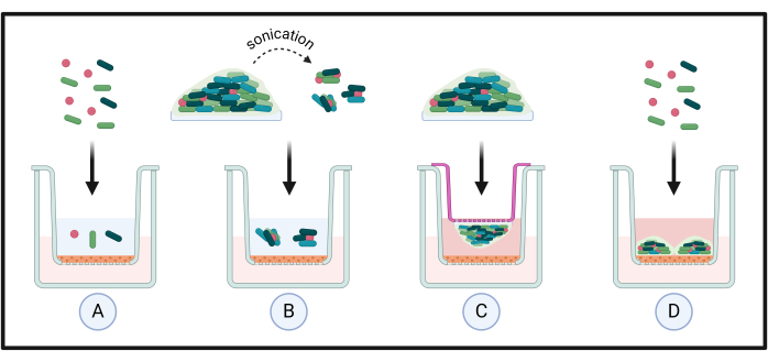
图1:类器官组织共培养模型的不同应用。 将标准化的微生物悬液直接施加于组织上,以研究微生物与宿主之间的相互作用(A)。此类方法常用于评估微生物在较短时间内(例如,<24 h)的定植情况。通过超声处理复杂生物膜,以研究分散的单细胞或生物膜聚集体对组织的影响(B)。这一点至关重要,因为已有研究表明,这些细胞相较于浮游状态或生物膜状态的对应细胞具有独特的表型;为此,可通过额外的小型插入装置将完整的生物膜直接添加至组织,从而实现与宿主的邻近暴露(C)。最后,在适当的培养基中将浮游细胞添加至组织,以评估微生物在组织上的定植、生物膜形成及生长动态(D)。 请点击此处查看该图的放大版本。
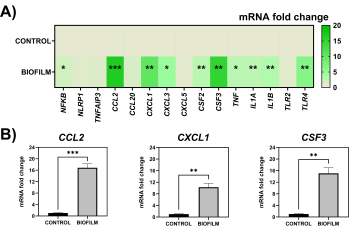
图2:HOE组织经7种微生物组成的生物膜超声裂解物刺激后的基因表达情况。 在对照组(未刺激)样本和暴露于100 µL 7种微生物生物膜超声裂解物的HOE组织中,共15种炎症相关基因的mRNA表达倍数变化,均以持家基因GAPDH为标准化参照(A)。其中mRNA表达变化最显著的三个基因(CCL2、CXCL1、CSF3)在(B)中单独展示。统计分析采用参数性非配对T检验,显著性差异表示为 *p < 0.05,**p < 0.01,***p < 0.001。数据通过统计与绘图软件进行绘制与分析。请点击此处查看该图的放大版本。
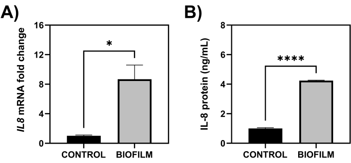
图3:IL8 基因表达及HOE组织产生的IL-8蛋白水平。通过基于SYBR Green的qPCR检测HOE组织中IL8的mRNA表达倍数变化(A),以及利用ELISA方法定量培养后组织培养基中的IL-8蛋白水平(B)。统计分析采用参数化的非配对T检验,显著性差异表示为 *p < 0.05 和 ****p < 0.0001。数据使用统计与绘图软件进行绘图和分析。请点击此处查看该图的放大版本。
讨论
本文描述了一种用于构建代表牙龈炎的复杂多物种生物膜模型的方法,该模型可与口腔上皮(HOE)组织共培养,以评估微生物刺激后宿主的免疫反应。本方案可进一步调整,用于研究浮游态细胞、单一种类、双物种或混合物种生物膜,以及生物膜分散细胞与来自人体不同生态位的上皮组织之间的宿主-病原体相互作用。相较于以往局限于二维单层细胞、且无法真实模拟体内生理环境的共培养系统,利用组织模型研究宿主-病原体相互作用代表了重要的技术进步。 体内 上皮组织包含多层细胞的情况16,17,18此外,与之相关的挑战包括 体外 多层组织模型的培养存在诸多挑战,使用多种细胞系和/或在气液界面培养细胞层时易受污染。 commercially available tissues 提供了可重复的实验平台,其模型质量稳定且高度一致,不同重复样本及批次间的可比性良好,如先前发表的研究所示。3,7.
对于 "共培养微生物群落的制备" 该部分中,将这些微生物纳入7种物种牙龈炎模型的情况如下 根据从口腔健康到疾病转变过程中常见的共生菌和病原菌进行选择。与所有复杂的生物膜模型系统一样 体外,微生物的纳入由针对特定健康或疾病生态位的微生物组研究决定。为此,我们及其他研究者已报道使用一种与口腔健康相关的生物膜模型,该模型包含共生微生物(例如, 链球菌 和 罗氏菌属 spp.)3,40,41此外,还可以向这些模型中添加其他病原体,以构建与不同疾病相关的生物膜。例如,向本文所述的七种微生物中加入三种微生物,可构建与牙周炎相关的疾病模型。3,而真菌病原体 Candida albicans 可添加以增加多微生物性,同时增加跨域相互作用的复杂性33. 事实上,促进真菌-细菌相互作用可能对利用此类生物膜模型获得的各种实验结果产生重大影响 体外 与仅含细菌的生物膜相比42最终,强烈建议在为这类研究建立新的多物种生物膜模型时应审慎考虑。尽管所选微生物应能通过广泛的文献检索轻易确定,但对于大多数生态位和/或疾病而言,这些微生物可能难以很好地整合到复杂的模型中。 体外 出于不同原因。以下列举了一些其他应考虑的因素:
生物膜形成动态
生物膜形成 体内 通常涉及 早期、中期和晚期特定微生物的定植。龈上和龈下牙菌斑的形成主要由先锋菌种在釉质表面的唾液薄膜上初始附着所决定(例如, 链球菌 物种),其中中晚期定植的病原体需要这些物质作为定植的支架43在细菌性阴道病期间的阴道环境中也提出了类似的现象,即 Gardnerella vaginalis 被认为是最初的定植菌,随后由其他厌氧菌继发定植44然而,需要注意的是,对于其他生态位中的其他疾病,情况可能并非如此。
每种微生物的接种密度
微生物在培养时会表现出不同的大小和/或生长动态 体外;因此,在标准化过程中必须考虑这一点。例如, C. albicans 是细菌细胞大小的100至150倍45,46因此,可能需要以较低浓度添加,例如 1 x 10⁶ 细胞/毫升,加入上述生物膜模型中。
种间拮抗作用
用于这些模型的某些微生物(包括同一种类但不同分离株、实验室和/或临床菌株)可能在生物膜形成过程中相互竞争,从而抑制某些微生物物种的生长47。Sadiq 等人的一项研究强调了不同微生物组合如何通过微生物协同作用(或拮抗作用)影响生物膜生物量48。尽管在生物膜模型中研究此类相互作用可能对科研团队具有重要意义(例如,测试益生菌或益生元治疗),但其他研究可能未考虑此类拮抗作用,这可能在构建模型时影响多物种体系的复杂性。
生物膜成熟时间
优化生物膜成熟的时间框架至关重要,因为这取决于所包含微生物的生长动态以及用于培养的模型系统(包括基质)。较长的成熟时间可能有利于后续病原菌的定植,但也会导致模型内细胞死亡增加,尤其是早期定植菌。相反,若希望研究未成熟生物膜模型对宿主的影响,则较短的孵育时间可能更为重要。Brown 等人的一项研究描述了同一十种微生物组成的伤口生物膜模型在成熟 24 小时、48 小时和 72 小时后,对人源 THP-1 细胞诱导出不同的炎症反应谱,提示生物膜越不成熟,其促炎性越强7。在多层组织模型中也可能观察到类似结果。此外,需要注意的是,定期每日更换培养基对于减少生物膜中的细胞死亡十分必要,尽管这一操作可根据正在进行的治疗方案进行调整,例如在评估长期抗菌干预效果时。
对于"类器官组织处理、实验设置及组织加工"部分,孵育时间可根据研究者的需求进行调整。宿主-病原体相互作用可在较早的时间点(例如 1–12 小时)至较晚的时间点(如 48 小时和 72 小时)进行研究,具体取决于研究问题。其次,所有维持培养基均含有抗生素;因此,需在订购时向公司提出申请,根据实验设计决定是否去除这些抗生素。尽管培养插件下方的培养基通常不会直接接触组织的上缘区域,除非模型中造成了伤口6,8,但在研究微生物侵入组织时,仍可考虑去除抗生素。
用于共培养刺激的微生物材料也可以根据上述讨论进行更换,以便评估浮游状态、用过的生物膜上清液(过滤和未过滤)、完整生物膜,或生物膜超声裂解物与组织的孵育效果。例如,先前的研究证据表明,EpiSkin 提供的组织模型可用于研究浮游真菌-宿主相互作用:C. albicans、Candida auris、Malassezia furfur 和 Trichophyton rubrum 细胞已被证明能够附着并相互作用于 HOE、重建人表皮(RHE)或人阴道上皮的外周组织层,其中一些研究还显示真菌感染后可激发宿主反应1,5,6,11,12,49。关于细菌-宿主相互作用也有类似发表,例如,常见于头皮微生物群并与头皮屑相关的病原体Cutibacterium acnes 在 RHE 表面的生物膜形成已被研究,包括单独培养以及与皮肤真菌Malassezia restricta 共培养的情况10。其他研究者还探讨了Staphylococcus spp. 在 RHE 上的附着能力,并测量了该细菌-宿主相互作用的理化和微生物学特征50。N'Diaye 及其合作者探究了人源神经肽降钙素基因相关肽(Calcitonin Gene-Related Peptide)如何影响 RHE 组织模型中Staphylococcus aureus 的毒力51。另有研究评估了益生菌干预对铜绿假单胞菌(Pseudomonas aeruginosa)感染人角膜上皮的保护作用14。同时,从多微生物的角度来看,不同研究团队已探讨了多种物种混合生物膜对不同组织基质的影响2,3,7,13。
总体而言,上述引用的方案和研究记录了类器官组织模型在研究宿主-病原体相互作用方面的广泛应用。尽管已有研究表明,EpiSkin 模型(尤其是 RHE 皮肤模型)在用于化妆品产品的腐蚀性/刺激性测试方面具有良好的适用性52,53,54,55,但这些模型与真实世界的 ex vivo 组织外植体或其他商业组织供应商提供的组织之间仍存在一些局限性56,57。一个明显的局限性在于,所有 commercially available 组织模型均是在无菌、“无菌”"germ-free"环境中由细胞系生成的,这意味着组织从未暴露于微生物扰动之下:这可能会加剧宿主的炎症反应,远超 in vivo 条件下或 ex vivo 组织外植体刺激后所观察到的程度57。相反,这些实验室模型系统不包含与 in vivo 组织相关的深层结缔组织层或血管系统,而这些特征在 ex vivo 组织外植过程中可以被保留,并且会影响炎症反应。事实上,最近一项系统综述强调,应谨慎考虑使用通过多种工程技术(包括生物材料)构建的具有适当血管系统的组织模型58。例如,最近一项研究利用嵌入牙龈成纤维细胞和微血管内皮细胞的纤维蛋白基质,构建了具有血管化的牙龈组织等效物。作者 描述了该模型在接触健康或疾病相关微生物后表现出差异性的炎症反应59。从商业角度来看,目前已出现更复杂的模型,例如包含全层组织结构的“T-Skin 模型”"T-Skin model",其真皮层由成纤维细胞构成,并覆盖有表皮层。比较此类复杂模型与仅含表皮系统的模型将十分有意义。
尽管没有任何模型是完美的,但类器官组织模型为临床前测试提供了颇具前景的替代方案,符合动物实验“替代、减少和优化”(3R原则)框架的要求。虽然动物模型能够为生物膜相关人类疾病的复杂病理生理学特性提供重要的研究见解,但其存在明显的缺点。这些三维模型易于操作,可在无需伦理审批的情况下对实验进行大规模或细微的调整。将这些模型与上述现有的复杂生物膜系统相结合,可极大地增进我们对宿主-病原体相互作用的理解,并在开展in vivo研究之前更准确地预测新型疗法的有效性。
披露
作者无任何利益冲突需要披露。
致谢
作者感谢Haleon为Saeed Alqahtani开展与本文所述组织模型相关的项目提供的资金支持,同时也感谢沙特阿拉伯教育部为Muhanna Alshehri提供的博士研究资助。
材料
| 姓名 | 公司 | 目录编号 | 评论 |
|---|---|---|---|
| 13 mm 羟基磷灰石(HA)圆片 | Plasma Biotal Limited | n/a(定制生产) | 用于模拟牙面,作为口腔生物膜形成的基质 |
| 19 G 针头(40 mm) | VWR | 613-2028 | 用于辅助从培养板中取出 HA 圆片,以及切割插入物中的组织 |
| 24 孔 TC 处理平底微孔板(带盖) | Scientific Laboratory Supplies | 3524(每包 100 个) | 用于生物膜培养及组织共培养实验 |
| 酸洗玻璃珠(425–600 μm) | Merck: Sigma-Aldrich | G8772 | 用于组织裂解,以提高 RNA 提取的得率 |
| Actinomyces naeslundii DSM 17233 | Leibniz Institute DSMZ | n/a | 用于构建复杂生物膜模型的微生物之一 |
| Bead Mill 24 匀浆器(或同等设备) | Fisher Scientific | 15-340-163 | 用于组织裂解,以提高 RNA 提取的得率 |
| Bijoux 管(7 mL) | Greiner Bio-One | 189170(每袋 700 个) | 用于生物膜超声处理 |
| cDNA 合成试剂盒(高容量) | Thermo Fisher Scientific | 4368814(200 次反应) | 用于从组织 RNA 合成 cDNA |
| 哥伦比亚血琼脂(CBA)基础培养基 | Thermo Fisher Scientific | CM0331B(500 克) | 用于制备 CBA 平板,以培养需氧微生物 |
| 去纤维马血 | E&O Laboratories | DHB | 用于补充 CBA 和 FAA 平板,以培养需氧和厌氧微生物 |
| 杜氏磷酸盐缓冲液(dPBS:不含 CaCl2 和 MgCl2) | Merck: Sigma-Aldrich | DB537 | 用于生物膜超声液与组织共培养的 dPBS |
| Fusobacterium nucleatum ATCC 10953 | American Type Culture Collection | n/a | 用于构建复杂生物膜模型的微生物之一 |
| Fusobacterium nucleatum 亚种(subspp.)vincentii DSM 19507 | Leibniz Institute DSMZ | n/a | 用于构建复杂生物膜模型的微生物之一 |
| GraphPad Prism(版本 10) | GraphPad Software, Inc | n/a | 用于图表绘制和数据分析 |
| 人 IL-8 ELISA 试剂盒(未包被 ELISA 试剂盒,含微孔板) | Thermo Fisher Scientific (Invitrogen) | 88-8086-86(10 × 96 次检测) | 用于检测使用过的组织培养基中蛋白含量的 ELISA 试剂盒及微孔板 |
| 人源口腔上皮组织(HOE) | Episkin | n/a(定制生产) | 共培养模型系统所使用的上皮组织 |
| 接种环(一次性,10 µL,或同等规格) | Fisher Scientific | 12870155(每包 1000 个) | 用于固体和液体培养基中的微生物接种 |
| MicroAmp 快速反应 96 孔 qPCR 板(0.1 mL) | Thermo Fisher Scientific | 4346907(10 块) | 用于组织基因表达谱的 qPCR 分析 |
| Microbank 保存管 | Pro-Lab Diagnostics | PL.170 | 用于微生物的保存和冷冻保藏 |
| 无核酸酶水 | Thermo Fisher Scientific | AM9935(10 × 1.5 mL) | 用于 qPCR 实验的 RT-PCR 级水 |
| 培养皿(90 mm) | Fisher Scientific (Sterilin) | 11309283(每包 500 个) | 用于制备琼脂平板以进行微生物培养 |
| 磷酸盐缓冲液(PBS) | Thermo Fisher Scientific | 18912014(每包 100 片) | 用于微生物标准化及生物膜洗涤步骤 |
| RNeasy 小量试剂盒 | Qiagen | 74106(250 次反应) | 基于柱法的组织 RNA 提取试剂盒 |
| Roswell Park Memorial Institute 1640 培养基 | Merck: Sigma-Aldrich | R7755-10L(10 L) | 用于生物膜培养的培养基 |
| RT2 分析者阵列 | Qiagen | 330171(定制) | 含有预置引物的 qPCR 阵列,用于基因表达谱分析 |
| Schaedler 厌氧琼脂基础培养基 | Merck: Sigma-Aldrich (Millipore) | 91019(500 克) | 用于制备 Fastidious Anaerobic Agar 平板 |
| Schaedler 厌氧肉汤 | Thermo Fisher Scientific | CM0497B(500 克) | 用于厌氧微生物生长的液体培养基 |
| 螺口 Beadbug O 型圈管 | Scientific Laboratory Supplies | Z763837-1000EA(每包 1000 个) | 用于 Bead Mill 24 匀浆器中裂解组织 |
| 超声波清洗仪 | Fisher Scientific (Fisherbrand) | n/a | 用于从 HA 圆片上超声洗脱生物膜 |
| Streptococcus intermedius DSM 20753 | Leibniz Institute DSMZ | n/a | 用于构建复杂生物膜模型的微生物之一 |
| Streptococcus mitis NCTC 12261 | National Collection of Type Cultures | n/a | 用于构建复杂生物膜模型的微生物之一 |
| Streptococcus oralis NTCC 11427 | National Collection of Type Cultures | n/a | 用于构建复杂生物膜模型的微生物之一 |
| SYBR Green(qPCRBIO SyGreen Mix Hi-ROX) | PCR Biosystems | PB20.12 | 用于 qPCR 的 SYBR Green 混合液 |
| Todd Hewitt 肉汤 | Merck: Sigma-Aldrich | T1438 | 用于生物膜培养的培养基 |
| 胰蛋白胨大豆肉汤(TSB)培养基 | Merck: Sigma-Aldrich | 22092(500 克) | 用于需氧微生物生长的液体培养基 |
| 镊子(150 mm,不锈钢,带齿) | RS-Pro | 545-187 | 用于移动 HA 圆片及组织插入物/组织提取物 |
| 通用管 (30 mL) | Greiner Bio-One | 201150(每袋 400 个) | 用于液体培养中微生物的培养容器 |
| Veillonella dispar NCTC 11831 | National Collection of Type Cultures | n/a` | 用于构建复杂生物膜模型的微生物之一 |
| β-巯基乙醇 | Merck: Sigma-Aldrich | M3148 | 用于组织样本中 RNase 的失活 |
参考文献
- Moyes, D. L., et al. A biphasic innate immune MAPK response discriminates between the yeast and hyphal forms of candida albicans in epithelial cells. Cell Host Microbe. 8 (3), 225-235 (2010).
- Morse, D. J., et al. Denture-associated biofilm infection in three-dimensional oral mucosal tissue models. J Med Microbiol. 67 (3), 364-375 (2018).
- Brown, J. L., et al. Biofilm-stimulated epithelium modulates the inflammatory responses in co-cultured immune cells. Sci Rep. 9 (1), 15779(2019).
- Yadev, N. P., Murdoch, C., Saville, S. P., Thornhill, M. H. Evaluation of tissue engineered models of the oral mucosa to investigate oral candidiasis. Microb Pathog. 50 (6), 278-285 (2011).
- Schaller, M., Zakikhany, K., Naglik, J. R., Weindl, G., Hube, B. Models of oral and vaginal candidiasis based on in vitro reconstituted human epithelia. Nat Protoc. 1 (6), 2767-2773 (2006).
- Brown, J. L., et al. Candida auris phenotypic heterogeneity determines pathogenicity in vitro. mSphere. 5 (3), e00371-e00420 (2020).
- Brown, J. L., et al. Assessing the inflammatory response to in vitro polymicrobial wound biofilms in a skin epidermis model. NPJ Biofilms Microbiomes. 8 (1), 19(2022).
- Butcher, M. C., et al. Antifungal-loaded calcium sulfate beads as a potential therapeutic in combating candida auris. Antimicrob Agents Chemother. 66 (1), e0171321(2022).
- Duckney, P., et al. The role of the skin barrier in modulating the effects of common skin microbial species on the inflammation, differentiation and proliferation status of epidermal keratinocytes. BMC Res Notes. 6, 474(2013).
- Meloni, M., Balzaretti, S., Collard, N., Desaint, S., Laperdrix, C. Reproducing the scalp microbiota community: Co-colonization of a 3D reconstructed human epidermis with c. Acnes and m. Restricta. Int J Cosmet Sci. 43 (2), 235-245 (2021).
- Pedrosa, A. F., et al. Malassezia colonisation on a reconstructed human epidermis: Imaging studies. Mycoses. 62 (12), 1194-1201 (2019).
- Schaller, M., Weindl, G. Models of oral and vaginal candidiasis based on in vitro reconstituted human epithelia for the study of host-pathogen interactions. Methods Mol Biol. 470, 327-345 (2009).
- Rosca, A. S., et al. Six bacterial vaginosis-associated species can form an in vitro and ex vivo polymicrobial biofilm that is susceptible to thymbra capitata essential oil. Front Cell Infect Microbiol. 12, 824860(2022).
- Paterniti, I., et al. Protective effect of probiotics against pseudomonas aeruginosa infection of human corneal epithelial cells. Int J Mol Sci. 25 (3), 1770(2024).
- Pellegatta, G., et al. Evaluation of human esophageal epithelium permeability in presence of different formulations containing hyaluronic acid and chondroitin sulphate. Med Devices (Auckl). 13, 57-66 (2020).
- Jensen, C., Teng, Is it time to start transitioning from 2D to 3D cell culture. Front Mol Biosci. 7, (2020).
- Mountcastle, S. E., et al. A review of co-culture models to study the oral microenvironment and disease. J Oral Microbiol. 12 (1), 1773122(2020).
- Cacciamali, A., Villa, R., Dotti, S. 3D cell cultures: Evolution of an ancient tool for new applications. Front Physiol. 13, 836480(2022).
- Langfeldt, D., et al. Health- and disease-associated species clusters in complex natural biofilms determine the innate immune response in oral epithelial cells during biofilm maturation. FEMS Microbiol Lett. 360 (2), 137-143 (2014).
- Ramage, G., et al. The epithelial cell response to health and disease associated oral biofilm models. J Periodontal Res. 52 (3), 325-333 (2017).
- Shang, L., et al. Multi-species oral biofilm promotes reconstructed human gingiva epithelial barrier function. Sci Rep. 8 (1), 16061(2018).
- Myers, S., et al. Immunomodulatory streptococci that inhibit cxcl8 secretion and NF-kappaB activation are common members of the oral microbiota. J Med Microbiol. 70 (3), 001329(2021).
- Tang, Y. L., Sim, T. S., Tan, K. S. Oral streptococci subvert the host innate immune response through hydrogen peroxide. Sci Rep. 12 (1), 656(2022).
- Peyyala, R., Kirakodu, S. S., Novak, K. F., Ebersole, J. L. Oral microbial biofilm stimulation of epithelial cell responses. Cytokine. 58 (1), 65-72 (2012).
- Yee, M., Kim, S., Sethi, P., Duzgunes, N., Konopka, K. Porphyromonas gingivalis stimulates il-6 and il-8 secretion in gsm-k, hsc-3 and h413 oral epithelial cells. Anaerobe. 28, 62-67 (2014).
- Groeger, S., Jarzina, F., Domann, E., Meyle, J. Porphyromonas gingivalis activates NF-kappaB and MAPK pathways in human oral epithelial cells. BMC Immunol. 18 (1), 1(2017).
- Groeger, S., Zhou, Y., Ruf, S., Meyle, J. Pathogenic mechanisms of fusobacterium nucleatum on oral epithelial cells. Front Oral Health. 3, 831607(2022).
- Li, X., et al. Saliva-derived microcosm biofilms grown on different oral surfaces in vitro. NPJ Biofilms Microbiomes. 7 (1), 74(2021).
- Krishnamoorthy, A. L., Lemus, A. A., Solomon, A. P., Valm, A. M., Neelakantan, P. Interactions between Candida albicans and Enterococcus faecalis in an organotypic oral epithelial model. Microorganisms. 8 (11), 1771(2020).
- Brown, J. L., et al. Generation of multi-species oral bacteria biofilm models. Methods Mol Biol. 2588, 187-199 (2023).
- Miles, A. A., Misra, S. S., Irwin, J. O. The estimation of the bactericidal power of the blood. J Hyg (Lond). 38 (6), 732-749 (1938).
- Lukic, D., Karygianni, L., Flury, M., Attin, T., Thurnheer, T. Endodontic-like oral biofilms as models for multi-species interactions in endodontic diseases. Microorganisms. 8 (5), 674(2020).
- Sherry, L., et al. Viable compositional analysis of an eleven-species oral polymicrobial biofilm. Front Microbiol. 7, 912(2016).
- Brown, J. L., et al. An in vitro evaluation of denture cleansing regimens against a polymicrobial denture biofilm model. Antibiotics (Basel). 11 (1), 113(2022).
- Ammann, T. W., Gmur, R., Thurnheer, T. Advancement of the 10-species subgingival zurich biofilm model by examining different nutritional conditions and defining the structure of the in vitro biofilms. BMC Microbiol. 12, 227(2012).
- Montelongo-Jauregui, D., Srinivasan, A., Ramasubramanian, A. K., Lopez-Ribot, J. L. An in vitro model for oral mixed biofilms of candida albicans and streptococcus gordonii in synthetic saliva. Front Microbiol. 7, 686(2016).
- Rumbaugh, K. P., Sauer, K. Biofilm dispersion. Nat Rev Microbiol. 18 (10), 571-586 (2020).
- Chua, S. L., et al. Dispersed cells represent a distinct stage in the transition from bacterial biofilm to planktonic lifestyles. Nat Commun. 5, 4462(2014).
- Buskermolen, J. K., Janus, M. M., Roffel, S., Krom, B. P., Gibbs, S. Saliva-derived commensal and pathogenic biofilms in a human gingiva model. J Dent Res. 97 (2), 201-208 (2018).
- Rosier, B. T., Buetas, E., Moya-Gonzalvez, E. M., Artacho, A., Mira, A. Nitrate as a potential prebiotic for the oral microbiome. Sci Rep. 10 (1), 12895(2020).
- Rosier, B. T., Moya-Gonzalvez, E. M., Corell-Escuin, P., Mira, A. Isolation and characterization of nitrate-reducing bacteria as potential probiotics for oral and systemic health. Front Microbiol. 11, 555465(2020).
- Young, T., et al. Candida albicans as an essential "keystone" component within polymicrobial oral biofilm models. Microorganisms. 9 (1), 59(2020).
- Marsh, P. D., Lewis, M. A., Rogers, H., Williams, D., Wilson, M. Marsh and Martin's oral microbiology-e-book: Marsh and Martin's oral microbiology-e-book. , Elsevier Health Sciences. (2016).
- Johnston, W., et al. In vitro bacterial vaginosis biofilm community manipulation using endolysin therapy. Biofilm. 5, 100101(2023).
- Janus, M. M., Willems, H. M., Krom, B. P. Candida albicans in multi-species oral communities; a keystone commensal. Adv Exp Med Biol. 931, 13-20 (2016).
- Delaney, C., et al. Fungi at the scene of the crime: Innocent bystanders or accomplices in oral infections. Curr Clin Microbiol Rep. 5, 190-200 (2018).
- Luo, A., Wang, F., Sun, D., Liu, X., Xin, B. Formation, development, and cross-species interactions in biofilms. Front Microbiol. 12, 757327(2021).
- Sadiq, F. A., De Reu, K., Burmolle, M., Maes, S., Heyndrickx, M. Synergistic interactions in multi-species biofilm combinations of bacterial isolates recovered from diverse food processing industries. Front Microbiol. 14, 1159434(2023).
- Liang, P. P., et al. A trichophyton rubrum infection model based on the reconstructed human epidermis - EpiSkin(r). Chin Med J (Engl). 129 (1), 54-58 (2016).
- Lerebour, G., Cupferman, S., Bellon-Fontaine, M. N. Adhesion of staphylococcus aureus and staphylococcus epidermidis to the EpiSkin reconstructed epidermis model and to an inert 304 stainless steel substrate. J Appl Microbiol. 97 (1), 7-16 (2004).
- N'diaye, A. R., et al. Skin-bacteria communication: Involvement of the neurohormone calcitonin gene-related peptide (cgrp) in the regulation of staphylococcus epidermidis virulence. Sci Rep. 6, 35379(2016).
- Géniès, C., et al. Comparison of the metabolism of 10 cosmetics-relevant chemicals in EpiSkin s9 subcellular fractions and in vitro human skin explants. J Appl Toxicol. 40 (2), 313-326 (2020).
- Alépée, N., Grandidier, M. -H., Cotovio, J. Usefulness of the EpiSkin reconstructed human epidermis model within integrated approaches on testing and assessment (IATA) for skin corrosion and irritation. Toxicol Vitro. 54, 147-167 (2019).
- Spielmann, H., et al. The ECVAM international validation study on in vitro tests for acute skin irritation: Report on the validity of the EpiSkin and epiderm assays and on the skin integrity function test. Alternatives to Laboratory Animals. 35 (6), 559-601 (2007).
- Cotovio, J., Grandidier, M. -H., Portes, P., Roguet, R., Rubinstenn, G. The in vitro acute skin irritation of chemicals: Optimisation of the EpiSkin prediction model within the framework of the ECVAM validation process. Alt Lab Anim. 33 (4), 329-349 (2005).
- Netzlaff, F., Lehr, C. M., Wertz, P. W., Schaefer, U. F. The human epidermis models EpiSkin, SkinEthic and epiderm: An evaluation of morphology and their suitability for testing phototoxicity, irritancy, corrosivity, and substance transport. Eur J Pharm Biopharm. 60 (2), 167-178 (2005).
- Larson, P. J., Chong, D., Fleming, E., Oh, J. Challenges in developing a human model system for skin microbiome research. J Invest Dermatol. 141 (1), 228-231.e4 (2021).
- Dechelette, C., Smirani, R., Medina, C., Naveau, A. Cellularized biomaterials used as gingival connective tissue substitutes in vivo: A systematic review. Tissue Eng Part B Rev. , (2024).
- Makkar, H., Lim, C. T., Tan, K. S., Sriram, G. Modeling periodontal host-microbe interactions using vascularized gingival connective tissue equivalents. Biofabrication. 15 (4), (2023).
重印与许可
