本文展示了从人体受试者的生物液体(如泪液和唾液)中收集的细胞外囊泡(EVs)的表征方法。该方法所使用的扫描仪能够检测仅1 µL样本中EVs的表型、大小及总颗粒数。
本文内容
摘要
摘要
细胞外囊泡(EVs)是由细胞产生的一类结构,通过将生物分子从一个细胞转运至另一个细胞,参与细胞间通讯。研究表明,EVs 可在体内进行短距离和长距离运输,并具有组织特异性。EVs 不仅存在于组织中,几乎所有的体液中均可发现其存在,例如泪液、唾液、脑脊液、血液等。尽管可通过非侵入性方式从泪液和唾液中收集 EVs,但单次可采集的体积较小,可能导致难以获得足够用于蛋白质分析的 EVs。本文所述的检测仪是一种纳米颗粒分析仪,可为该问题提供解决方案 这一问题,使我们能够仅用1 µL生物液体即可表征和研究细胞外囊泡(EVs)的表型、大小及总颗粒数。本方案将拓展从小体积、难获取的患者样本中获得的EVs相关知识,有助于提高患者的舒适度,并可能为多种疾病和障碍发现新的治疗靶点。
引言
细胞通过多种信号机制与邻近细胞进行通讯,其中包括释放细胞外囊泡(EVs),这些囊泡在细胞间通讯中发挥关键作用。EVs通过将遗传物质(如DNA、RNA和蛋白质)从一个细胞传递到另一个细胞来参与细胞间通讯1,2,3,4,5。目前,EVs可分为三类:外泌体、微囊泡和凋亡小体,其分类依据主要是大小。外泌体是最小的一类,直径为30–150 nm6,7,8,由内体膜系统形成9,10,11(图1)。微囊泡比外泌体更大,大小范围为100–1000 nm12,13,14,由质膜出芽形成11,12,13(图1)。凋亡小体是EVs中最大的一类,大小范围为1000–5000 nm12,14,15,同样由质膜出芽形成12,16,17(图1)。除了大小之外,EVs还可根据其他生物物理特性进行分类,包括密度、分子标志物(如CD63、CD81、CD9)以及其生成机制18。EVs可在相邻细胞之间进行短距离传播,也可在体内进行长距离传播19,20,21,22,23。EVs存在于多种生物体液中,例如血液24,25,26、脑脊液27,28,29、泪液30,31,32和唾液33,34,35等。
迄今为止,超速离心法是分离样本中外泌体(EVs)最广为人知的方法之一36,37,38。该方法需要进行多轮离心和超速离心,可通过逐步提高转速的方式,从细胞及细胞碎片中分离出外泌体。该流程通常从低速离心开始,沉淀细胞,随后进行中速离心以去除较大的囊泡,最后通过超速离心步骤沉淀外泌体18。尽管超速离心法被认为是外泌体分离的最佳方法,但仍存在一些局限性,例如会改变外泌体的形态结构39,40,41。另一种用于分离外泌体的技术是流式细胞术,其优势在于可实现多个时间点和终点评估,并支持高通量的单个外泌体分析。然而,流式细胞术的局限性包括但不限于孔道堵塞和信号微弱等问题。另一种常用方法是密度梯度离心法,该方法利用不同密度的介质与外泌体一同离心,从而实现比超速离心更优的分离效果。尽管该技术提高了分离纯度,但操作繁琐、耗时较长,且可能导致样品大量损失。此外,沉淀法和过滤法也可用于外泌体的分离。这两种方法操作简便、快速,但均可能导致样品污染。尽管目前存在多种外泌体分离技术,但每种方法均有其优缺点,如下所列(表1) 42,43,44,45,46,47,48,49,50,51,52,53,54,55, 56,57,58,59,60,61,62,63,64,65,66,67。
外泌体分离完成后,可进行分子检测以表征其特性。Western blot 是检测外泌体表面蛋白及携带蛋白表达的常用方法。68,69,70 以及采用聚合酶链式反应(PCR)检测miRNA表达水平71,72,73 用于EVs74,75,76这些检测方法已较为成熟,能够产生引人关注的结果。然而,这些方法的局限性在于需要从细胞外囊泡(EVs)中获取大量蛋白质或RNA才能获得可检测信号。77,这对于起始体积较小或细胞外囊泡浓度较低的样本而言是一个问题。
本文讨论的纳米颗粒分析仪使用户能够克服表1和表2 78,79,80,81,82,83,84,85,86,87,88,89,90中所述的诸多限制。该方法无需使用分离技术,有助于克服外泌体(EVs)得率降低的问题。该方法还允许用户仅用低至1 µL的样本体积,即可分析外泌体的表面蛋白和内容物蛋白、总外泌体数量以及外泌体大小。该过程通过使用该公司提供的四跨膜蛋白芯片实现,该芯片采用含有四跨膜蛋白抗体(CD63、CD81和CD9)的抗体微阵列,以识别溶液中的外泌体,如图2所示。荧光标记抗体可确认外泌体的存在,同时防止污染颗粒干扰实验结果。
该技术的总体目标是提供一种更省时的方法来分析细胞外囊泡(EVs),并能够利用少量样本进行EV分析。使用这种纳米颗粒分析仪,用户仅需1 µL样本即可分析颗粒的大小、总粒子数量以及表面蛋白,非常适合泪液和唾液等生物液体的分析。
方案
所有所述研究均遵循《赫尔辛基宣言》。在纳入研究前,已从每位受试者处获得书面知情同意。根据联邦和机构指南,已获得奥胡斯大学医院(1-10-72-77-14)和迪恩·麦吉研究所(1576837-2)的机构审查委员会(IRB)批准。在处理前,所有泪液和唾液样本均已去标识化。所有研究均经北德克萨斯地区机构审查委员会(#2020-030)审查并批准。以下方案遵守所有相关指南,并如上所述获得批准。
1. 第1天:样品制备与孵育
- 采集受试者的生物流体样本(如泪液和唾液),或从冰箱中取出样本进行解冻。
- 泪液:使用玻璃毛细管从外侧半月处被动收集泪液。
- 将玻璃毛细管置于下眼睑上,注意不要触碰到角膜。
- 在10分钟内收集10 μL泪液体积。
- 立即使用泪液,或在 -20 保存 °C.
- 唾液:用 1.5 mL 微量离心管收集被动滴流的唾液
- 让供体在口中积聚唾液。
- 让供体通过漏斗将唾液滴入试管中。
- 立即使用唾液,或在 -20 保存 °C.
- 泪液:使用玻璃毛细管从外侧半月处被动收集泪液。
- 将四次跨膜蛋白芯片放入芯片清洗板中静置15分钟,使其升至室温,然后再加入样品。放置芯片时,确保芯片编号朝下且正对上方,并避免芯片与孔壁接触。
- 在笔记本中记录ID编号和芯片编号,以便追踪芯片。
- 插入试剂盒附带的通用串行总线(USB),并将带有相应试剂盒编号的文件夹提取并保存至您选择的指定位置。
- 在1.5 mL离心管中加入99 μL的溶液B。
- 将1 μL生物流体加入上一步准备好的1.5 mL离心管中。
- 离心收集管中的样品。
- 从步骤1.6中取70 μL溶液,用移液器将溶液加至四跨膜蛋白芯片上。
- 将薄膜置于芯片清洗板上,以防止样品蒸发。
- 在室温(RT)下振荡孵育 16 小时–自由工作台
2. 第2天:芯片清洗
- 打开芯片清洗机。
- 从孔板上取下步骤 1.9 中的封膜。
- 将孔板放入芯片清洗机中,确保其牢固锁定,并盖上盖子。
注意:若已正确锁定,孔板将不易移动。 - 在芯片清洗机上按下 CW-TETRA v0 选项。
- 选择起始行及需要清洗的行数。
- 按下芯片清洗机上的 继续 按钮。
- 在芯片清洗过程中,按以下步骤在离心管中配制含抗体的封闭液:
- 每张芯片准备 300 µL 封闭液。
- 每张芯片加入 0.6 µL 的每种抗体(加入封闭液前需涡旋振荡并短暂离心抗体)。
- 涡旋振荡封闭液与抗体混合液。
注意:需避光保存封闭液与抗体混合液,以确保抗体稳定性。该溶液必须在使用当天配制。
- 当芯片清洗机发出提示音时,向芯片表面加入 250 µL 封闭液与抗体混合液。
- 盖上盖子,按下 继续 按钮。
注意:芯片清洗机将开始进行 1 小时的孵育,并随后进行多轮清洗和漂洗。 - 当芯片清洗机发出提示音时,取出芯片清洗板,将芯片沿斜坡向上移动。
- 将孔板重新放回芯片清洗机中,确保其牢固锁定。盖上盖子并按下 继续 按钮。
- 当芯片清洗机完成程序后,取出孔板并将芯片放置在纸巾上。避免光照芯片。
3. 第2天:扫描芯片
- 打开扫描仪并启动扫描软件。
注意:扫描仪将进行自检,以提示是否存在任何错误或是否可以继续扫描。 - 选择 保存文件夹,并指定文件保存位置。
- 选择 ChipFile 文件夹,并选择步骤 1.4 中保存的文件夹。
- 通过下拉选项选择每张芯片,并在卡盘上的 芯片位置 处定位。对所有需要扫描的芯片重复此步骤。
- 单击 芯片位置 下方的三个方块以开启荧光,方块将分别变为黄色、蓝色和红色。
- 按下盖子并将其拉出,以取下卡盘盖。
- 将芯片放置在卡盘上,确保其牢固固定。
- 将卡盘盖重新安装,将定位销插入孔中,向下按压,并滑动盖子使其锁定到位。
- 将卡盘放置到载物台上,确保其锁定到位。
- 选择 扫描芯片。
- 选择 确定。
- 当载物台完全进入仪器内部后,关闭门。
注意:扫描单张芯片大约需要 12 分钟。 - 程序完成芯片扫描后,打开门以弹出载物台,检查扫描是否成功。
- 如果扫描成功,丢弃芯片,并将下一组芯片放置到卡盘上,或把空卡盘放回载物台。然后退出软件,并在完成后关闭仪器。
- 如果扫描失败,请查看错误代码并纠正错误。
4. 数据处理
- 打开分析软件。
- 选择 预扫描数据 并选择包含第2天步骤3.2扫描结果的文件夹。
- 选择 下一步。
- 标记芯片 样本名称 通过单击单元格并输入样本编号,在列中填写样本名称。
- 接下来,标记四跨膜蛋白中的 频道名称 柱状图。红色 = CD63,绿色 = CD81,蓝色 = CD9。
- 选择 下一步.
- 选择 高变异系数 来自 选择斑点组 检查下拉框,并通过禁用一个或两个位点来关闭每个芯片的高变异系数(CVs)。
- 接下来,选择 高计数 并尝试关闭高计数警告。
注意:某些样本的计数值可能过高,但仍可进行分析。 - 选择 所有斑点 从下拉菜单中选择 斑点拼接图,并目视检查每张芯片是否存在可能的变形或划痕。
- 选择 下一步.
- 在 CD63 通道中,将最小值设为 300 以去除红细胞 平均百分比 % 包含该行;黄色细胞正常,继续进行CD81通道检测。若将最小值设为300后,红色细胞仍未变为黄色或白色,请转至步骤4.14进行故障排查。
- 在 CD81 通道中,将最小值设为 300 以去除红细胞 平均百分比 包含该行;黄色细胞正常,继续进行CD9通道检测。若将最小值设为300后,红色细胞仍未变为黄色或白色,请转至步骤4.14进行故障排查。
- 在 CD9 通道中,将最小值设为 400 以去除红细胞 平均百分比 % 包含该行;黄色单元格正常,继续执行步骤4.15。若将最小值设为400后红色单元格仍未变为黄色或白色,请转至步骤4.14进行故障排查。
- 故障排除
- 将最小值增加100个单位。
- 如果步骤 4.14.1 未成功,则选择芯片中的 挑选芯片 进行切片区段并选择芯片。选择 颗粒计数 并禁用粒子计数较高的区域。选择 强度/大小,选择 所有芯片, 并查看此操作是否解决了问题。
- 如果步骤 4.14.2 未成功,请选择芯片中的 挑选芯片 进行切片区段并选择芯片。选择 单芯片截断值 并将最小值增加100个单位。然后选择 所有芯片 观察红细胞是否已变为黄色。如果此步骤无效,重复操作直至达到预期效果。 平均百分比 包含的细胞现在呈黄色或白色。
- 选择 下一步.
- 取消选择 IM 通道。
- 将右下角的热图值设置为包含所有颗粒,然后选择 将图表添加到报告.
- 在下拉框中调整最大数值,并手动输入一个值。
- 接下来,选择顶部中央下方的下拉菜单 捕获探针 并选择 CD63.
- 选择添加 绘图以报告 将总颗粒数添加到报告中。
- 选择下拉菜单–下拉菜单下方 分析模式,选择 共定位,以及 将图表添加到报告.
- 接下来,选择 大小,选择 切换视图为图表,并选择 将图表添加到报告.
- 最后,选择 图像 并选择 将图表添加到报告.
- 对 CD81 和 CD9 重复步骤 4.18–4.22,每次确保取消选择 IM 通道。
- 所有物品添加到报告后,选择 导出报告,确保其保存到正确的文件,然后选择 文件夹.
注意:Excel 文件和图像已生成,现在可用于进一步分析。
结果
分析芯片上的信号点时,应通过观察 CD63、CD81 和 CD9 通道中荧光标记的 EVs 来确定 EVs 的最佳浓度,而 MIgG 通道30,91 应保持无信号,因其为对照通道,如图 3A所示。对于生物体液,CD81 信号会有所降低。若在 CD63、CD81 和 CD9 通道中均未检测到荧光信号,则表明缺乏 EVs(图 3B)。需注意避免芯片信号过饱和(图 3C),否则分析软件将难以准确计算 EVs 的粒径、总颗粒数及表型特征。在最佳浓度的信号点基础上,分析软件可测定 EVs 的粒径(50–200 nm)、总颗粒数以及四跨膜蛋白的表达情况(图 4),并导出至单独的电子表格中以供进一步分析。四跨膜蛋白分析将包括 EVs 表面蛋白的共定位情况,共定位以“/”表示,例如“CD63/CD81”。此处“/”并非表示 CD63 与 CD81 相结合,而是指 CD63 和 CD81 均存在于 EVs 的表面。
这些结果将为健康样本与病变样本之间的差异提供有价值的见解。我们将能够确定健康样本或病变样本是否产生更多的细胞外囊泡(EVs)、更大或更小的细胞外囊泡,以及细胞外囊泡的表型特征。这些特征中任何一项或全部都可能在疾病的发生和/或进展中发挥作用。通过这些结果,我们将能够观察到四跨膜蛋白(tetraspanin)水平是否存在缺失或升高,从而深入了解细胞过程及细胞外囊泡的形成机制。

图 1:不同细胞外囊泡(EVs)的类型与大小。 请点击此处查看本图的放大版本。
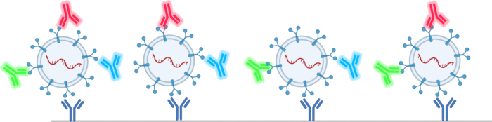
图 2:四跨膜蛋白芯片捕获和检测细胞外囊泡(EVs)的示意图。 请点击此处查看该图的放大版本。
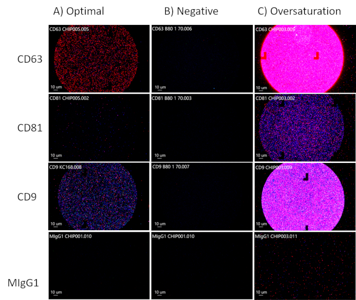
图 3:优化细胞外囊泡(EV)浓度。(A)EV 的最佳浓度。(B)阴性结果:无 EV 存在。(C)EV 过饱和。请点击此处查看此图的放大版本。
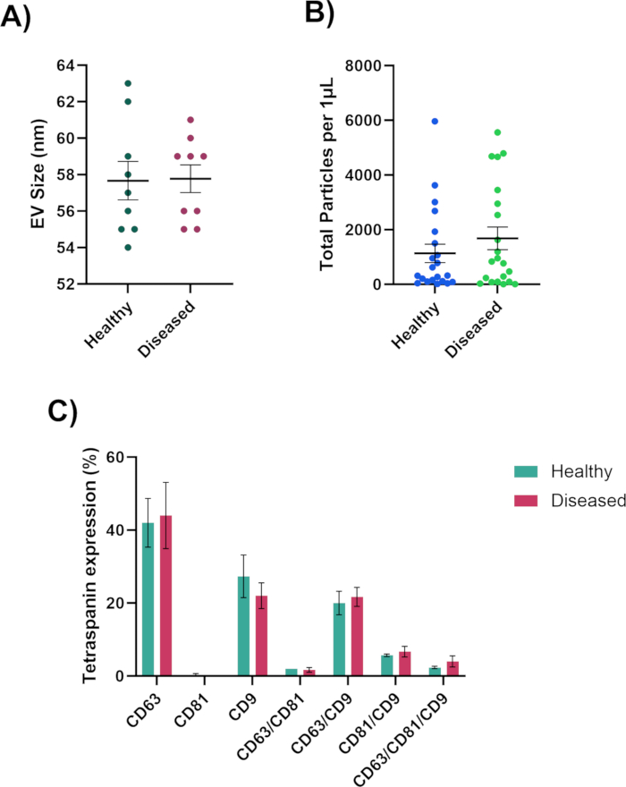
图4:纳米颗粒分析仪生成的数据。(A)以纳米为单位测量的EV粒径。(B)EV的总粒子数。(C)EV共定位表型。请点击此处查看该图的高清版本。
| 技术 | 优点 | 局限性 |
| 超速离心 | 高纯度42,43 | 改变细胞外囊泡(EV)形态42,44 |
| 均一性42,45 | 需要大体积样品42,44,46 | |
| 功能完整性42,43 | 成本昂贵42,43 | |
| 流式细胞术 | 可在多个时间点评估样品47 | 信号弱48,49 |
| 多终点检测47,50 | 孔道堵塞51 | |
| 高通量单个EV分析52,53 | ||
| 密度梯度离心 | 可获得高纯度样品54 | 操作繁琐55,56 |
| 适用于多种样品类型55,57 | 耗时较长55,58 | |
| 得率显著降低54 | ||
| 沉淀法 | 操作简单59,60 | 易受污染36,44,61 |
| 快速61,62 | 非特异性沉淀63 | |
| 高得率44,59,62 | ||
| 中性pH36 | ||
| 过滤法 | 操作简单64 | 截留外泌体65 |
| 快速64 | 损伤大体积囊泡36 | |
| 成本低64 | 滤饼形成66,67 | |
| 外泌体堵塞65 |
表1:EV分离技术的优点与局限性。 各种EV分离方法的列表,包括其优点和局限性。
| 技术 | 优点 | 局限性 |
| 动态光散射 | 可测量尺寸范围为 1 nm 至 6 µM 的颗粒78 | 不适用于粒径分布范围较大的复杂外泌体样品的测量79 |
| 下限为 10 nm,适用于单一分散体系79 | 无法区分外泌体与污染蛋白79 | |
| 透射电子显微镜(TEM) | 可观察外泌体的形态79,80 | 样品制备过程复杂79 |
| 可观察内部结构81 | 由于荧光信号被放大,无法根据大小和形状区分外泌体82 | |
| 纳米颗粒追踪分析 | 可测量 10 nm 至 2 µM 范围内外泌体的浓度、尺寸及尺寸分布78 | 无法区分细胞外囊泡(EVs)与蛋白聚集体及其他污染物83 |
| 样品制备和检测速度快78,84 | NTA 仪器价格昂贵85 | |
| 样品可回收并保持天然状态84 | 对振动敏感85 | |
| Western Blot(WB) | 可对外泌体标志蛋白进行定性和定量分析79,86 | 操作复杂且耗时79 |
| 适用于分析细胞培养基中外泌体79 | 分离得到的 EV 可能含有脂蛋白及其他污染物86 | |
| 流式细胞术 | 具有更高的灵敏度和高分辨率成像能力, | 检测过程耗时且繁琐,检测下限为 400 nm79,88 |
| 可区分染色的外泌体与污染物87 | ||
| 所需样品浓度低79 | 光学信号会干扰检测的准确性和分辨率88 | |
| Exoview | 可检测外泌体上的四跨膜蛋白(Tetraspanins,如 CD9、CD63 和 CD81)89 | 无法检测较大尺寸的细胞外囊泡(EV)75 |
| 可检测 EV 的 cargo 蛋白90 |
表2:EV表征技术的优点与局限性。
讨论
本方案中最关键的步骤是确保达到细胞外囊泡(EVs)的最佳浓度。EVs 的数量需足够以获得有效读数,但又不能过多,以免导致芯片过饱和。确定最佳 EV 浓度的最佳方法是使用 1 µL 样本进行一次优化实验,观察是否需要调整浓度。另一个关键步骤是检查样本中是否存在大量细胞碎片,这可通过分析软件中芯片上出现的大块物质来判断。如果样本中含有细胞碎片,可通过简单的离心或过滤处理来解决此问题。
另一个关键步骤是在将芯片沿斜坡放置时,确保芯片不接触孔壁,并避免触碰芯片的中心区域。扫描仪将从芯片中央的方块区域读取信号,因此必须避免用镊子接触该区域,以防干扰外泌体(EVs)。
该方法确实存在一些局限性,例如仪器检测时囊泡浓度过低。可通过干燥样品或使用浓缩管来提高浓度。另一局限性在于分析软件仅能测量50-200 nm范围内的囊泡,因此部分微囊泡及所有凋亡小体均被排除在粒径测量之外75。
分离细胞外囊泡(EV)的方法有多种(表1 42,43,44,45,46,47,48,49,50,51,52, 53,54,55,56,57,58,59,60, 61,62,63,64,65,66,67),分析EV的方法也多种多样(表2 78,79,80,81,82,83,84,85,86,87,88,89,90),目前EV分离的金标准是超速离心法36,37,38,用于将细胞沉淀下来,以便进行如Western印迹68,69,70和PCR71,72,73,74,75,76等检测。尽管该方法适用于大体积样本42,44,46,但获取生物体液往往较为困难,通常每次只能收集到少量样本,这并不适合超速离心法。相比之下,使用这种纳米颗粒分析仪仅需1 µL样本即可获得有关EV大小、总颗粒数及表型等有价值的数据,因此特别适用于生物体液的分析。我们将能够利用难以提取的微量样本进一步拓展对EV的认识,这不仅可提高患者的舒适度,还可能发现多种疾病和障碍的潜在治疗靶点。
披露
作者声明无竞争性财务利益或其他利益冲突。
致谢
我们感谢美国国立卫生研究院(NIH)提供的资助(EY031316 和 EY034714)。我们还要感谢北德克萨斯健康科学中心(UNTHSC)和 NTERI 提供的实验室空间。
材料
| 姓名 | 公司 | 目录编号 | 评论 |
|---|---|---|---|
| ChipWasher 100 | NanoView | EV-CW100 | 用于孵育、洗涤、漂洗并干燥四跨膜蛋白芯片。此型号目前已不再供应。购买时价格:$9,995.00 |
| ExoView Analyzer 软件 | NanoView | N/A | 分析芯片信息并生成可用于进一步分析的 Excel 文件。此产品目前已不再供应。 |
| ExoView R100 | NanoView | EV-R100 | 用于在三个不同波长下扫描四跨膜蛋白芯片。此型号目前已不再供应。购买时价格: $110,000.00 |
| ExoView Scanner 软件 | NanoView | N/A | 在三个不同波长下扫描芯片。此产品目前已不再供应。 |
| 人源四跨膜蛋白检测试剂盒 | Unchained Labs | EV-TETRA-C | 包含 8 个四跨膜蛋白芯片、孵育液、封闭液、CD63 抗体、CD81 抗体、CD9 抗体、溶液 A、溶液 B、USB 数据线和板盖。 |
参考文献
- Abels, E. R., Breakefield, X. O. Introduction to extracellular vesicles: Biogenesis, RNA cargo selection, content, release, and uptake. Cell Mol Neurobiol. 36 (3), 301-312 (2016).
- Radler, J., Gupta, D., Zickler, A., Andaloussi, S. E. Exploiting the biogenesis of extracellular vesicles for bioengineering and therapeutic cargo loading. Mol Ther. 31 (5), 1231-1250 (2023).
- Gupta, D., Zickler, A. M., El Andaloussi, S. Dosing extracellular vesicles. Adv Drug Deliv Rev. 178, 113961(2021).
- Dixson, A. C., Dawson, T. R., Di Vizio, D., Weaver, A. M. Context-specific regulation of extracellular vesicle biogenesis and cargo selection. Nat Rev Mol Cell Biol. 24 (7), 454-476 (2023).
- Keshtkar, S., Azarpira, N., Ghahremani, M. H. Mesenchymal stem cell-derived extracellular vesicles: novel frontiers in regenerative medicine. Stem Cell Res Ther. 9 (1), 63(2018).
- Zhang, H., et al. Exosome-induced regulation in inflammatory bowel disease. Front Immunol. 10, 1464(2019).
- Barzin, M., et al. Application of plant-derived exosome-like nanoparticles in drug delivery. Pharm Dev Technol. 28 (5), 383-402 (2023).
- Zhou, C., et al. Stem cell-derived exosomes: emerging therapeutic opportunities for wound healing. Stem Cell Res Ther. 14 (1), 107(2023).
- Kalluri, R., LeBleu, V. S. The biology, function, and biomedical applications of exosomes. Science. 367 (6478), aau6977(2020).
- Mondal, J., et al. Hybrid exosomes, exosome-like nanovesicles and engineered exosomes for therapeutic applications. J Control Release. 353, 1127-1149 (2023).
- van Niel, G., D'Angelo, G., Raposo, G. Shedding light on the cell biology of extracellular vesicles. Nat Rev Mol Cell Biol. 19 (4), 213-228 (2018).
- Paul, N., Sultana, Z., Fisher, J. J., Maiti, K., Smith, R. Extracellular vesicles- crucial players in human pregnancy. Placenta. 140, 30-38 (2023).
- Zaldivia, M. T. K., McFadyen, J. D., Lim, B., Wang, X., Peter, K. Platelet-derived microvesicles in cardiovascular diseases. Front Cardiovasc Med. 4, 74(2017).
- Tamasi, V., Nemeth, K., Csala, M. Role of extracellular vesicles in liver diseases. Life (Basel). 13 (5), 1117(2023).
- Fu, Y., et al. Emerging understanding of apoptosis in mediating mesenchymal stem cell therapy. Cell Death Dis. 12 (6), 596(2021).
- Santavanond, J. P., Rutter, S. F., Atkin-Smith, G. K., Poon, I. K. H. Apoptotic bodies: Mechanism of formation, isolation and functional relevance. Subcell Biochem. 97, 61-88 (2021).
- Yu, L., et al. Apoptotic bodies: bioactive treasure left behind by the dying cells with robust diagnostic and therapeutic application potentials. J Nanobiotechnology. 21 (1), 218(2023).
- Momen-Heravi, F., Getting, S. J., Moschos, S. A. Extracellular vesicles and their nucleic acids for biomarker discovery. Pharmacol Ther. 192, 170-187 (2018).
- Liu, Y. J., Wang, C. A review of the regulatory mechanisms of extracellular vesicles-mediated intercellular communication. Cell Commun Signal. 21 (1), 77(2023).
- Grange, C., Bussolati, B. Extracellular vesicles in kidney disease. Nat Rev Nephrol. 18 (8), 499-513 (2022).
- Cocucci, E., Meldolesi, J. Ectosomes and exosomes: shedding the confusion between extracellular vesicles. Trends Cell Biol. 25 (6), 364-372 (2015).
- Iannotta, D. A. A., Kijas, A. W., Rowan, A. E., Wolfram, J. Entry and exit of extracellular vesicles to and from the blood circulation. Nat Nanotechnol. 19 (1), 13-20 (2024).
- Saint-Pol, J., Gosselet, F., Duban-Deweer, S., Pottiez, G., Karamanos, Y. Targeting and crossing the blood-brain barrier with extracellular vesicles. Cells. 9 (4), 851(2020).
- Alberro, A., Iparraguirre, L., Fernandes, A., Otaegui, D. Extracellular vesicles in blood: Sources, effects, and applications. Int J Mol Sci. 22 (15), 8163(2021).
- Nieuwland, R., Siljander, P. R. A beginner's guide to study extracellular vesicles in human blood plasma and serum. J Extracell Vesicles. 13 (1), e12400(2024).
- Thangaraju, K., Neerukonda, S. N., Katneni, U., Buehler, P. W. Extracellular vesicles from red blood cells and their evolving roles in health, coagulopathy and therapy. Int J Mol Sci. 22 (1), 153(2020).
- Li, C., et al. Cerebrospinal fluid-derived extracellular vesicles after spinal cord injury promote vascular regeneration via PI3K/AKT signaling pathway. J Orthop Translat. 39, 124-134 (2023).
- Deng, Y., et al. Phosphoproteome analysis of cerebrospinal fluid extracellular vesicles in primary central nervous system lymphoma. Analyst. 148 (15), 3594-3602 (2023).
- Hirschberg, Y., et al. Proteomic comparison between non-purified cerebrospinal fluid and cerebrospinal fluid-derived extracellular vesicles from patients with Alzheimer's, Parkinson's and Lewy body dementia. J Extracell Vesicles. 12 (12), e12383(2023).
- Hefley, B. S., et al. Revealing the presence of tear extracellular vesicles in Keratoconus. Exp Eye Res. 224, 109242(2022).
- Cross, T., et al. RNA profiles of tear fluid extracellular vesicles in patients with dry eye-related symptoms. Int J Mol Sci. 24 (20), 15390(2023).
- Ma, H., et al. Metabolic signatures of tear extracellular vesicles caused by herpes simplex keratitis. Ocul Surf. 31, 21-30 (2024).
- Han, P., Bartold, P. M., Ivanovski, S. The emerging role of small extracellular vesicles in saliva and gingival crevicular fluid as diagnostics for periodontitis. J Periodontal Res. 57 (1), 219-231 (2022).
- Han, P., Li, X., Wei, W., Ivanovski, S. Saliva diagnosis using small extracellular vesicles and salivaomics. Methods Mol Biol. 2588, 25-39 (2023).
- Reseco, L., et al. Characterization of extracellular vesicles from human saliva: Effects of age and isolation techniques. Cells. 13 (1), 95(2024).
- Tiwari, S., Kumar, V., Randhawa, S., Verma, S. K. Preparation and characterization of extracellular vesicles. Am J Reprod Immunol. 85 (2), e13367(2021).
- Tomiyama, E., Fujita, K., Nonomura, N. Urinary extracellular vesicles: Ultracentrifugation method. Methods Mol Biol. 2292, 173-181 (2021).
- Xu, K., Jin, Y., Li, Y., Huang, Y., Zhao, R. Recent progress of exosome isolation and peptide recognition-guided strategies for exosome research. Front Chem. 10, 844124(2022).
- Brennan, K., et al. A comparison of methods for the isolation and separation of extracellular vesicles from protein and lipid particles in human serum. Sci Rep. 10 (1), 1039(2020).
- Gao, J., et al. Recent developments in isolating methods for exosomes. Front Bioeng Biotechnol. 10, 1100892(2022).
- Mol, E. A., Goumans, M. J., Doevendans, P. A., Sluijter, J. P. G., Vader, P. Higher functionality of extracellular vesicles isolated using size-exclusion chromatography compared to ultracentrifugation. Nanomedicine. 13 (6), 2061-2065 (2017).
- Ansari, F. J., et al. Comparison of the efficiency of ultrafiltration, precipitation, and ultracentrifugation methods for exosome isolation. Biochem Biophys Rep. 38, 101668(2024).
- Patel, G. K., et al. Comparative analysis of exosome isolation methods using culture supernatant for optimum yield, purity and downstream applications. Sci Rep. 9 (1), 5335(2019).
- Stam, J., Bartel, S., Bischoff, R., Wolters, J. C. Isolation of extracellular vesicles with combined enrichment methods. J Chromatogr B Analyt Technol Biomed Life Sci. 1169, 122604(2021).
- Le Roy, A., et al. AUC and small-angle scattering for membrane proteins. Methods Enzymol. 562, 257-286 (2015).
- Chhoy, P., Brown, C. W., Amante, J. J., Mercurio, A. M. Protocol for the separation of extracellular vesicles by ultracentrifugation from in vitro cell culture models. STAR Protoc. 2 (1), 100303(2021).
- McDorman, K. S., Chan, C., Rojko, J., Satterwhite, C. M., Morrison, J. P. Chapter 7 - Special Techniques in Toxicologic Pathology. Haschek and Rousseaux's Handbook of Toxicologic Pathology (Third Edition). , Academic Press. (2013).
- Woud, W. W., et al. An imaging flow cytometry-based methodology for the analysis of single extracellular vesicles in unprocessed human plasma. Commun Biol. 5 (1), 633(2022).
- Gul, B., Syed, F., Khan, S., Iqbal, A., Ahmad, I. Characterization of extracellular vesicles by flow cytometry: Challenges and promises. Micron. 161, 103341(2022).
- Black, C. B., Duensing, T. D., Trinkle, L. S., Dunlay, R. T. Cell-based screening using high-throughput flow cytometry. Assay Drug Dev Technol. 9 (1), 13-20 (2011).
- Safford, H. R., Bischel, H. N. Flow cytometry applications in water treatment, distribution, and reuse: A review. Water Res. 151, 110-133 (2019).
- Kobayashi, H., et al. Precise analysis of single small extracellular vesicles using flow cytometry. Sci Rep. 14 (1), 7465(2024).
- Welsh, J. A., et al. A compendium of single extracellular vesicle flow cytometry. J Extracell Vesicles. 12 (2), e12299(2023).
- Carnino, J. M., Lee, H. Extracellular vesicles in respiratory disease. Adv Clin Chem. 108, 105-127 (2022).
- Sun, Y., Sethu, P. Low-stress microfluidic density-gradient centrifugation for blood cell sorting. Biomed Microdevices. 20 (3), 77(2018).
- Strachan, B. C., Xia, H., Voros, E., Gifford, S. C., Shevkoplyas, S. S. Improved expansion of T cells in culture when isolated with an equipment-free, high-throughput, flow-through microfluidic module versus traditional density gradient centrifugation. Cytotherapy. 21 (2), 234-245 (2019).
- Malvezzi, H., Sharma, R., Agarwal, A., Abuzenadah, A. M., Abu-Elmagd, M. Sperm quality after density gradient centrifugation with three commercially available media: a controlled trial. Reprod Biol Endocrinol. 12, 121(2014).
- Sun, Y., Sethu, P. Microfluidic adaptation of density-gradient centrifugation for isolation of particles and cells. Bioengineering (Basel). 4 (3), 67(2017).
- Karttunen, J., et al. Precipitation-based extracellular vesicle isolation from rat plasma co-precipitate vesicle-free microRNAs. J Extracell Vesicles. 8 (1), 1555410(2019).
- De Sousa, K. P., et al. Isolation and characterization of extracellular vesicles and future directions in diagnosis and therapy. Wiley Interdiscip Rev Nanomed Nanobiotechnol. 15 (1), e1835(2023).
- Sidhom, K., Obi, P. O., Saleem, A. A review of exosomal isolation methods: Is size exclusion chromatography the best option. Int J Mol Sci. 21 (18), 6466(2020).
- Coughlan, C., et al. Exosome isolation by ultracentrifugation and precipitation and techniques for downstream analyses. Curr Protoc Cell Biol. 88 (1), e110(2020).
- Konoshenko, M. Y., et al. Isolation of extracellular vesicles from biological fluids via the aggregation-precipitation approach for downstream miRNAs detection. Diagnostics (Basel). 11 (3), 384(2021).
- Drozdz, A., et al. Low-vacuum filtration as an alternative extracellular vesicle concentration method: A comparison with ultracentrifugation and differential centrifugation. Pharmaceutics. 12 (9), 872(2020).
- Akbar, A., Malekian, F., Baghban, N., Kodam, S. P., Ullah, M. Methodologies to isolate and purify clinical grade extracellular vesicles for medical applications. Cells. 11 (2), 186(2022).
- Chen, J., et al. Review on strategies and technologies for exosome isolation and purification. Front Bioeng Biotechnol. 9, 811971(2021).
- Helling, A., et al. Investigation of microbial cell deformability by filter cake compressibility using ultrafiltration membranes. Colloids Surf B Biointerfaces. 185, 110626(2020).
- Bagci, C., et al. Overview of extracellular vesicle characterization techniques and introduction to combined reflectance and fluorescence confocal microscopy to distinguish extracellular vesicle subpopulations. Neurophotonics. 9 (2), 021903(2022).
- Thery, C., et al. Minimal information for studies of extracellular vesicles 2018 (MISEV2018): a position statement of the International Society for Extracellular Vesicles and update of the MISEV2014 guidelines. J Extracell Vesicles. 7 (1), 1535750(2018).
- van Maanen, J. C., et al. A combined western and bead-based multiplex platform to characterize extracellular vesicles. Tissue Eng Part C Methods. 29 (11), 493-504 (2023).
- Xu, D., et al. MicroRNAs in extracellular vesicles: Sorting mechanisms, diagnostic value, isolation, and detection technology. Front Bioeng Biotechnol. 10, 948959(2022).
- Lee, H., He, X., Le, T., Carnino, J. M., Jin, Y. Single-step RT-qPCR for detection of extracellular vesicle microRNAs in vivo: a time- and cost-effective method. Am J Physiol Lung Cell Mol Physiol. 318 (4), L742-L749 (2020).
- Kim, J. A., et al. Small RNA sequencing of circulating small extracellular vesicles microRNAs in patients with amyotrophic lateral sclerosis. Sci Rep. 13 (1), 5528(2023).
- Fan, Y., et al. Differential proteomics argues against a general role for CD9, CD81 or CD63 in the sorting of proteins into extracellular vesicles. J Extracell Vesicles. 12 (8), e12352(2023).
- Silva, A. M., et al. Quantification of protein cargo loading into engineered extracellular vesicles at single-vesicle and single-molecule resolution. J Extracell Vesicles. 10 (10), e12130(2021).
- Salunkhe, S., Basak Dheeraj, M., Chitkara, D., Mittal, A. Surface functionalization of exosomes for target-specific delivery and in vivo imaging, tracking: Strategies and significance. J Control Release. 326, 599-614 (2020).
- Silva, A. K. A., et al. Development of extracellular vesicle-based medicinal products: A position paper of the group "Extracellular Vesicle translatiOn to clinicaL perspectiVEs - EVOLVE France". Adv Drug Deliv Rev. 179, 114001(2021).
- Gurunathan, S., Kang, M. H., Jeyaraj, M., Qasim, M., Kim, J. H. Review of the isolation, characterization, biological function, and multifarious therapeutic approaches of exosomes. Cells. 8 (4), 307(2019).
- Zhang, Y., et al. Exosome: A review of its classification, isolation techniques, storage, diagnostic and targeted therapy applications. Int J Nanomedicine. 15, 6917-6934 (2020).
- Colombo, M., Raposo, G., Thery, C. Biogenesis, secretion, and intercellular interactions of exosomes and other extracellular vesicles. Annu Rev Cell Dev Biol. 30, 255-289 (2014).
- Pisitkun, T., Shen, R. F., Knepper, M. A. Identification and proteomic profiling of exosomes in human urine. Proc Natl Acad Sci U S A. 101 (36), 13368-13373 (2004).
- Zaborowski, M. P., Balaj, L., Breakefield, X. O., Lai, C. P. Extracellular vesicles: Composition, biological relevance, and methods of study. Bioscience. 65 (8), 783-797 (2015).
- Kowkabany, G., Bao, Y. Nanoparticle tracking analysis: An effective tool to characterize extracellular vesicles. Molecules. 29 (19), 4672(2024).
- Li, H. Metastatic characteristics of SY86B human gastric carcinoma in athymic nude mice. Zhonghua Zhong Liu Za Zhi. 10 (6), 421-423 (1988).
- Zhao, Z., Wijerathne, H., Godwin, A. K., Soper, S. A. Isolation and analysis methods of extracellular vesicles (EVs). Extracell Vesicles Circ Nucl Acids. 2, 80-103 (2021).
- Gandham, S., et al. Technologies and standardization in research on extracellular vesicles. Trends Biotechnol. 38 (10), 1066-1098 (2020).
- van der Vlist, E. J., Nolte-'t Hoen, E. N., Stoorvogel, W., Arkesteijn, G. J., Wauben, M. H. Fluorescent labeling of nano-sized vesicles released by cells and subsequent quantitative and qualitative analysis by high-resolution flow cytometry. Nat Protoc. 7 (7), 1311-1326 (2012).
- Pospichalova, V., et al. Simplified protocol for flow cytometry analysis of fluorescently labeled exosomes and microvesicles using dedicated flow cytometer. J Extracell Vesicles. 4, 25530(2015).
- Breitwieser, K., et al. Detailed characterization of small extracellular vesicles from different cell types based on tetraspanin composition by ExoView R100 platform. Int J Mol Sci. 23 (15), 8544(2022).
- An, H. J., Cho, H. K., Song, D. H., Kee, C. Quantitative analysis of exosomes in the aqueous humor of Korean patients with pseudoexfoliation glaucoma. Sci Rep. 12 (1), 12875(2022).
- Hefley, B. S., McKay, T. B., Hutcheon, A. E. K., Ciolino, J. B., Karamichos, D. Corneal epithelial-stromal constructs to study differences associated with diabetes mellitus. Exp Eye Res. 248, 110100(2024).
重印与许可
