目前尚缺乏一种用于评估实验室环境中朊病毒污染常见区域及有效去污染方法的检测手段。本文所述方案为实施实验室朊病毒安全拭子检测提供了关键基础,且该方法可轻松调整,以满足不同实验室的具体需求。
本文内容
摘要
摘要
医源性朊病毒疾病的传播曾发生于使用受污染的神经外科器械、移植材料以及职业性接触受朊病毒污染的实验工具。朊病毒通过模板化机制使正常的细胞型朊蛋白 PrPC 发生错误折叠,转化为错误折叠且具有致病性的 PrPSc 形式,从而引发疾病,且此类疾病 invariably 致命。减少医源性和职业性朊病毒传播具有挑战性。首先,朊病毒可结合并长期存在于各种表面。其次,朊病毒对灭活处理具有高度抗性。因此,在去污不彻底的情况下,表面可能长期保持感染性。这不仅可能对从事朊病毒研究的实验人员构成潜在的职业风险,还可能污染使用高灵敏度朊病毒扩增技术的实验室实验。本文所述的朊病毒安全实验室拭子检测方案,包括识别和记录实验室高使用频率区域的步骤、推荐的采样对照以确保结果有效性、针对表面拭子检测阳性位点采取适当应对措施的流程、典型的朊病毒拭子检测结果,以及可能出现的假阳性结果。总体而言,朊病毒安全实验室拭子检测可作为更广泛的朊病毒安全计划的一部分,用于评估表面去污效果、监测公共区域的朊病毒污染情况,并实现朊病毒去污状态的文件化记录。
引言
朊病毒病是一类致命的神经退行性疾病,目前尚无已知的治疗方法或治愈手段。朊病毒病由PrPSc引起,它是正常细胞型朊病毒蛋白PrPC的错误折叠且具有致病性的形式1,2,3,4,5。已知朊病毒病可影响人类及多种其他动物物种。其中一种人类朊病毒病——克雅氏病(Creutzfeldt-Jakob Disease, CJD)具有三种已知的病因:散发性、遗传性和获得性。获得性CJD可由意外传播(医源性和职业性)导致,巴布亚新几内亚福雷族人中的库鲁病(Kuru)被认为即由此引起6。
朊病毒的传播与受朊病毒污染的医疗器械及移植材料有关7,8,9,10,11,12,13,14,15,16,17。医源性克雅氏病(CJD)可通过血液、组织或受朊病毒污染的表面传播18,19,20。例如,在接受过脑电图检查的患者中,若所使用的电极曾用于处于CJD临床前期的个体,之后该个体死于CJD,则该患者可能发展为医源性CJD21。更近期的实验室职业暴露传播案例中,一名实验室工作人员在使用镊子处理感染羊适应性BSE动物的脑切片时,因皮肤被刺破而感染了朊病毒病22,23。此类传播情形可能发生在临床、实验室以及诊断实验室等处理朊病毒样本的环境中。
朊病毒对常见的消毒技术具有抗性,可在物体表面长期存留并保持感染性24,25,26,27,28,29。乙醇、酚类清洁剂、过氧化氢、各种形式的辐射以及甲醛等常用消毒方法均无法有效灭活朊病毒,导致物体表面仍具有感染性30,31,32,33,34,35,36,37。这些特性促进了朊病毒在医源性暴露和职业暴露过程中的传播。
环境朊病毒的检测方法最近才得以开发。通过环境拭子采样结合实时震荡诱导转化(RT-QuIC)技术,可评估环境表面以及实验室常见表面在未有效消毒后残留的朊病毒传染性38,39,40,41,42。本文描述了该技术如何整合到更广泛的朊病毒安全计划中。总体而言,该方法可用于监测实验室依赖的消毒方案,调查并准确记录污染状况,从而通过减少交叉污染来确保实验结果的有效性;同时可用于评估共用空间的朊病毒污染情况,并根据常见污染区域对人员进行有针对性的再培训。
方案
所有涉及动物的实验程序均经克瑞顿大学机构动物护理和使用委员会批准,并符合《实验动物护理和使用指南》的要求。
注:图1展示了朊病毒安全实验室拭子检测的示意图。
1. 拭子采样位点的选择与表面采样准备
- 确定并标记适合取样的监测区域。参考所提供的模板进行记录和核对(图2 和 补充图1)。
- 为每个已确定的区域准备两个1.5 mL的微量离心管。向第一组微量离心管中加入250 µL的杜氏磷酸盐缓冲液,用于步骤3.2。第二组微量离心管保持空置,用于步骤4.3。
- 从无朊病毒污染的区域取出储存的泡沫头取样拭子。
- 准备一个装有超纯水(MQ H2O)的挤压瓶,用于步骤2.1。
2. 阳性对照与阴性对照棉签的制备
- 使用相应的对照类型制备阳性和阴性拭子对照的稀释液。示例:对于阳性对照,将朊病毒感染的脑匀浆液以1%的比例稀释于杜氏磷酸盐缓冲液(DPBS)中;对于阴性对照,将未感染(UN)同种动物的脑匀浆液(BH)以0.1%的比例稀释于DPBS中。
- 使用洁净手套,从洁净包装中取出相应数量的拭子,将手柄端朝下放入试管架中,注意间隔放置泡沫拭子,避免拭子头部接触其他拭子或表面。每个对照样品准备三支拭子。每次取样之间更换手套。
- 用干净的泡沫拭子手柄夹住拭子,涂抹50 µ将相应阳性和阴性对照样品各 L 加到泡沫棉签头部。确保棉签头部两侧均加样,以保证完全吸收。
注意:每次从试管架中取出洁净棉签前,务必更换手套。 - 使用剪刀剪去棉签柄部多余的部分(约 ½ 将棉签插入预装的微量离心管中,泡沫头朝下(插入深度约为棉签长度的一半)。确保泡沫头完全浸入DPBS中,并适当剪短棉签柄,以便能够完全盖紧微量离心管盖。
- 更换手套后,继续应用对照样品,直至所有样品均完成施加。
3. 表面拭子取样
- 用手指握住干净的泡沫头拭子手柄,用超纯水润湿泡沫头2将棉签浸入溶液中,轻轻甩去多余液体。将湿润的棉签泡沫端置于选定的监测区域,来回擦拭约十次,同时在表面上同步旋转棉签头部。
- 使用剪刀剪去棉签手柄的多余部分(约 ½ 将棉签插入预装的微量离心管中,泡沫头朝下,插入深度约为棉签长度的一半。确保泡沫头完全浸没在DPBS中,并适当剪短棉签柄,以便能够完全盖紧微量离心管的管盖。
- 每次对不同的采样位点进行拭子取样前,应丢弃手套并戴上新手套,以最大限度降低交叉污染的可能性。
- 重复该步骤,直至所有选定的监测区域均完成拭子采样。
4. 擦拭样本提取与真空浓缩
注意:使用前30分钟打开真空浓缩仪,以使仪器预热。
- 将微量离心管放入圆形试管架中,然后将试管架置于杯状探头超声波水浴中。确保微量离心管内含DPBS的泡沫拭子完全浸没于水下(手柄部分无需浸没)。设置以下参数:总运行时间15秒(开启5秒,关闭5秒),功率约为75–85瓦。
- 超声处理后,将试管离心约15秒,使DPBS液体聚集于管底,以便后续转移。
- 使用P1000移液器设定250 µL,小心吸取微量离心管底部的所有液体(拭子提取液),并转移至第二组对应编号的空预标记微量离心管中。用移液器吸头挤压泡沫头以挤出残留液体。将含有拭子的空管弃去。
- 设置真空浓缩仪参数如下:温度:45 °C,加热时间:15分钟,运行时间:2小时,真空度:5.1。
- 将含有拭子提取液的微量离心管放入真空浓缩仪中,确保管子平衡放置且所有管盖均处于打开状态。
- 程序结束后,确认样品已完全浓缩(仅剩沉淀物)。将沉淀物储存于-80 °C,待用于RT-QuIC检测。
注意:在某些情况下,可能需要额外的真空浓缩时间以确保完全浓缩。取出所有已浓缩的样品,仅保留仍含有液体的管子。重新平衡浓缩仪内的剩余管子,并以每小时为增量继续运行,直至所有样品完全浓缩。
5. 准备用于RT-QuIC的拭子对照样本
注意:在检测环境拭子提取物之前,应先进行RT-QuIC对照实验,以确保在拭子采样、提取或浓缩过程中未引入污染。示例布局参见图3和图4。
- 通过将未感染的脑匀浆(UN BH)稀释至适当的浓度于组织稀释液(N2-0.1%SDS/PBS)中,制备合适的阴性RT-QuIC板对照。通过将感染性朊病毒脑匀浆以10-3稀释度稀释于组织稀释液中,制备阳性板对照稀释液。
- 从-80 °C取出先前储存的对照拭子提取物沉淀,用50 µL MQ H2O通过反复吹打约10次重悬,随后短暂涡旋振荡。在执行后续步骤期间,将样品置于室温(RT)下静置。
- 向RT-QuIC板孔中加入2 µL指定的板对照,包括阴性板对照(仅组织稀释液和未感染脑匀浆)和阳性板对照(感染性脑匀浆)。
- 将每个阳性与阴性拭子提取物的技术重复样本各取2 µL,分别加入至少四个重复的RT-QuIC板孔中。
注意:若阴性拭子对照在RT-QuIC检测中显示出超过实验室阳性判定标准的种子活性,则表明在拭子采样、提取及浓缩过程中已引入污染,应重新进行拭子实验
6. RT-QuIC 实验用样本的制备
- 通过将未感染的脑匀浆、未感染的淋巴结等适当稀释至组织稀释液(N2-0.1%SDS/PBS)中的适当浓度,制备相应的阴性板对照。通过将感染性脑匀浆用组织稀释液稀释至10-3浓度,制备阳性板对照。
- 从-80 °C取出先前储存的拭子提取物沉淀,根据疑似污染程度加入20–50 µL组织稀释液,用移液器上下吹打约十次使其重悬,随后短暂涡旋振荡。在进行后续步骤时,将样品置于室温下静置。
- 向RT-QuIC板孔中加入2 µL指定的板对照样品,包括阴性板对照(仅组织稀释液和未感染脑匀浆)以及阳性板对照(感染性脑匀浆)。
- 将每种拭子提取物各2 µL分别加入四个复孔中。
7. RT-QuIC 分析与结果
- 根据各实验室的实验方案进行RT-QuIC检测(可选方案38,40)。
- 确定判定样本阳性所需参数(参见代表性结果,图5)。
注:这些参数由各实验室自行定义。 - 将结果记录于提供的表格中,并根据实验室最佳实践采取相应措施(表1 和 补充图2)。
结果
阳性与阴性结果的文字描述(包括阳性和阴性平板及拭子对照)
在监测拭子采集中,需包含阴性对照拭子,以监测在拭子采集、提取和浓缩过程中可能引入的朊病毒污染。针对特定月份监测所进行的第一块RT-QuIC板,应包含阳性和阴性拭子对照。成功的阴性对照不会跨越阳性荧光阈值(图6A),该结果表明实验操作过程中未引入污染。成功的阳性对照拭子提取物在给定样本的所有重复孔中均应显示阳性接种信号(阳性对照拭子被仓鼠适应性传染性水貂脑病高毒株(HY TME)脑匀浆污染)。包含阳性对照的系列稀释样本,可用于确定特定实验中朊病毒检测的灵敏度(图6A)。
在检测表面拭子提取样本时,若表面未显示出高于预设阳性荧光阈值的扩增信号,则可视为不含朊病毒(图6B)。相反,被朊病毒污染的表面拭子提取物会表现出高于阳性荧光阈值的扩增能力,尽管其最大扩增点比率(MPR)和达到荧光信号的时间可能与同批实验中的阳性对照孔存在差异(图6B)。该方法评估消毒效果的能力可通过经漂白剂处理的朊病毒污染表面得到体现:这些表面样本不再引发RT-QuIC扩增反应(图6B)。
重要的是,尽管本实验室将阳性样本定义为在至少一半的重复孔中达到预设阳性荧光阈值的样本,但每个实验室都必须建立自己的标准。淀粉样蛋白形成速率(RAF)和荧光出现时间也可用于帮助建立实验室特有的阳性判定阈值。
我们观察到一些表面污染物的结果虽然超过了阳性荧光阈值,但其动力学曲线发生改变,且荧光出现时间明显长于阳性对照样本(图6C)。这些结果应谨慎解读,因其可能由表面存在的灰尘或残留化学物质引起。这些发现凸显了保持实验室整体清洁的重要性,以及区分真正阳性与假阳性信号的标准的必要性。
固体和液体生物危害性废物的处置应遵循相关机构现行的生物危害物处理指南。常见的朊病毒消毒方法包括使用氢氧化钠、次氯酸钠(漂白剂)处理,或在134 °C下高压灭菌18分钟43,44,45,46。
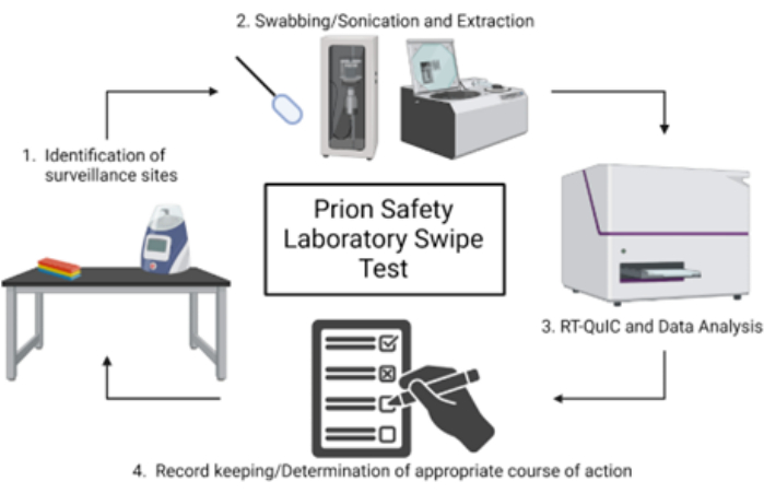
图1:朊病毒安全实验室拭子检测的示意图概述。 请点击此处查看此图的放大版本。
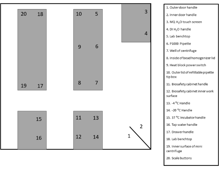
图 2:样本拭子采样点布局。 请点击此处查看此图的放大版本。
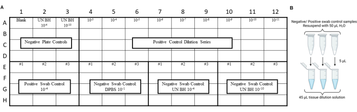
图3:包含朊病毒安全实验室擦拭测试的拭子对照样本实验设计。(A)阴性与阳性拭子提取物对照的样本布局。(B)阴性与阳性拭子提取物对照应使用 50 µL H2O 重悬。将重悬后的拭子提取物加入组织稀释液中,制备 10 倍稀释液。请点击此处查看该图的放大版本。
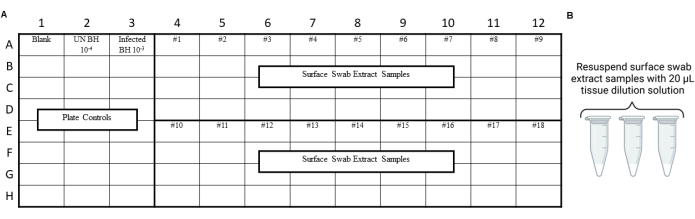
图 4:朊病毒安全实验室擦拭测试中表面拭子提取物的示例实验设计。 (A)表面拭子提取物的样品布局。(B)表面拭子提取物样品应使用 20 µL 组织稀释液重新悬浮。请点击此处查看该图的放大版本。
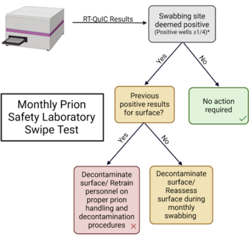
图5:朊病毒安全实验室月度拭子检测。用于解读结果并确定适当应对措施的流程图。请点击此处查看此图的放大版本。

图6:典型的表面拭子采样实验。(A)表面拭子对照平板,包含三份阴性拭子提取物对照(DPBS、未感染仓鼠脑匀浆 10-4 和 10-10)以及三份阳性拭子提取物对照(HY 脑匀浆 10-3)。(B)实验台、玻璃和不锈钢(S.S.)表面的代表性拭子提取物,分别在污染前、接种 HY 10-3 污染后以及污染表面经漂白剂处理后进行采样。(C)未感染脑匀浆 10-4、HY 脑匀浆 10-3 以及覆盖有一层细尘膜的实验台表面拭子提取物的荧光变化比较。A 和 B 面板的阴性对照平板包括空白对照(组织稀释液)和未感染仓鼠脑匀浆 10-4。B 面板还额外加入了未感染脑匀浆 10-4 以及阳性对照平板 HY 脑匀浆 10-3。阳性荧光阈值(以红色虚线表示)设定为 2。报告的最大点比值(MPR)为最大荧光值除以酶标仪测得的初始荧光读数。每个数据点代表给定样本类型的单个技术重复孔。数据以均值 ± 标准差表示。请点击此处查看该图的放大版本。
表1:样本拭子采集位点月度记录表。 请点击此处下载该表格。
补充图1:拭子采样位点布局模板。 请点击此处下载该文件。
补充图2:拭子采样位点月度记录表。 请点击此处下载该文件。
讨论
所述的朊病毒安全拭子检测方法可用于加强现有的朊病毒安全防护措施。该方法可用于监测朊病毒实验室空间和设备,以及共用的实验室区域,以发现潜在的朊病毒污染。重要的是,该方法可被调整用于测试实验室特定的消毒技术,以验证被朊病毒污染表面的去污效果。由于不同朊病毒株对消毒方法的敏感性各异,该方法可确认这些消毒技术对当前实验室实验的有效性,例如氢氧化钠或次氯酸钠(漂白剂)处理43,44。
该方法的关键步骤包括确定合适的拭子采样位点,以获取高频率接触区域的样本,同时监测可能涉及实验下游交叉污染的区域。此外,实验室应使用与特定实验室或诊所常规操作相匹配的阳性和阴性对照。例如,若实验涉及啮齿类朊病毒,则阳性和阴性对照应与该物种一致。最后,应持续更新并系统整理监测数据,以便易于识别阳性结果的变化趋势,从而在特定区域持续出现阳性结果时,及时采取干预措施并对实验室人员进行再培训。
本方案的一个局限性是可能产生错误的RT-QuIC结果。由于RT-QuIC具有高度敏感性,实验台面上残留的去垢剂、盐类及其他物质可能会影响反应结果39。因此,考虑到这一现象,保持实验表面不含有干扰RT-QuIC的物质(如残留盐分、去垢剂和灰尘)将具有显著优势。随着RT-QuIC技术方法的不断改进,我们预期当前RT-QuIC的许多局限性将逐步得到解决。因此,我们建议最终使用者密切关注相关文献,以及时采用RT-QuIC技术的最新改进成果。
该方法对于研究朊病毒的实验室而言,一个关键优势在于能够最大限度地减少可能影响敏感扩增检测结果的污染。剖检工具在重复用于后续剖检前通常会进行消毒处理。本文所述的拭子取样方法提供了一种评估剖检工具上是否存在残留朊病毒传染性的手段,从而避免对后续实验结果造成干扰。该方法可为实验增加一层严谨性,以排除组织中检测到朊病毒是源于剖检工具污染的可能性。
披露
J.C.B. 和 Q.Y. 是一项关于朊病毒表面拭子技术的专利申请的发明人。
致谢
本工作由克罗伊茨费尔特-雅各布病基金会的资助支持。资助方在研究设计、数据收集和解释,以及决定提交论文发表方面均未发挥作用。
材料
| 姓名 | 公司 | 目录编号 | 评论 |
|---|---|---|---|
| Fisherbrand PurSwab 泡沫拭子 | Fisher brand | 目录号 #14-960-3E | |
| Milli-Q IQ 7005 超纯水系统 | MilliporeSigma | Q7005T0C | |
| 微型离心机,6000 rpm | Southern Labware | MLX-306 | |
| Omega 酶标仪 | BMG Labtech | FLUOstar Omega 酶标仪 | |
| Q700 超声破碎仪 | QSonica | Q700-110 | |
| 重组仓鼠朊病毒蛋白 | MNPRO | MNPROtein-Hamster | 叙利亚仓鼠,氨基酸 90-231 |
| Savant speedvac | Thermo Scientific | SPD1030-230 | |
| 二氧化硅纳米球(50 nm) | nano Composix | SISN50-25M |
参考文献
- Prusiner, S. B. Novel proteinaceous infectious particles cause scrapie. Science. 216 (4542), 136-144 (1982).
- Bolton, D. C., McKinley, M. P., Prusiner, S. B. Identification of a protein that purifies with the scrapie prion. Science. 218 (4579), 1309-1311 (1982).
- Oesch, B., et al. A cellular gene encodes scrapie PrP 27-30 protein. Cell. 40 (4), 735-746 (1985).
- Caughey, B., Raymond, G. The scrapie-associated form of PrP is made from a cell surface precursor that is both protease- and phospholipase-sensitive. J Biol Chem. 266 (27), 18217-18223 (1991).
- Deleault, N. R., Harris, B. T., Rees, J. R., Supattapone, S. Formation of native prions from minimal components in vitro. Proc Natl Acad Sci U S A. 104 (23), 9741-9746 (2007).
- Gajdusek, D. C., Zigas, V. Degenerative disease of the central nervous system in New Guinea: the endemic occurrence of "kuru" in the native population. N Engl J Med. 257, 974-978 (1957).
- Centers for Disease Control. Fatal degenerative neurologic disease in patients who received pituitary-derived human growth hormone. MMWR Morb Mortal Wkly Rep. 34, 359-361 (1985).
- Duffy, P., Wolf, J., Collins, G., DeVoe, A. G., Streeten, B., Cowen, D. Letter: possible person-to-person transmission of Creutzfeldt-Jakob disease. N Engl J Med. 290 (12), 692-693 (1974).
- Brown, P., et al. Iatrogenic Creutzfeldt-Jakob disease at the millennium. Neurology. 55 (8), 1075-1081 (2000).
- Huillard d'Aignaux, J., et al. Incubation period of Creutzfeldt-Jakob disease in human growth hormone recipients in France. Neurology. 53 (6), 1197-1201 (1999).
- Marzewski, D. J., Towfighi, J., Harrington, M. G., Merril, C. R., Brown, P. Creutzfeldt-Jakob disease following pituitary-derived human growth hormone therapy: a new American case. Neurology. 38, 1131-1133 (1988).
- Hannah, E. L., et al. Creutzfeldt-Jakob disease after receipt of a previously unimplicated brand of dura mater graft. Neurology. 56 (8), 1080-1083 (2001).
- Shimizu, S., et al. Creutzfeldt-Jakob disease with florid-type plaques after cadaveric dura mater grafting. Arch Neurol. 56 (3), 357-362 (1999).
- Antoine, J. C., et al. Creutzfeldt-Jakob disease after extracranial dura mater embolization for a nasopharyngeal angiofibroma. Neurology. 48 (5), 1451-1453 (1997).
- Esmonde, T., Lueck, C. J., Symon, L., Duchen, L. W., Will, R. G. Creutzfeldt-Jakob disease and lyophilised dura mater grafts: report of two cases. J Neurol Neurosurg Psychiatry. 56, 999-1000 (1993).
- Willison, H. J., Gale, A. N., McLaughlin, J. E. Creutzfeldt-Jakob disease following cadaveric dura mater graft. J Neurol Neurosurg Psychiatry. 54, 940(1991).
- Diringer, H., Braig, H. R. Infectivity of unconventional viruses in dura mater. Lancet. 334, 439-440 (1989).
- Peden, A. H., Head, M. W., Ritchie, D. L., Bell, J. E., Ironside, J. W. Preclinical vCJD after blood transfusion in a PRNP codon 129 heterozygous patient. Lancet. 364 (9433), 527-529 (2004).
- Will, R. G., Matthews, W. B. Evidence for case-to-case transmission of Creutzfeldt-Jakob disease. J Neurol Neurosurg Psychiatry. 45, 235-238 (1982).
- el Hachimi, K. H., Chaunu, M. P., Cervenakova, L., Brown, P., Foncin, J. F. Putative neurosurgical transmission of Creutzfeldt-Jakob disease with analysis of donor and recipient: agent strains. C R Acad Sci III. 320 (4), 319-328 (1997).
- Alper, T., et al. Does the agent of scrapie replicate without nucleic acid. Nature. 214, 764-766 (1967).
- Casassus, B. France halts prion research amid safety concerns. Science. 373 (6554), 475-476 (2021).
- Brandel, J. P., et al. Variant Creutzfeldt-Jakob disease diagnosed 7.5 years after occupational exposure. N Engl J Med. 383 (1), 83-85 (2020).
- Flechsig, E., Hegyi, I., Enari, M., Schwarz, P., Collinge, J., Weissmann, C. Transmission of scrapie by steel-surface-bound prions. Mol Med. 7 (10), 679-684 (2001).
- Zobeley, E., Flechsig, E., Cozzio, A., Enari, M., Weissmann, C. Infectivity of scrapie prions bound to a stainless steel surface. Mol Med. 5 (4), 240-243 (1999).
- Brown, P., Gajdusek, D. C. Survival of scrapie virus after 3 years' interment. Lancet. 337 (8736), 269-270 (1991).
- Seidel, B., et al. Scrapie agent (strain 263K) can transmit disease via the oral route after persistence in soil over years. PLoS One. 2 (5), e435(2007).
- Maddison, B. C., et al. Environmental sources of scrapie prions. J Virol. 84 (21), 11560-11562 (2010).
- Pritzkow, S., Morales, R., Lyon, A., Concha-Marambio, L., Urayama, A., Soto, C. Efficient prion disease transmission through common environmental materials. J Biol Chem. 293 (9), 3363-3373 (2018).
- Alper, T., Cramp, W., Haig, D., Clarke, M. Does the agent of scrapie replicate without nucleic acid. Nature. 214 (90), 764-766 (1967).
- Bernoulli, C., et al. Danger of accidental person-to-person transmission of Creutzfeldt-Jakob disease by surgery. Lancet. 309 (8009), 478-479 (1977).
- Brown, P., Rohwer, R. G., Green, E. M., Gajdusek, D. C. Effect of chemicals, heat, and histopathologic processing on high- infectivity hamster-adapted scrapie virus. J Infect Dis. 145 (5), 683-687 (1982).
- Brown, P., et al. Chemical disinfection of Creutzfeldt-Jakob disease virus. N Engl J Med. 306, 1279-1282 (1982).
- Dickinson, A. G., Taylor, D. M. Resistance of scrapie agent to decontamination. N Engl J Med. 299, 1413-1414 (1978).
- Hunter, G. D., Millson, G. C. Studies on the heat stability and chromatographic behavior of the scrapie agent. J Gen Microbiol. 37, 251-258 (1964).
- Pattison, I. H. Resistance of the scrapie agent to formalin. J Comp Pathol. 75, 159-164 (1965).
- Fraser, H., Farquhar, C., McConnell, I., Davies, D. The scrapie disease process is unaffected by ionising radiation. Prog Clin Biol Res. 317, 653-658 (1989).
- Yuan, Q., et al. Sensitive detection of chronic wasting disease prions recovered from environmentally relevant surfaces. Environ Int. 166, 107347(2022).
- Simmons, S. M., et al. Rapid and sensitive determination of residual prion infectivity from prion-decontaminated surfaces. mSphere. 9, e00504-e00524 (2024).
- Orru, C. D., et al. Sensitive detection of pathological seeds of alpha-synuclein, tau and prion protein on solid surfaces. PLoS Pathog. 20 (4), e1012175(2024).
- Wilham, J. M., et al. Rapid end-point quantitation of prion seeding activity with sensitivity comparable to bioassays. PLoS Pathog. 6 (12), e1001217(2010).
- Srivastava, A., et al. Enhanced quantitation of pathological alpha-synuclein in patient biospecimens by RT-QuIC seed amplification assays. PLoS Pathog. 20 (9), e1012554(2024).
- Hughson, A. G., et al. Inactivation of prions and amyloid seeds with hypochlorous acid. PLoS Pathog. 12 (9), e1005914(2016).
- Williams, K., Hughson, A. G., Chesebro, B., Race, B. Inactivation of chronic wasting disease prions using sodium hypochlorite. PLoS One. 14 (10), e0223659(2019).
- Taylor, D. M. Autoclaving standards for Creutzfeldt-Jakob disease agent [letter]. Ann Neurol. 22 (4), 557-558 (1987).
- Brown, P., Rohwer, R. G., Gajdusek, D. C. Newer data on the inactivation of scrapie virus or Creutzfeldt-Jakob disease virus in brain tissue. J Infect Dis. 153 (6), 1145-1148 (1986).
重印与许可
