This prospective cohort study demonstrates that modified posterior scleral reinforcement combined with comprehensive therapy is associated with slower axial elongation and refractive progression in high and pathologic myopia, with a favorable safety profile over 24 months.
研究文章
Modified Posterior Scleral Reinforcement with Comprehensive Therapy in High and Pathologic Myopia: A Prospective Cohort Study
本文内容
摘要
摘要
This prospective cohort study aimed to assess the efficacy and safety of modified posterior scleral reinforcement (PSR) combined with comprehensive therapy versus conventional optical correction for controlling myopia progression in patients with high and pathologic myopia. A total of 203 patients aged 8–18 years were allocated, based on preference, to either the combination therapy group (n = 111; receiving modified PSR plus low-concentration atropine and/or peripheral defocus optical correction) or the conventional control group (n = 92; fitted with rigid gas-permeable contact lenses only). After accounting for 10 withdrawals during the study period, 193 patients completed the 24-month follow-up (combination group, n = 102; control group, n = 91) and were included in the final analysis. The primary outcomes were changes in axial length (AL) and spherical equivalent (SE). Secondary outcomes included best-corrected visual acuity (BCVA), posterior segment structural lesions, and adverse events. After 24 months, the combination therapy group showed significantly less AL elongation (0.33 ± 0.13 mm vs 0.70 ± 0.23 mm, p < 0.001) and SE progression (−0.75 ± 0.27 dioptres [D] vs −1.48 ± 0.57 D, p < 0.001) than the control group. The combination therapy group also demonstrated greater improvement in BCVA (p < 0.001) and lower rates of progression of myopic tractional maculopathy (2.9% vs 12.1%, p = 0.014) and aggravated tessellated fundus (6.9% vs 17.6%, p = 0.022) than the control group. The incidence of adverse events did not differ significantly between the groups (4.9% vs 4.4%, p = 1.000). In real-world clinical practice, personalized combination therapy centered on modified PSR is associated with slower axial elongation and refractive error progression and may help to stabilize posterior segment structures in patients with high and pathologic myopia, demonstrating more favorable clinical outcomes than conventional optical correction alone.
引言
Myopia, particularly high myopia, has emerged as a major global public health challenge, with its prevalence increasing markedly in recent years, especially across East Asia. High myopia is typically defined as a refractive error exceeding -6.00 dioptres (D) or an axial length (AL) greater than 26.5 mm. It is characterized by pathological alterations in the posterior segment, including posterior staphyloma, macular atrophy, and choroidal neovascularisation, which may ultimately progress to pathologic myopia1,2. As a leading cause of irreversible vision loss and blindness among the working-age population, pathologic myopia arises from fundamental anatomical changes, namely excessive axial elongation accompanied by progressive thinning and expansion of the posterior sclera3,4. Consequently, controlling axial elongation and slowing refractive error progression are pivotal strategies for preventing or delaying the transition from high myopia to pathologic myopia, representing a major clinical challenge and research priority in ophthalmology.
To address this challenge, a range of clinical interventions has been explored and implemented. In terms of optical correction, specially designed spectacles (e.g., progressive addition lenses and peripheral defocus-modifying spectacles) and contact lenses (e.g., orthokeratology lenses and multifocal soft contact lenses) suppress axial elongation by manipulating peripheral defocus signals5,6. Pharmacological intervention with low-concentration atropine eye drops has been validated in multiple randomized controlled trials as an effective means of slowing myopia progression. Although its mechanism of action may involve specific retinal and scleral receptors, concerns remain regarding long-term adherence, adverse effects (e.g., photophobia and impaired accommodation), and rebound effects following treatment discontinuation7. Nevertheless, for individuals experiencing rapid progression or with established high or pathologic myopia, these conventional approaches often exhibit limited efficacy or a ‘ceiling effect’, rendering them insufficient to effectively halt further axial elongation and deterioration of posterior segment lesions.
Against this backdrop, posterior scleral reinforcement (PSR) offers an alternative approach to managing pathologic myopia progression through mechanical reinforcement of the weakened posterior sclera. The theoretical basis of this procedure dates back more than half a century, whereby implanted biological or synthetic materials enhance posterior scleral strength, restrict pathological expansion, and may stabilize posterior segment structures by improving local circulation8,9. Although conventional PSR has demonstrated promising long-term efficacy, its widespread adoption has been limited by substantial surgical trauma and a relatively high risk of complications. More recently, advances in minimally invasive surgical concepts and techniques have led to the development of modified PSR. This refined approach optimizes surgical incisions, material processing, and fixation methods to reduce surgical trauma while improving safety without compromising efficacy in controlling axial elongation10. Consequently, the clinical acceptability and applicability of this technique have increased substantially.
Notably, the pathogenesis of myopia involves complex interactions among genetic, environmental, and visual feedback factors. As single interventions typically target only one pathway, researchers have increasingly explored combination strategies to evaluate their potential synergistic effects and enhance treatment efficacy. Theoretically, modified PSR structurally reinforces the posterior ocular wall, whereas low-concentration atropine or specialized optical corrections target axial elongation through biochemical signaling and optical mechanisms. Their integration may establish a more comprehensive management framework. The novelty of this study lies in evaluating a dynamically adjusted, multi-targeted strategy that integrates modified PSR with individualized optical and pharmacological interventions to simultaneously control axial elongation and stabilize posterior structural lesions, thereby addressing the relative scarcity of evidence regarding such surgery-centered combination therapies over a 24-month period in real-world clinical settings. Although high-quality evidence predominantly originates from rigorously conducted randomized controlled trials, the generalisability of their findings to heterogeneous routine clinical populations may be limited. Real-world studies seek to reflect actual therapeutic outcomes and safety profiles under routine care conditions, where patients select treatments based on personal preferences and clinical circumstances, thereby providing complementary evidence to support clinical decision-making in heterogeneous populations.
Consequently, this study employs a prospective cohort design to systematically evaluate, in a real-world clinical setting, the efficacy and safety of personalized combination therapy centered on modified PSR compared with conventional optical correction alone for controlling AL elongation and spherical equivalent (SE) progression in patients with high or pathologic myopia. The findings provide important clinical evidence to inform the management of high and pathologic myopia, assisting clinicians and patients in making more informed therapeutic decisions when managing complex disease progression.
方案
This study was conducted in accordance with the Declaration of Helsinki and received ethical approval from the Institutional Review Board of Shijiazhuang People’s Hospital (Approval ID: 2024-043). Written informed consent was obtained from all participants and/or their legal guardians prior to enrolment. An overall schematic of the study design is provided in Figure 1.
Study design and participants
This prospective cohort study aimed to evaluate the efficacy of a modified PSR-based combination therapy compared with conventional optical correction in slowing axial elongation in pediatric patients with progressive high or pathologic myopia. Consecutive patients meeting the eligibility criteria were recruited from the Pediatric Ophthalmology Clinic at Shijiazhuang People’s Hospital between October 2022 and October 2023. All participants and/or their legal guardians provided written informed consent after receiving a detailed explanation of the study protocol, potential risks, benefits, and alternative treatment options.
Inclusion and exclusion criteria
The inclusion criteria were as follows: age 8–18 years; diagnosis of high or pathologic myopia, defined as an SE refraction of ≤-6.00 D and/or an AL of ≥26.5 mm; documented active myopia progression within the 6 months preceding enrolment, retrospectively verified using historical medical records and previous optical biometry or refraction data from the institution or referring clinics, as evidenced by an SE progression rate of ≥-0.50 D/year or AL elongation of ≥0.3 mm/year; best-corrected visual acuity (BCVA) of ≥0.1 in decimal notation (equivalent to ≤1.0 on a logarithm of the minimum angle of resolution [logMAR] chart) to exclude patients with severe amblyopia; and provision of written informed consent and willingness to complete the 2-year follow-up schedule.
The exclusion criteria were as follows: coexisting ocular pathologies that could affect AL or refraction (e.g. congenital glaucoma, keratoconus, significant cataract, uveitis or retinal detachment); systemic disorders affecting visual function (e.g. albinism, Marfan syndrome or connective tissue disorders); a history of ocular surgery or trauma; documented hypersensitivity to any materials or medications used in the study (e.g. surgical materials or atropine); severe amblyopia or an inability to cooperate with the required ophthalmic examinations; and an anticipated inability to complete the study due to personal circumstances.
Sample size calculation
The sample size was calculated based on the primary outcome of the between-group difference in cumulative AL elongation over 24 months. Preliminary data indicated an anticipated mean AL elongation of 0.70 ± 0.23 mm in the control group and 0.33 ± 0.13 mm in the combination therapy group. Using a two-sided α level of 0.05 and a power (1 − β) of 80%, with an estimated pooled standard deviation (σ) of 0.20 mm and an expected effect size (Δ) of 0.37 mm, the required sample size per group was calculated using the following formula for comparing two independent means:
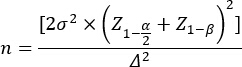
This calculation yielded a required sample size of 77 participants per group. To account for potential attrition, a total of 203 participants were enrolled (combination therapy group, n = 111; conventional control group, n = 92), exceeding the minimum required sample size. Following the withdrawal of 10 participants, a final cohort of 193 patients (102 in the combination group and 91 in the control group) completed the 24-month follow-up and provided complete datasets for analysis.
Group allocation and interventions
Group allocation was based on the autonomous choice of patients and/or their legal guardians following comprehensive counseling by the attending ophthalmologists. The counseling covered the potential benefits, risks, costs, and postoperative requirements of the modified PSR-based combination therapy, together with a detailed explanation of conventional optical correction.
Combination therapy group (n = 102)
Participants underwent a modified PSR procedure performed by a single experienced attending surgeon using standardized allogeneic scleral materials and refined surgical techniques. Postoperatively, patients received a structured, highly individualized, comprehensive therapy regimen. Specifically, the clinical decision algorithm generally began with peripheral defocus-modifying optical correction (required wear time ≥10 h/day). If consecutive follow-up assessments indicated AL elongation >0.15 mm/6 months or SE progression >0.25 D/6 months, 0.01% low-concentration atropine eye drops (one drop nightly) were added incrementally. Structured vision therapy exercises (20 min/session, 2–3 times/week) were prescribed for patients demonstrating specific accommodative dysfunctions. Reflecting real-world clinical practice, the specific adjunctive treatments, including 0.01% low-concentration atropine eye drops, peripheral defocus-modifying optical correction, and structured vision therapy exercises, were not assigned statistically. Instead, they were dynamically tailored and adjusted (escalated or modified) for each patient based on continuous clinical feedback, including serial anatomical measurements and refractive changes over the 24-month follow-up period. Although strict adherence is inherently difficult to control in a real-world setting, patient compliance was monitored qualitatively at each follow-up visit using structured patient- or parent-reported diaries and medication check-ins, acknowledging that these subjective measures could not completely eliminate adherence-related confounding. Owing to the frequent crossover and dynamic nature of these regimens, stratifying the cohort into mutually exclusive subgroups for statistical comparison was not feasible.
Conventional control group (n = 91)
Participants who declined surgical intervention received refractive correction alone. This group was uniformly fitted with rigid gas-permeable (RGP) contact lenses for optical management. They did not receive systematic low-concentration atropine or other specific myopia control agents.
Surgical technique for modified posterior scleral reinforcement
All surgical procedures were performed under general anesthesia with endotracheal intubation by the same experienced surgical team. The modified PSR procedure was standardized as follows:
Incision and exposure
A conjunctival peritomy of about 180° was made in the inferotemporal quadrant, approximately 3 mm from the corneal limbus. The Tenon’s capsule was bluntly dissected to expose the bare sclera. The lateral rectus and inferior rectus muscles were isolated and secured with 5–0 silk traction sutures to optimize surgical exposure of the posterior pole.
Material preparation
The reinforcement material consisted of lyophilized allogeneic human sclera, pretreated and preserved by glycerol dehydration and γ-irradiation (25 kGy). Intraoperatively, the graft was rehydrated in sterile normal saline for 15 min and trimmed into a single-strip configuration. The standard graft dimensions were approximately 35 mm in length and 9 mm in width, with the final size tailored according to the patient’s preoperative AL and the extent of the posterior staphyloma.
Graft placement and fixation
A specialized scleral depressor (macular retractor) was used to introduce the prepared graft into the posterior sub-Tenon’s space. The graft was carefully advanced posteriorly along the temporal equator, wrapping around the macular region and posterior pole while avoiding compression of the optic nerve and vortex veins. The inferior oblique muscle was identified and gently retracted to allow the graft to pass beneath it. The graft was then tensioned appropriately to provide mechanical support to the thinned staphyloma, with its position confirmed to be closely opposed to the sclera without wrinkles or distortion. Fixation was achieved by anchoring the ends of the graft to the host sclera at the superotemporal and inferonasal quadrants near the equator using interrupted 5–0 non-absorbable polyester sutures. The inferior rectus and lateral rectus muscles were each secured to the overlying graft with one additional suture to prevent graft migration.
Following meticulous verification of hemostasis and optimal graft positioning without undue tension on the globe, the muscle traction sutures were removed. The conjunctival incision was then closed with continuous 8–0 absorbable sutures. Postoperatively, standard topical antibiotics (levofloxacin 0.5%, four times daily for 1 week) and corticosteroids (prednisolone acetate 1%, tapered over 4 weeks) were administered.
Baseline assessment and outcome measures
At the baseline visit, comprehensive data were collected for all participants, including demographic characteristics (age and sex), age at myopia onset, cycloplegic SE refraction, AL, and BCVA. Best-corrected visual acuity testing was assessed using a standard Early Treatment Diabetic Retinopathy Study (ETDRS) chart at a distance of 4 m under uniform lighting conditions (approximately 85 cd/m2). The ETDRS letter score was calculated as the total number of letters read correctly at 4 m plus 30. For patients unable to read any letters at 4 m, testing was repeated at 1 m. Cycloplegia was induced using 0.5% compound tropicamide eye drops (one drop every 5 min for three doses), and measurements were obtained 30 min after the final drop. Axial length was measured using a partial coherence interferometry-based optical biometer, with the final AL value recorded as the mean of five consecutive valid measurements for each eye.
The primary outcomes were cumulative change in AL (mm) from baseline11 and cumulative progression of SE (D) from baseline12. Both were assessed at each follow-up visit. Secondary outcomes included the following: change in BCVA (logMAR)13; incidence (%) of progression of myopic tractional maculopathy (worsening macular curvature), assessed by optical coherence tomography (OCT) according to the established atrophy-traction-neovascularisation (ATN) grading system for myopic maculopathy and strictly defined as an increase of at least one severity grade in the traction (T) component from baseline (e.g., from T0 to T1 or T1 to T2)14; and incidence (%) of aggravated tessellated fundus changes, graded according to the international Meta-Analysis for Pathologic Myopia (META-PM) Study Group classification15. Progression of tessellated fundus was defined as the transition to a higher META-PM category (e.g., from Category 1 tessellated fundus to Category 2 diffuse chorioretinal atrophy or Category 3 patchy chorioretinal atrophy) or clinically significant expansion of the affected atrophic area, determined by side-by-side comparison of baseline and follow-up fundus photographs. Both structural outcomes were evaluated independently by two retinal specialists masked to group allocation. Inter-rater reliability was assessed using Cohen’s kappa coefficient, yielding a high level of agreement (κ = 0.85). In cases of disagreement, a third senior retinal specialist was consulted to reach a consensus. Baseline macular morphological status was documented on spectral-domain OCT based on posterior pole curvature (posterior staphyloma or macular curvature index ≥ 1.5)16 alongside ATN tractional staging, and baseline chorioretinal status was categorized by the META-PM system, ensuring standardized longitudinal comparison.
Follow-up schedule
All participants were scheduled for follow-up visits at 1, 3, 6, 12, 18, and 24 months after enrolment (or postoperatively for the surgical group). Core parameters (SE, AL, and BCVA) were reassessed at every visit. Comprehensive posterior segment evaluations, including OCT and fundus photography, were performed at the 6-, 12-, and 24-month follow-up visits to monitor structural changes. In addition, comprehensive safety assessments were conducted at every follow-up visit. Any complications or adverse events were meticulously documented, including the type of event, time of onset, severity (categorized as mild, moderate, or severe), and the clinical management strategies implemented until resolution.
Statistical analysis
Data were analyzed using statistical software. The normality of the data distribution was assessed using the Shapiro–Wilk test. Normally distributed continuous variables are presented as mean ± standard deviation and were compared using independent-samples t-tests. Non-normally distributed continuous variables are presented as median (interquartile range) and were compared using Mann–Whitney U tests. Categorical data are presented as frequencies (percentages) and were compared using the χ2 test or Fisher’s exact test, as appropriate. All tests were two-sided, with a p-value <0.05 considered statistically significant. Between-group differences for the primary outcomes are reported with their corresponding 95% confidence intervals (CIs). As all baseline demographic and clinical characteristics were highly comparable and well balanced between the two cohorts, with no statistically significant differences, propensity score matching was not considered necessary because it would have unnecessarily discarded patient data and reduced statistical power without addressing unmeasured confounding. To appropriately address potential selection bias, we utilized a linear mixed-effects model to analyze the longitudinal repeated-measures data for the primary outcomes (AL and SE) across the multiple follow-up time points (1, 3, 6, 12, 18, and 24 months). The model included treatment group, time point, and their interaction (group × time) as fixed effects and accounted for individual baseline variations and intra-subject correlation by including random intercepts for each participant.
结果
This study ultimately enrolled 203 patients with high or pathologic myopia, who were naturally allocated to two cohorts according to their treatment preferences. In the combination therapy group, 9 participants withdrew, resulting in 102 individuals completing the 24-month follow-up. In the conventional control group, 1 participant withdrew, leaving 91 patients who completed the full follow-up period. All participants who completed the 24-month follow-up provided complete datasets for analysis.
Baseline patient characteristics
Comprehensive baseline comparisons were conducted to evaluate between-group comparability prior to the intervention. The combination therapy and conventional control groups showed no statistically significant differences in demographic characteristics, including age (12.53 ± 2.71 vs 12.03 ± 3.28 years; p = 0.252) and sex distribution (men/women: 48/54 vs 47/44; p = 0.524). Key ocular parameters were also well balanced between the groups, including SE (−8.35 ± 1.81 D vs −7.82 ± 2.10 D; p = 0.060), AL (27.74 ± 0.81 mm vs 27.72 ± 0.95 mm; p = 0.847), age at myopia onset (6.03 ± 1.54 vs 5.80 ± 1.75 years; p = 0.344) and BCVA (0.11 ± 0.03 vs 0.10 ± 0.03 logMAR; p = 0.314). These findings indicate comparable baseline disease severity based on the measured parameters (Table 1). However, given the preference-based, non-randomized allocation, the potential influence of unmeasured confounding factors, such as family treatment motivation, cost tolerance, or adherence behavior, cannot be completely excluded.
Baseline structural characteristics of the posterior pole, including macular curvature abnormalities and tessellated fundus changes, were also compared between the two groups (Table 1). Macular curvature abnormalities (defined as posterior staphyloma or a macular curvature index ≥ 1.5 on spectral-domain OCT16, corresponding to early ATN stage T0–T1) were observed in 41 of 102 participants (40.2%) in the combination therapy group and 37 of 91 participants (40.7%) in the conventional control group. Tessellated fundus changes (defined as Category ≥ 1 according to the Meta-Analysis for Pathologic Myopia Study Group classification15) were present in 68 of 102 participants (66.7%) in the combination therapy group and 60 of 91 participants (65.9%) in the conventional control group. No statistically significant between-group differences were observed for the baseline prevalence of either macular curvature abnormalities or tessellated fundus changes (both P > 0.05; Table 1), providing further evidence of baseline comparability between the two cohorts.
Primary outcomes: efficacy in controlling axial length and refractive progression
Follow-up assessments of the primary outcomes demonstrated that combination therapy provided significantly better control of axial elongation and myopic refractive progression than conventional treatment from an early stage, with this advantage increasing progressively over time. For AL control, the combination therapy group exhibited a slightly smaller cumulative increase in AL (0.02 ± 0.01 mm) than the conventional control group (0.03 ± 0.01 mm) at 1 month after the intervention, with a between-group difference of -0.01 mm (95% CI: -0.04 to 0.02; p = 0.618). The between-group difference became statistically significant and progressively more pronounced at 3, 6, 12, and 18 months. By the 24-month follow-up, cumulative AL elongation was 0.33 ± 0.13 mm in the combination therapy group compared with 0.70 ± 0.23 mm in the conventional control group, yielding a between-group difference of -0.37 mm (95% CI: -0.41 to -0.34; p < 0.001) (Table 2).
Regarding SE progression, at 1 month after the intervention, the difference between the two groups (combination therapy group: -0.05 ± 0.05 D; conventional control group: -0.06 ± 0.02 D) was not statistically significant (p = 0.788). However, from the 6-month follow-up onwards, the advantage of the combination therapy group became statistically significant and remained consistent throughout the study period. By the end of the 24-month follow-up, cumulative SE progression in the combination therapy group was -0.75 ± 0.27 D, indicating an average increase in myopia of approximately 0.75 D, whereas the conventional control group showed a cumulative progression of -1.48 ± 0.57 D, corresponding to a worsening of almost 1.50 D. The between-group difference was 0.73 D (95% CI: 0.66 to 0.79), representing an approximately 50% reduction in myopic refractive progression over 2 years in the combination therapy group (p < 0.001) (Table 3).
The modified PSR technique used in this study, employing a single-strip allogeneic scleral graft, is illustrated sequentially in Supplementary Figure 1.
Secondary outcomes and safety evaluation
Regarding the secondary efficacy outcomes at 24 months, the combination therapy group demonstrated greater improvement in BCVA and significantly lower incidence rates of tractional maculopathy progression and aggravated tessellated fundus than the conventional control group (Table 4). Within the combination therapy group (n = 102), the distribution of dynamically adjusted adjunctive therapies over the 24-month follow-up was as follows: 33 patients received peripheral defocus optical correction alone, 16 patients received 0.01% low-concentration atropine alone, 38 patients received a combination of peripheral defocus optical correction and atropine and 15 patients required the addition of structured vision therapy exercises alongside their optical or pharmacological regimens. Due to the frequent crossover, varied duration, and dynamic escalation of these adjunctive therapies tailored to individual clinical responses, a valid subgroup statistical comparison of efficacy among these specific regimens could not be performed. Regarding safety, the incidence of complications and adverse events was low in both groups. Five adverse events were reported in the combination therapy group (4.9%), compared with four (4.4%) in the conventional control group. Fisher’s exact test showed no statistically significant difference in the incidence of adverse events between the two groups (p = 1.000).
To provide a comprehensive safety profile, a detailed breakdown of these events was analyzed and is summarised in Table 5. In the combination therapy group, the 5 adverse events consisted of 2 cases of transient intraocular pressure (IOP) elevation occurring within 1 week postoperatively (mild severity), which were managed effectively with topical hypotensive eye drops for 1 week; 2 cases of mild photophobia secondary to atropine use, beginning at 1 month, which were managed by advising appropriate outdoor eye protection; and 1 case of mild conjunctival chemosis, which resolved spontaneously within 2 weeks. In the conventional control group, the 4 adverse events comprised 3 cases of mild corneal epitheliopathy related to RGP contact lens wear, presenting between 3 and 6 months, which resolved after temporary discontinuation of lens wear and treatment with artificial tears, and 1 case of mild allergic conjunctivitis, which was managed with topical antihistamines. All adverse events in both cohorts were classified as mild to moderate in severity and resolved with symptomatic management, with no cases of severe or permanent functional impairment. Importantly, no serious surgery-related complications, such as retinal detachment, scleral perforation, severe infection, or graft rejection, were observed throughout the 24-month follow-up period. Regarding treatment adherence, based on the self-reported diaries and medication check-ins, overall compliance was acceptable in the combination therapy group, with approximately 85% of patients adhering to their prescribed optical or pharmacological regimens on at least 5 days per week.
DATA AVAILABILITY:
The original de-identified dataset supporting the conclusions of this article is included within the article and its supplementary information files (Supplementary File 1).
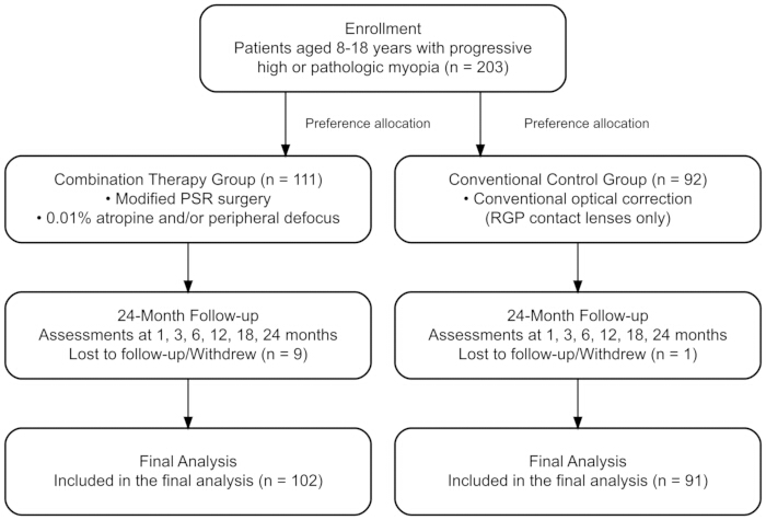
Figure 1: Overall schematic of the study design. Flowchart detailing the enrollment, preference-based allocation, follow-up, and final analysis of patients with high and pathologic myopia comparing the modified posterior scleral reinforcement (PSR) combination therapy versus conventional optical correction over a 24-month period. Please click here to view a larger version of this figure.
| Variable | Combination therapy group(n = 102) | Conventional control group (n = 91) | t/χ² value | P value |
| Age (year) | 12.53 ± 2.71 | 12.03 ± 3.28 | 1.15 | 0.252 |
| Gender (male/female) | 48/54 | 47/44 | 0.405 | 0.524 |
| Spherical equivalent (D) | -8.35 ± 1.81 | -7.82 ± 2.10 | -1.895 | 0.06 |
| Axial length(mm) | 27.74 ± 0.81 | 27.72 ± 0.95 | 0.193 | 0.847 |
| Age at myopia onset (year) | 6.03 ± 1.54 | 5.80 ± 1.75 | 0.948 | 0.344 |
| Best corrected visual acuity (logMAR) | 0.11 ± 0.03 | 0.10 ± 0.03 | 1.009 | 0.314 |
| Macular curvature abnormality (n, %) | 41 (40.2%) | 37 (40.7%) | 0.004 | 0.948 |
| Tessellated fundus change (n, %) | 68 (66.7%) | 60 (65.9%) | 0.012 | 0.914 |
Table 1: Baseline demographic and ocular characteristics of the study cohorts. Data are presented as mean ± standard deviation for continuous variables and n (%) for categorical variables. All parameters were evaluated prior to the intervention. The denominators for all variables were 102 in the combination therapy group and 91 in the conventional control group. Baseline macular curvature abnormalities were defined on optical coherence tomography as posterior staphyloma or a macular curvature index ≥ 1.5, corresponding to early ATN stage T0–T1. Baseline tessellated fundus changes were graded according to the Meta-Analysis for Pathologic Myopia (META-PM) Study Group classification.D: dioptre; logMAR: logarithm of the minimum angle of resolution. All p-values were calculated using independent-samples t-tests or Pearson’s chi-square tests, as appropriate.
| Follow-up Time Point | Combination Therapy Group (n = 102) | Conventional Control Group (n = 91) | Between-group Difference (95% CI) | P value |
| 1 month | 0.02 ± 0.01 | 0.03 ± 0.01 | -0.01 (-0.04, 0.02) | 0.618 |
| 3 months | 0.05 ± 0.03 | 0.10 ± 0.04 | -0.04 (-0.08, -0.01) | 0.008 |
| 6 months | 0.09 ± 0.05 | 0.22 ± 0.09 | -0.13 (-0.16, -0.10) | <0.001 |
| 12 months | 0.17 ± 0.07 | 0.39 ± 0.11 | -0.21 (-0.25, -0.18) | <0.001 |
| 18 months | 0.25 ± 0.12 | 0.60 ± 0.20 | -0.35 (-0.38, -0.32) | <0.001 |
| 24 months | 0.33 ± 0.13 | 0.70 ± 0.23 | -0.37 (-0.41, -0.34) | <0.001 |
Table 2: Cumulative change in axial length (mm). Data are presented as mean ± standard deviation. The denominators at all time points remained constant, reflecting the final cohort of patients who completed the 24-month follow-up (n = 102 for the combination therapy group; n = 91 for the conventional control group). AL: axial length; CI: confidence interval. The p-values for the between-group differences at each time point, together with the overall main effect of group and the group × time interaction, were derived from a longitudinal linear mixed-effects model accounting for repeated measurements over time.
| Follow-up Time Point | Combination Therapy Group (n = 102) | Conventional Control Group (n = 91) | Between-group Difference (95% CI) | P value |
| 1 month | -0.05 ± 0.05 | -0.06 ± 0.02 | 0.01 (-0.06, 0.08) | 0.788 |
| 3 months | -0.11 ± 0.08 | -0.18 ± 0.10 | 0.07 (-0.00, 0.13) | 0.051 |
| 6 months | -0.20 ± 0.10 | -0.37 ± 0.18 | 0.17 (0.10, 0.23) | <0.001 |
| 12 months | -0.36 ± 0.10 | -0.72 ± 0.22 | 0.37 (0.30, 0.43) | <0.001 |
| 18 months | -0.53 ± 0.20 | -0.98 ± 0.38 | 0.45 (0.38, 0.52) | <0.001 |
| 24 months | -0.75 ± 0.27 | -1.48 ± 0.57 | 0.73 (0.66, 0.79) | <0.001 |
Table 3: Cumulative progression of spherical equivalent (D). Data are presented as mean ± standard deviation. The denominators at all time points remained constant, reflecting the final cohort of patients who completed the 24-month follow-up (n = 102 for the combination therapy group; n = 91 for the conventional control group). SE: spherical equivalent; D: dioptre; CI: confidence interval. The p-values for the between-group differences at each time point, together with the overall main effect of group and the group × time interaction, were derived from a longitudinal linear mixed-effects model accounting for repeated measurements over time.
| Measure | Combination Therapy Group (n = 102) | Conventional Control Group (n = 91) | P value |
| Change in Best Corrected Visual Acuity (logMAR) | -0.04 ± 0.01 | -0.02 ± 0.01 | <0.001 |
| Progression of tractional maculopathy (ATN T component, n, %) | 3 (2.9%) | 11 (12.1%) | 0.014 |
| Aggravation of Tessellated Fundus (n, %) | 7 (6.9%) | 16 (17.6%) | 0.022 |
| Complications/Adverse Events (n, %) | 5 (4.9%) | 4 (4.4%) | 1.000 |
Table 4: Secondary outcome measures and safety evaluation. All secondary outcomes and cumulative adverse events were assessed at the final 24-month follow-up visit. The denominators were n = 102 for the combination therapy group and n = 91 for the conventional control group. Continuous variables are presented as mean ± standard deviation, and categorical variables as n (%). Progression of myopic tractional maculopathy was assessed using the atrophy-traction-neovascularisation (ATN) grading system, while aggravated tessellated fundus was defined according to the Meta-Analysis for Pathologic Myopia Study Group classification.BCVA: best-corrected visual acuity; logMAR: logarithm of the minimum angle of resolution. For BCVA, a negative change indicates an improvement in visual function. All p-values were calculated using independent-samples t-tests for continuous variables and Fisher’s exact test for categorical variables.
| Type of Adverse Event | Cases (n) | Timing of Onset | Severity | Management Strategy | Outcome |
| Transient intraocular pressure (IOP) elevation | 2 | Within 1 week postoperatively | Mild | Topical hypotensive drops for 1 week | Resolved effectively |
| Photophobia (secondary to atropine) | 2 | Starting at 1 month | Mild | Adjusting outdoor optical protection | Resolved |
| Conjunctival chemosis | 1 | Unspecified | Mild | Observation | Resolved spontaneously within 2 weeks |
| Corneal epitheliopathy (related to RGP lens) | 3 | Between 3 and 6 months | Mild | Temporary suspension of lens wear and artificial tears | Resolved |
| Allergic conjunctivitis | 1 | Unspecified | Mild | Topical antihistamines | Resolved |
Table 5: Summary of adverse events by study group. This table details the type of adverse event, number of cases, time of onset, severity, clinical management strategy, and final clinical outcome for all adverse events reported during the 24-month follow-up period in both the combination therapy (n = 102) and conventional control (n = 91) groups.
Supplementary Figure 1: Sequential stages of the modified posterior scleral reinforcement (PSR) procedure using a single-strip allogeneic scleral graft. (A) Isolation of the extraocular muscles and exposure of the posterior sclera. (B) Introduction of the prepared scleral graft into the posterior sub-Tenon’s space. (C) Advancement of the graft posteriorly along the temporal equator. (D) Positioning and appropriate tensioning of the graft over the macular region. (E) Fixation of the graft extremities to the host sclera. (F) Final verification of optimal graft position and muscle securement before closure.Please click here to download this file.
Supplementary File 1: Raw data.Please click here to download this file.
讨论
This 24-month prospective cohort study, conducted in a real-world clinical setting, evaluated the efficacy and safety of a combination therapy centered on modified PSR surgery for controlling the progression of high and pathologic myopia. The results demonstrated that, compared with conventional optical correction alone, the combination therapy was associated with significant and sustained reductions in axial elongation and myopic refractive progression, together with better visual outcomes and greater preservation of posterior segment structures, without an increased risk of complications.
The most notable finding of this study is that the combination therapy achieved superior long-term control of the core indicators of myopia progression, as demonstrated by the substantial differences observed in the primary outcomes. At 24 months, axial elongation in the combination therapy group (0.33 mm) was less than half that in the control group (0.70 mm), whereas SE progression (-0.75 D) was also significantly lower than that in the control group (-1.48 D). These findings were not only highly statistically significant but also clinically meaningful. Previous studies have shown that AL is a stronger predictor of fundus complications in pathologic myopia, with each 1 mm increase in AL significantly increasing the risk of myopic macular degeneration17,18. Therefore, maintaining a lower annual rate of axial elongation (approximately 0.165 mm/year in the combination therapy group in this study) is crucial for preventing or delaying irreversible visual impairment and structural fundus complications, as supported by our secondary structural outcome assessments. Our findings are consistent with recent reports on PSR surgery19. For example, a meta-analysis reported that PSR surgery effectively slows axial elongation in patients with pathologic myopia, with an average annual increase of approximately 0.18 mm, significantly lower than the natural rate of progression20. The modified surgical technique employed in this study may inhibit the underlying anatomical progression of pathologic myopia by mechanically reinforcing the posterior sclera, thereby limiting its passive expansion under IOP and providing a structural basis for improved choroidal blood flow.
Patients in the combination therapy group received individualized postoperative management, including low-dose atropine and peripheral defocus optical correction, among other interventions. Low-dose atropine has been shown in multiple large-scale randomized controlled trials, including the Atropine for the Treatment of Myopia and Low-Concentration Atropine for Myopia Progression studies, to effectively slow myopia progression through antimuscarinic mechanisms21,22. Peripheral defocus optical correction, by contrast, reduces the stimulus for axial elongation by eliminating or reducing hyperopic defocus in the peripheral retina. Modified PSR surgery mechanically reinforces the posterior ocular wall at the macroscopic level, whereas pharmacological and optical interventions act at the cellular level and on signaling pathways to influence scleral remodeling and visual feedback, respectively. Together, these three components may constitute a multi-targeted, three-dimensional synergistic control strategy, which may explain the significantly greater efficacy of the combination therapy compared with conventional optical correction alone23. This approach is also consistent with the current trend towards diversified and individualized combination strategies for myopia management.
Regarding safety, the incidence of complications in the combination therapy group did not differ significantly from that in the control group, and all adverse events were mild and resolved without sequelae. These findings indicate that modified PSR surgery, when performed by experienced surgeons using established techniques, has a manageable safety profile. Compared with the conventional procedure, the modified technique may also reduce surgical trauma and lower the risks of rejection and postoperative complications24. Furthermore, the advantages of the combination therapy in the secondary outcomes are noteworthy. Greater improvement in BCVA, together with lower rates of tractional maculopathy progression and aggravated tessellated fundus, suggests that this regimen may provide better control of refractive progression and reduce the risk of progression of pathologic myopia-related fundus lesions. This is particularly important for preserving long-term visual function and quality of life25,26.
Several limitations of this study should be acknowledged. First, the non-randomized, preference-based treatment allocation inherently introduced the possibility of unmeasured confounding. Variables such as family support, health beliefs, treatment motivation, and long-term treatment adherence could not be fully controlled and may have influenced the outcomes. Second, the intervention in the study group was intentionally designed as a comprehensive, highly individualized treatment strategy. Reflecting real-world clinical practice, postoperative adjunctive therapies were dynamically adjusted according to continuous clinical feedback. Because of the heterogeneity and dynamic crossover of these adjunctive treatments, including varying use of 0.01% atropine, defocus-modifying lenses, and vision therapy, it was not possible to isolate the independent contribution of the modified PSR procedure. Specifically, the preference-based allocation and the sample size limitations within these dynamically changing subgroups precluded a statistically powered, unconfounded analysis of the isolated effect of PSR. Future studies using factorial designs are needed to disentangle the effects of the individual treatment components. Third, the control group received conventional RGP contact lenses, which primarily provide standard optical correction. RGP lenses were specifically selected as the control intervention because they offer optimal visual rehabilitation and are a well-established, safe standard of care for extreme refractive errors, whereas active myopia control interventions, such as orthokeratology, are highly effective in low-to-moderate myopia but are generally contraindicated or used off-label in patients with extreme refractive errors (SE ≤ -6.00 D) represented in our cohort. Nevertheless, we acknowledge that comparing the comprehensive treatment package with standard optical correction alone may have overestimated the relative benefit of the combination therapy. Finally, the 24-month follow-up remains relatively short for evaluating the lifelong progression of pathologic myopia, and the study was conducted at a single center with a relatively modest sample size. Despite these limitations, the robust longitudinal analysis provides compelling real-world evidence that reflects routine clinical practice.
披露
All authors have completed the ICMJE Uniform Disclosure Form and declare no competing interests relevant to the subject matter or materials discussed in this manuscript. No artificial intelligence (AI) tools were used in the preparation of the manuscript or figures.
致谢
This work was supported by the Shijiazhuang Health Commission Guidance Plan Project (Project No. 20251147).
材料
| 姓名 | 公司 | 目录编号 | 评论 |
|---|---|---|---|
| 0.01% low-concentration atropine eye drops | Compounded by Department of Pharmacy, Shijiazhuang People's Hospital, Shijiazhuang, China | https://www.sjzsdyyy.cn | compounded preparation |
| 5-0 non-absorbable polyester (Mersilene) suture | Ethicon, Inc. (Johnson and Johnson), Somerville, NJ, USA | Catalog #D6216 | Mersilene 5-0 |
| 5-0 silk traction suture | Ethicon, Inc. (Johnson and Johnson), Somerville, NJ, USA | Catalog #K889H | Silk 5-0 |
| 8-0 absorbable polyglactin 910 (Vicryl) suture | Ethicon, Inc. (Johnson and Johnson), Somerville, NJ, USA | Catalog #J570G | Vicryl 8-0 |
| Compound tropicamide eye drops (0.5%) | Santen Pharmaceutical Co., Ltd., Osaka, Japan | https://www.santen.com | Mydrin-P (tropicamide 0.5% + phenylephrine HCl 0.5%); NMPA Approval No. H20050462 |
| Levofloxacin 0.5% eye drops | Santen Pharmaceutical Co., Ltd., Osaka, Japan | https://www.santen.com | Cravit; NMPA Approval No. H20020215 |
| Lyophilised allogeneic human sclera | Hospital-prepared (Shijiazhuang People's Hospital Tissue Bank, Shijiazhuang, China) | https://www.sjzsdyyy.cn | glycerol-dehydrated and gamma-irradiated at 25 kGy |
| Partial coherence interferometry-based optical biometer | Carl Zeiss Meditec AG, Jena, Germany | https://www.zeiss.com/meditec/en/products/optical-biometers/iolmaster-700.html | IOLMaster 700 |
| Prednisolone acetate 1% eye drops | Allergan Pharmaceuticals Ireland, Westport, Ireland | https://www.allergan.com | Pred Forte; NMPA Approval No. H20130136 |
| Rigid gas permeable (RGP) contact lenses | Menicon Co., Ltd., Nagoya, Japan | https://www.menicon.com/professional/products/specialty-lenses/menicon-z | Menicon Z |
| Spectral-domain optical coherence tomography (OCT) device | Heidelberg Engineering GmbH, Heidelberg, Germany | RRID:SCR_019362 ; https://www.heidelbergengineering.com | Spectralis HRA+OCT |
| Standard ETDRS chart | Precision Vision, Inc., Woodstock, IL, USA | RRID:SCR_025206; https://precision-vision.com | ETDRS 2000 Series Chart 1 |
| SPSS | IBM Corp. | version 26.0 | Statistical software |
参考文献
- Baird PN, Saw SM, Lanca C. Myopia. Nat Rev Dis Primers. 2020;6(1):99.
- Ohno-Matsui K, et al. IMI Pathologic Myopia. Invest Ophthalmol Vis Sci. 2021;62(5):5.
- Jonas JB, et al. IMI Prevention of Myopia and Its Progression. Invest Ophthalmol Vis Sci. 2021;62(5):6.
- Haarman AEG, et al. The Complications of Myopia: A Review and Meta-Analysis. Invest Ophthalmol Vis Sci. 2020;61(4):49.
- Chamberlain P, et al. A 3-year Randomized Clinical Trial of MiSight Lenses for Myopia Control. Optom Vis Sci. 2019;96(8):556–67.
- Bao J, et al. One-year myopia control efficacy of spectacle lenses with aspherical lenslets. Br J Ophthalmol. 2022;106(8):1171–6.
- Yam JC, et al. Low-Concentration Atropine for Myopia Progression (LAMP) Study: A Randomized, Double-Blinded, Placebo-Controlled Trial of 0.05%, 0.025%, and 0.01% Atropine Eye Drops in Myopia Control. Ophthalmology. 2019;126(1):113–24.
- Dong X, Liu J, Bu J. The efficacy of modified posterior scleral reinforcement with round scleral patches in Chinese children with high myopia. Graefes Arch Clin Exp Ophthalmol. 2020;258:1543–7.
- Gao S, et al. Clinical observation of posterior scleral reinforcement in the treatment of progressive high myopia in Chinese children: a retrospective study. BMC Ophthalmol. 2022;22:147.
- Ye H, et al. Clinical outcomes of posterior scleral reinforcement in Chinese high myopia children. Sci Rep. 2024;14:16479.
- Wolffsohn JS, et al. IMI - Clinical Myopia Control Trials and Instrumentation Report. Invest Ophthalmol Vis Sci. 2019;60(3):M132–M60.
- McCullough SJ, O'Donoghue L, Saunders KJ. Six Year Refractive Change among White Children and Young Adults: Evidence for Significant Increase in Myopia among White UK Children. PLoS One. 2016;11(1):e0146332.
- Ferris FL, 3rd, et al. Clinical classification of age-related macular degeneration. Ophthalmology. 2013;120(4):844–51.
- Ruiz-Medrano J, et al. Myopic maculopathy: Current status and proposal for a new classification and grading system (ATN). Prog Retin Eye Res. 2019;69:80–115.
- Ohno-Matsui K, et al. International photographic classification and grading system for myopic maculopathy. Am J Ophthalmol. 2015;159(5):877-83.e7.
- Li H, et al. Microcirculatory parameters as risk factors for predicting progression of posterior staphyloma in highly myopic eyes: a case-control study. Eye Vis (Lond). 2024;11:45.
- Tideman JW, et al. Association of Axial Length With Risk of Uncorrectable Visual Impairment for Europeans With Myopia. JAMA Ophthalmol. 2016;134(12):1355–63.
- Verkicharla PK, Ohno-Matsui K, Saw SM. Current and predicted demographics of high myopia and an update of its associated pathological changes. Ophthalmic Physiol Opt. 2015;35(5):465–75.
- Wang YH, et al. Posterior scleral reinforcement surgery effectively slows the rate of high myopic progression in children. J Fr Ophtalmol. 2024;47(7):104213.
- Chen CA, Lin PY, Wu PC. Treatment effect of posterior scleral reinforcement on controlling myopia progression: A systematic review and meta-analysis. PLoS One. 2020;15(5):e0233564.
- Yam JC, et al. Three-Year Clinical Trial of Low-Concentration Atropine for Myopia Progression (LAMP) Study: Continued Versus Washout: Phase 3 Report. Ophthalmology. 2022;129(3):308–21.
- Chia A, et al. Atropine for the treatment of childhood myopia: safety and efficacy of 0.5%, 0.1%, and 0.01% doses (Atropine for the Treatment of Myopia 2). Ophthalmology. 2012;119(2):347–54.
- Walline JJ, et al. Interventions to slow progression of myopia in children. Cochrane Database Syst Rev. 2020;1(1):CD004916.
- Miao Z, et al. Modified Posterior Scleral Reinforcement as a Treatment for High Myopia in Children and Its Therapeutic Effect. Biomed Res Int. 2019;2019:5185780.
- Wong TY, et al. Epidemiology and disease burden of pathologic myopia and myopic choroidal neovascularization: an evidence-based systematic review. Am J Ophthalmol. 2014;157(1):9–25.e12.
- Guo Y, et al. PROGRESSION OF MYOPIC MACULOPATHY IN CHINESE CHILDREN WITH HIGH MYOPIA: A Long-Term Follow-Up Study. Retina. 2021;41(7):1502–11.