本文综述了儿科肺隔离技术,重点阐述基于年龄的特定策略:双腔管适用于儿童 >8岁及以上使用管腔内阻塞器,2至8岁(气管导管内径≥4.5 mm)使用管腔内阻塞器,婴幼儿使用管腔外阻塞器或支气管插管 <2年。该研究强调了3D打印与保护性通气如何优化安全性。
需要JoVE订阅才能观看此内容。 请登录或开始免费试用
本文内容
摘要
摘要
在儿科患者中实施单肺通气(OLV)由于年龄相关的生理和解剖限制,面临独特的临床挑战。随着微创胸外科手术在儿科领域的扩展,对精确肺隔离的需求日益增加。本叙述性综述评估了当前的技术方法——单腔气管导管(SLT)、双腔气管导管(DLT)和支气管阻塞器(BB),重点关注其安全性特征和临床适应证。研究结果表明,DLT仍是较大儿童的金标准,但对于年龄小于8–10岁或身高低于130 cm的儿童,因解剖限制而不适用。对于2–8岁(气管导管内径≥4.5 mm)的儿童,BB是主要替代方案,但传统装置如Arndt-Fogarty阻塞器存在设计上的局限性。对于2岁以下的婴儿,主要策略仍为单腔导管主支气管插管或腔外阻塞器置入,但两者均存在右上叶(RUL)阻塞的风险。新兴技术,特别是3D打印的解剖定制型阻塞器,被视为解决婴儿器械“适配”难题的潜在方案。围术期管理目前尚无统一标准,但基于证据的建议强调采用肺保护性通气策略以减少肺部并发症。约26%的儿科OLV病例会发生低氧血症,需优先采取包括纤维支气管镜确认、分泌物清除以及优化PEEP/CPAP应用在内的管理框架。通过整合现有证据,本综述提出了一种决策算法,以指导最佳器械选择并改善这一脆弱儿科人群的安全结局。
引言
对于麻醉科医生而言,儿童单肺通气(OLV)的管理比成人面临更大的挑战。随着微创胸腔镜和机器人手术技术的不断发展和普及,小儿胸外科手术中对单肺通气的需求日益增加。与此同时,对儿童生理学和下呼吸道解剖结构认识的深入,以及支气管阻塞装置及相关技术的开发,推动了小儿肺隔离技术的进步。
在生理条件下,人体维持着通气与血流灌注(V/Q)之间的稳态平衡。在成人胸科麻醉中,侧卧位利用重力和静水压梯度来优化依赖肺内的氧合。相比之下,小儿患者在单肺通气期间表现出更高的通气/血流不匹配及继发性低氧血症风险1,2。多种因素导致了这种差异。首先,小儿胸壁顺应性高,在侧卧位时对依赖肺的支撑不足,导致功能残气量(FRC)降低,并在麻醉诱导后使通气肺更容易发生气道闭合和肺不张3。其次,儿童膈肌的机械优势较成人弱。此外,体型较小导致静水压梯度减小,从而使通气肺的血流灌注相对减少。最后,FRC降低与代谢率显著升高(约为成人的三倍,6–8 mL/kg/min vs. 2–3 mL/kg/min)相结合,明显增加了去饱和的风险。
在肺隔离的临床背景下,儿童与成年患者之间的解剖学差异同样至关重要。儿童气道直径从婴儿期到青春期呈线性增长4,新生儿时约为5 mm,至10岁时扩大至8 mm5,最终达到成人的15–20 mm范围6。临床上,左主支气管比右侧更窄,因此在进行左侧插管时需使用更小的气管导管。这一限制在0–3个月大的婴儿中尤为明显,其左主支气管通常过小,甚至无法容纳直径3.0 mm的无套囊导管2。此外,在年幼患者中,隔离装置精确定位的“安全余地”极为有限。对于8岁以下儿童,隆突至右上叶(RUL)开口的距离通常小于1 cm。若装置意外越过该开口,可能导致仅右中叶和右下叶通气,从而引发严重低氧血症或妨碍手术视野7。相比之下,隆突至左上叶开口的距离通常约为前三倍,因此在进行左主支气管隔离时具有更大的容错空间8。
因此,本叙述性综述的目的是整合当前关于小儿肺隔离技术的证据,评估其临床特征,并为围手术期管理提供一个结构化的框架。
访问受限。请登录或开始试用以查看此内容。
综述与观点
小儿肺隔离技术
尽管双腔管(DLT)在成人胸科手术单肺通气(OLV)中被视为确定性的金标准,但小儿肺隔离尚无普遍接受的金标准9。目前的临床实践根据患者的年龄和解剖结构,个体化选择使用单腔气管导管(SLT)、双腔管(DLT)或支气管阻塞器(BB)10(表1)。
| 器械类别 | 器械名称 / 型号 | 外径 / 规格 | 套囊特性 | 中央管腔可用性 | 临床适应证与局限性 | ||
| 支气管阻塞器 (BB) | Arndt 阻塞器 | 5F(最小) | 低压 / 高容(安全) | 是(内置导丝环) | 适用于 2–6 岁儿童(若为管腔内使用)。 | ||
| • 规格要求:管腔内使用需气管导管内径 ≥ 4.5 mm。 | |||||||
| • 风险:操作复杂;存在导丝嵌顿可能。 | |||||||
| Fogarty 导管 | 4F, 5F | 高压 / 低容(有风险) | 否 | 用于 < 2 岁婴儿(超说明书使用)。 | |||
| • 局限性:无法吸痰或提供持续气道正压;无引导通道。 | |||||||
| • 风险:支气管黏膜缺血/损伤风险高。 | |||||||
| Univent 导管 | 3.5, 4.5, 5.5(内径) | 高压 / 低容(有风险) | 是(独立通道) | 仅限用于较大儿童(> 6–8 岁)。 | |||
• 规格要求:3.5 内径 Univent 导管外径较大( 8.0 mm),相当于 6.0 气管导管。 8.0 mm),相当于 6.0 气管导管。 | |||||||
| • 风险:气流阻力高;管体较粗可能损伤声门。 | |||||||
| 5F Fuji 阻塞器 | 5F | 低压 / 高容(安全) | 否 | 适用于年幼儿童的替代选择。 | |||
| • 特点:远端预塑弯曲有助于放置后稳定。 | |||||||
| • 局限性:无中央管腔用于吸痰或持续气道正压。 | |||||||
| EZ-Blocker | 7F | 低压 / 高容(安全) | 否 | 仅限用于 > 6 岁儿童。 | |||
| • 规格要求:需气管导管内径 ≥ 5.5 mm(最低要求)。 | |||||||
| • 特点:Y 形结构可在隆突处提供稳定性。 | |||||||
| 单腔导管 (SLT) | 标准气管导管 | 2.5 – 6.0 mm(内径) | 带套囊 / 无套囊 | 是(主腔) | 适用于急诊或 < 2 岁婴儿。 | ||
| • 适应证:当气道过小无法容纳其他器械时使用。 | |||||||
| • 局限性:肺隔离密封效果差;无法对术侧肺进行吸痰。 | |||||||
| • 风险:若插入过深,可能阻塞右上叶支气管。 | |||||||
| 双腔导管 (DLT) | 最小号双腔导管 | 26F(8.7 mm 外径) | 低压 / 高容(安全) | 是(双管腔) | 仅限用于较大儿童(> 8–10 岁)。 | ||
| • 规格要求:相当于 6.5 mm 内径气管导管。 | |||||||
| • 优势:肺隔离与独立通气的金标准。 | |||||||
| • 风险:管径较大,在小气道中可能造成声门损伤。 | |||||||
表1:小儿肺隔离装置的物理参数与临床应用比较。 本表总结了常用肺隔离装置的物理参数,包括外径、套囊特性及中央管腔的可用性,以及临床应用特点,涵盖支气管阻塞器(Arndt、Fogarty、Univent、Fuji 和 EZ-Blocker)、单腔管和双腔管。请点击此处下载该表格。
单腔气管插管
使用单腔气管导管(SLT)进行支气管内插管是实现小儿单肺通气(OLV)的最简单方法,尤其适用于2岁以下儿童11。当缺乏合适的双腔气管导管(DLT)或支气管阻塞器(BB)时,由于SLT长度足够,常被选用12。Koo等(2020年)报道的一例病例显示,在缺乏小儿专用设备的情况下,使用成人纤维支气管镜(FOB)引导SLT成功实现了4岁儿童的单肺通气13。通常情况下,导管会自然进入右主支气管。在进行右侧支气管插管时,需注意避免插入过深,以免阻塞右上叶支气管14。对于左侧支气管插管,可将导管斜面旋转180°,并将患者头部转向右侧,以利于导管正确就位15。对于8岁以下儿童,行左侧支气管插管时应选用内径(ID)比常规小0.5 mm的导管2。
尽管在急救环境中有效且操作简便,单腔双腔管(SLT)存在显著局限性1˒2˒12:可能无法形成足够的密封,难以实现充分的肺萎陷,且不能对非通气侧肺进行吸引或施加持续气道正压(CPAP)。此外,气管导管易被分泌物和出血性碎屑阻塞。在临床实践中,曾尝试采用两根常规气管导管进行选择性支气管插管以实现独立肺通气的技术。尽管该方法在一定程度上克服了SLT的部分局限,特别是在吸引和CPAP输送方面,但由于存在显著缺点(包括气道阻力增加、气道清除困难以及声带和下呼吸道损伤风险)而极少被使用16。
近期的创新包括Mohan等人(2023年)报道的“管中管”技术,该技术将一根较细的导管通过较粗的气管插管同轴插入左主支气管。然而,该方法的有效隔离依赖于内层导管能否成功穿过外层导管17。
双腔管(DLT)
为克服单腔管(SLT)的功能局限性,在成人胸外科手术中,双腔管(DLT)因其能够实现独立肺通气和吸痰而被视为金标准。DLT 设计包含两个腔道:一个弯曲的较长腔道用于插入主支气管,另一个较短腔道则置于气管内12。DLT 有左侧型和右侧型两种构型,其中左侧型 DLT 更为常用,以避免阻塞右上叶支气管。
在成人中,插入深度与患者身高相关;然而,目前尚未建立适用于儿童的相应标准化方法1。当定位仅依赖听诊而非纤维支气管镜时,准确性显著降低,导致插入深度在很大程度上依赖于临床经验。
双腔管(DLT)具有多项优势,包括插管相对简便、在正确就位时可实现高质量的肺隔离、能够对术侧肺实施持续气道正压(CPAP)和吸引,以及可在单肺通气与双肺通气之间快速切换。然而,由于其体积较大,解剖学上不适合大多数儿科患者。目前最小的双腔管为26F,相当于6.5 mm的气管导管2˒18,通常不适用于8至10岁以下的儿童16。
一种专为新生儿和婴儿设计的双腔管(Marraro),由两根不同长度的无套囊气管导管独立组成,虽已成功应用,但尚未得到广泛采用9,19。对于接近成人体型的较大儿童和青少年,成人文献中存在基于身高和性别的导管尺寸推荐(例如 35F–41F)2,20,21;然而,这些推荐超出了常规儿科临床实践的实际适用范围。因此,双腔管的较大尺寸在年幼患者气道管理中造成了关键性的空白, necessitating alternative strategies2。设备选择的系统性概述见图1。
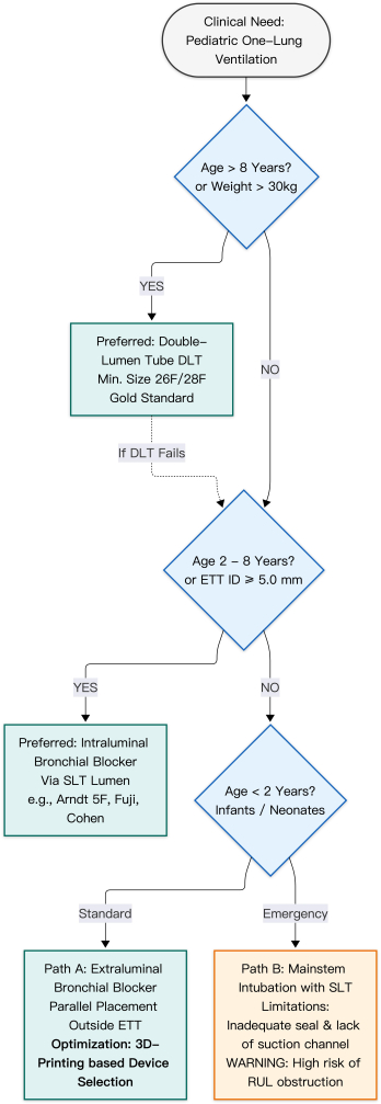
图1儿科肺隔离装置选择的决策算法 该流程图展示了一种基于患者年龄、体重和气道解剖结构的逐步临床决策策略。对于年龄较大的儿童(>8岁),双腔管(DLTs)是肺隔离的金标准。对于2至8岁儿童(或气管内导管(ETT)内径(ID)≥5.0 mm者),首选管腔内支气管阻塞器 通过 单腔管。适用于婴儿(<2年),可选方案有限。路径A表示腔外阻塞器置入,其中以3D打印装置选择作为优化策略,以确保精确的解剖适配。路径B代表传统的主支气管插管,需注意右上叶(RUL)阻塞的高风险。尽管基于年龄的分类提供了通用框架,但装置的选择更准确地依据气道尺寸,特别是气管导管(ETT)的内径(ID)以及儿科患者中狭窄的解剖“安全范围”。缩写:ETT,气管导管;ID,内径;RUL,右上叶;DLT,双腔导管。 请点击此处以查看此图的放大版本。
支气管阻塞器
支气管阻塞器(BBs)是6岁以下儿童的首选替代方案,可实现有效的肺隔离,同时减少气道损伤12。传统装置包括Fogarty取栓导管和Arndt阻塞器。Fogarty导管缺乏用于肺萎陷或持续气道正压(CPAP)的中央腔道22,且因其高压气囊存在导致支气管损伤的风险23。相比之下,Arndt阻塞器采用大容量、低压气囊,并配有用于纤维支气管镜(FOB)引导定位的金属导丝环2,适用于小儿胸科手术24。然而,由于尺寸和设计上的局限性,这两种装置均不理想,凸显了开发适用于不同年龄段、具有超薄膜结构的专用装置的必要性25。
较新型的器械包括5F Fuji封堵器、Univent导管和EZ-Blocker。5F Fuji封堵器具有预成型的远端弯曲结构和更坚硬的轴杆,但缺乏中央腔道26˒27。当其在4.5 mm气管插管内使用时,可能增加气道阻力;在此情况下,建议转换为压力控制通气-容量保证(PCV-VG)模式,以在限制峰值吸气压的同时维持潮气量。若经优化后峰值压力仍持续超过30–35 cmH₂O,则建议改用管外途径,以防止气压伤。
EZ-Blocker 具有“Y”形设计,需要至少 5.5 mm 内径的气管导管,限制了其在年幼儿童中的应用28。Univent 导管将阻塞器集成于气管导管内部,但其外径较大(例如,3.5 mm 内径对应 8 mm 外径),限制了其在较大儿童患者中的使用2。总体而言,目前的器械在儿科应用中仍不够理想。
对于具有解剖学定制设计的精密工程支气管阻塞器(BBs),目前存在明确的需求。Xiaomin D 等人于2022年的一项研究描述了一种基于3D打印技术、专为婴儿和幼儿开发的支气管阻塞器29。尽管该装置已获得专利并正在开展监管注册,但其在临床广泛使用仍面临诸多障碍,包括耗时成本高以及对材料安全性的严格要求。
目前,3D打印最实用的应用在于术前模拟。基于患者特异性CT数据构建的气道模型可用于测试现有器械的适配情况 体外减少术中试错,提高复杂病例的手术安全性。
对于2岁以下的婴儿,较小的气管插管直径通常无法同时容纳支气管镜和封堵器通过。在此类情况下,管外放置——即将封堵器置于气管插管外侧——往往是唯一可行的方法。该技术可保留气道直径以维持通气。技术改进包括对封堵器尖端进行塑形以实现旋转引导30,以及使用导丝辅助放置31。尽管将声门上气道工具(SADs)与支气管封堵器(BBs)联合使用可能减少气道损伤和住院时间,但移位和误吸的风险仍然显著32。
小儿单肺通气的围手术期管理
尽管目前尚无标准化的指南,但越来越多的证据表明,肺保护性通气策略可减少术后肺部并发症3。Zhu 等人于2022年进行的一项临床试验表明,与传统的容量控制通气相比,压力控制-容量保障通气模式(PCV-VG)可降低气道峰压并改善肺顺应性33。
低氧血症是一种常见并发症,在约26%的儿科单肺通气(OLV)病例中发生7。推荐采用优先分级、逐步处理的管理策略。初始干预措施包括通知外科团队、提高吸入氧浓度(FiO₂)以及清除气道分泌物。准确验证器械位置至关重要。当纤维支气管镜检查受限时,床旁超声(POCUS)可提供一种可靠的替代方法。术侧肺滑动征消失并伴有肺搏动征,是判断隔离成功的一个实用指标。呼气末二氧化碳(EtCO₂)的变化,包括突然下降或波形改变,可能提示器械位置不当。表2总结了不同技术下并发症及其相关危险因素。
| 技术 | 常见并发症 | 临床危险因素 / 病因 |
| 单腔管支气管内插管(SLT) | 黏膜损伤;无法吸除术侧肺的分泌物或血液;用于右侧肺隔离时效果欠佳。 | SLT远端管尖易造成组织创伤;术侧气道无法进行吸引;婴儿隆突至右上叶支气管距离极短,易导致右上叶阻塞及肺萎陷失败。 |
| 双腔管(DLT) | 喉部及气管黏膜损伤;声带水肿。 | DLT外径较大且本身较硬,易对狭窄而娇嫩的小儿气道造成损伤。 |
| 管腔内支气管阻塞器(BB 经气管导管内) | 气道阻力增高;低氧血症;气囊位置不当、移位或脱落。 | 与小直径气管导管合用时显著增加通气阻力;体位改变或手术操作可诱发气囊移位或脱落。 |
| 管腔外支气管阻塞器(BB 经气管导管外) | 置入失败;装置位置不当;气囊移位或脱落。 | 小型柔性阻塞器通过声门存在困难;体位改变或手术牵拉可导致装置移位。 |
| 向术侧半胸腔内灌注CO2 | 低氧血症;高碳酸血症;心肌抑制。 | 侧卧位时通气/血流(V/Q)比例失调;功能残气量(FRC)降低;缺乏持续的气囊压力监测。 |
表2:儿科肺隔离技术中常见并发症的总结与比较。 本表对各种隔离方式相关的主要并发症进行了比较性综述,并概述了潜在的解剖学和生理学危险因素,以协助临床医生进行术前风险评估和技术选择。请点击此处下载该表格。
如果在正确体位下低氧血症仍持续存在,可采取的继发性干预措施包括对通气肺施加呼气末正压(PEEP)或对非通气肺施加持续气道正压(CPAP)2˒20。作为最后的抢救措施,可能需要间歇性双肺通气或转为开胸手术34。持续保持警惕,并对恢复双肺通气保持较低的干预阈值,对患者安全至关重要35。
访问受限。请登录或开始试用以查看此内容。
结论
小儿单肺通气(OLV)的管理仍是现代麻醉学中最具挑战性的任务之一,由于生理和解剖结构的独特限制,其与成人实践存在根本性差异。本文综述指出,尽管小儿胸外科手术的需求日益增加,但目前仍缺乏统一的“金标准”肺隔离技术。器械的选择取决于年龄和气道尺寸:双腔管(DLT)在较大儿童中可提供理想的隔离效果,但在婴儿中因解剖限制而无法使用。因此,支气管阻塞器已成为幼儿和学龄儿童的主要选择,而对于2岁以下的婴儿,管外阻塞器置入或单腔管(SLT)使用仍是主要的——尽管并不完善——策略。
这些局限性的临床意义重大。小儿气道的安全范围较窄,尤其是隆突至右上叶支气管的距离较短,使得器械稳定性成为一个关键问题36。即使腔外阻塞器发生轻微的位置移动,也可能导致从手术视野受阻到严重低氧血症甚至完全气道阻塞等一系列并发症。此外,小儿气管黏膜对压力性损伤较为敏感,因此在气囊管理方面必须采取谨慎策略。目前依赖的器械往往并非专为儿科设计(例如血管取栓导管),这常常要求临床医生在实现有效肺隔离的需求与医源性损伤的风险之间进行权衡。
访问受限。请登录或开始试用以查看此内容。
披露
作者声明无利益冲突。
致谢
本工作获得首都卫生发展科研基金(编号:2022-2Z-2098,项目负责人:Xin Zhao)、北京市医院管理中心临床医学发展专项资助(编号:XMLX202145,项目负责人:Xin Zhao)以及北京市科学技术委员会 & 技术委员会概念验证平台项目(编号:2024030119),以及北京市科学技术委员会 & 科技委员会技术转移办公室(编号:20250481320)。
访问受限。请登录或开始试用以查看此内容。