This protocol describes endoscopic ultrasound-guided internal drainage of postoperative pancreatic fistula-related collections after pancreaticoduodenectomy. Intended for experienced interventional endoscopists working in expert multidisciplinary centers, it is supported by a retrospective two-center clinical series.
方法文章
Endoscopic Ultrasound-Guided Drainage of Post-Pancreaticoduodenectomy Pancreatic Fistula-Related Collections
本文内容
摘要
摘要
Clinically relevant postoperative pancreatic fistula (POPF)-related collections following pancreaticoduodenectomy may require invasive drainage. Endoscopic ultrasound (EUS)-guided internal drainage offers a minimally invasive alternative to percutaneous drainage, although well-defined technical approaches in this specific postoperative setting remain limited. This retrospective two-center series included 20 consecutive patients who underwent EUS-guided drainage between March 2019 and February 2026. All procedures were performed using a transgastric approach with double-pigtail plastic stents. The median interval between surgery and drainage was 19.5 days (interquartile range [IQR], 12.7–26.2 days), and the median collection diameter was 70 mm (IQR, 48.7–76.2 mm). One stent was placed in 13 patients (65%) and two stents in 7 patients (35%). Technical success was achieved in 20/20 patients (100%), while clinical success was achieved in 18/20 patients (90%). One patient experienced an early procedure-related death following a perforation, while another failed to achieve clinical resolution despite technically successful drainage and subsequently required surgery. Among the 18 patients who achieved clinical success, recurrence occurred in 3 (16.7%). This article presents a structured step-by-step protocol intended for experienced interventional endoscopists. The findings support the technical feasibility of this approach in selected patients while emphasizing the need for careful patient selection and awareness of potentially severe adverse events.
引言
Postoperative pancreatic fistula (POPF) is a clinically important complication of pancreaticoduodenectomy, occurring in approximately 10%–30% of patients1. According to the 2016 International Study Group of Pancreatic Surgery (ISGPS) definition, clinically relevant grade B and C POPF (occurring in approximately 13%–16% of patients) require therapeutic intervention or a substantial modification of postoperative management, whereas a biochemical leak does not require a clinically relevant change in care2,3,4. Established risk factors include a soft pancreatic texture, a small pancreatic duct, and increased body mass index1,5,6. When associated with fluid collections, infection, persistent external drainage, or organ dysfunction, POPF can substantially prolong hospitalization and increase postoperative morbidity and mortality7,8.
Percutaneous drainage has traditionally been used to manage POPF-related collections9,10. Although effective and widely available, it requires external drainage and may be associated with patient discomfort, catheter-related infection, fluid and electrolyte loss, repeated interventions, and persistent pancreatocutaneous fistula9,11,12. Endoscopic ultrasound (EUS)-guided drainage enables internal drainage into the gastrointestinal tract and may facilitate the removal of an existing percutaneous drain13,14. It therefore represents a minimally invasive alternative in appropriately selected patients14,15,16. Because this approach requires advanced EUS expertise and immediate access to multidisciplinary support, it is intended for experienced interventional endoscopists working in expert pancreatic centers9,13.
Evidence supporting EUS-guided drainage of postoperative collections is increasing, with generally high technical and clinical success rates14,17. However, most available studies are retrospective, include small and heterogeneous populations, and employ variable procedural techniques14,17,18. Published cohorts predominantly involve collections following distal pancreatectomy, whereas patients undergoing pancreaticoduodenectomy remain underrepresented15,17,18. Post-pancreaticoduodenectomy anatomy, particularly after antrectomy, may impair stable endoscopic positioning and limit access to collections located near the pancreatic anastomosis13,18. Consequently, results derived predominantly from distal pancreatectomy cannot be directly extrapolated to this population14,15.
Patient selection depends on both clinical indication and anatomical suitability18,19. EUS-guided drainage may be considered for POPF-related collections associated with infection, persistent systemic inflammation, pain, gastric or intestinal obstruction, ongoing pancreatocutaneous fistula, or failure or unsuitability of conservative or percutaneous management13,18,19,20,21. The collection must be clearly identifiable and accessible from the gastrointestinal lumen, without intervening vessels or bowel loops, and sufficiently contained to permit safe internal drainage18,22,23. This approach may not be appropriate in the presence of hemodynamic instability, suspected pseudoaneurysm, an uncontained or anatomically inaccessible collection, or a clinical condition requiring immediate radiological or surgical source control18,19,22.
The timing of intervention should be individualized according to the patient’s clinical condition, the accessibility and containment of the collection, and the expertise available within the treating institution17,18,24,25. Early drainage may be feasible in selected patients; however, procedures performed within 14 days of surgery require particular caution because insufficiently organized or poorly contained collections may increase the risk of peritoneal leakage17,18,21. The decision to proceed should therefore be made by a multidisciplinary pancreatic team with immediate access to interventional radiology and surgical support22,26,27.
This article presents a structured, step-by-step protocol for EUS-guided internal drainage of POPF-related collections following pancreaticoduodenectomy using double-pigtail plastic stents. The protocol is accompanied by a procedural video and supported by a retrospective two-center clinical series. Its objective is to provide practical guidance on patient selection, procedural preparation, drainage technique, postprocedural management, troubleshooting, and the recognition and prevention of potentially severe adverse events.
Study design and outcome definitions
This retrospective pooled series included all consecutive patients who underwent EUS-guided drainage for a POPF-related collection following pancreaticoduodenectomy at the two participating centers between March 2019 and February 2026. Fourteen patients were treated in Toruń and six at Erasme Hospital. No patient who underwent the procedure during the study period was excluded from the analysis. Center-level denominators for all pancreaticoduodenectomies, clinically relevant POPF cases, and patients managed using alternative drainage strategies were not reliably available from the retrospective records.
Technical success was defined as successful access to the collection or fistulous tract followed by transmural stent placement. Clinical success was defined as clinical improvement with radiological resolution of the collection or fistula, removal of the percutaneous drain when present, and avoidance of surgical intervention. Recurrence was defined as reappearance of a POPF-related collection after initial clinical and radiological resolution during follow-up. Adverse events were classified by timing and reported according to their management and clinical consequences; formal retrospective severity grading was not consistently available.
方案
The retrospective study was conducted in accordance with the institutional guidelines and ethical requirements of both participating centers. The study protocol was approved by the Ethics Committee of Nicolaus Copernicus University in Toruń under reference KB 379/2023/114 and by the Ethics Committee of Erasme Hospital. Clinical data were anonymized and processed in accordance with the General Data Protection Regulation. All procedural footage and clinical images included in this article were fully de-identified, and no identifiable patient information is shown. The consumables and equipment used are listed in the Table of Materials.
1. Patient selection and preprocedural assessment
- Confirm that the patient underwent pancreaticoduodenectomy and developed a clinically relevant postoperative pancreatic fistula (POPF) according to the International Study Group of Pancreatic Surgery criteria. Confirm the presence of an associated collection using contrast-enhanced computed tomography (CT) or magnetic resonance imaging (MRI).
- Confirm an indication for drainage based on infection, persistent fever or systemic inflammation, abdominal pain, gastric or intestinal obstruction, persistent external pancreatic fistula, inadequate control by an existing drain, or failure of conservative management.
- Review the case within a multidisciplinary pancreatic team involving interventional endoscopy, pancreatic surgery, and interventional radiology. Select internal endoscopic drainage when it is considered preferable to prolonged external drainage or surgery and when the collection is anatomically suitable for a transmural approach.
- Review recent contrast-enhanced CT imaging before the procedure. Evaluate the size, configuration, content, degree of containment, and relationship of the collection with the gastric or duodenal wall.
- Identify intervening bowel loops, vascular structures, surgical drains, and percutaneous drains on cross-sectional imaging. Assess the postoperative anatomy, including the presence of antrectomy, the size of the gastric remnant, and the location of the pancreatic anastomosis.
- Select EUS-guided drainage when the collection is clearly identifiable, sufficiently contained, and accessible through a short and stable transmural route. Exclude the procedure when stable apposition cannot be achieved or when an intervening vessel or bowel loop prevents safe puncture.
- Do not proceed in the presence of hemodynamic instability, active bleeding, suspected pseudoaneurysm, uncontrolled coagulopathy, or a condition requiring immediate radiological or surgical source control.
- Review the complete blood count, platelet count, coagulation profile, renal function, inflammatory markers, and available microbiological results. Correct clinically significant coagulation abnormalities and manage antithrombotic therapy according to institutional guidelines for high-risk endoscopic procedures.
- Administer intravenous antibiotics before the procedure. Select empirical coverage according to previous microbiological results, prior antibiotic exposure, the clinical context, and local bacterial ecology.
2. Patient preparation
- Obtain informed consent after explaining the expected benefits, available alternatives, and potential adverse events. Include bleeding, infection, pulmonary aspiration, stent migration or occlusion, peritoneal leakage, perforation, need for radiological or surgical intervention, and death.
- Withhold solid food for at least 6 h before the procedure. Permit clear liquids until 2 h before anesthesia unless gastric retention, intestinal obstruction, or the institutional anesthesia protocol requires a longer fasting period.
- Assess the risk of delayed gastric emptying and aspiration. Aspirate retained gastric contents through a nasogastric tube before the procedure when substantial gastric retention is suspected.
- Use general anesthesia with endotracheal intubation when gastric retention, altered postoperative anatomy, prolonged procedure duration, or increased aspiration risk is present. Use deep sedation only when airway protection and stable procedural conditions can be maintained.
- Position the patient in the left lateral or supine position according to the anesthesia plan and fluoroscopic configuration. Confirm that the selected position permits stable echoendoscope manipulation and unobstructed fluoroscopic visualization.
- Continuously monitor oxygen saturation, electrocardiography, blood pressure, respiratory rate, and end-tidal carbon dioxide. Complete a procedural safety check confirming the target collection, planned access route, antibiotic administration, antithrombotic management, and availability of surgical and radiological support.
3. Equipment preparation
- Prepare a therapeutic linear-array echoendoscope with a working channel compatible with all planned accessories. Prepare fluoroscopic equipment and confirm appropriate endoscopic and fluoroscopic image quality.
- Prepare a 19-G fine-needle aspiration (FNA) needle, diluted water-soluble contrast medium, sterile syringes, and sterile containers for microbiological sampling. Verify the absence of a relevant contrast allergy and review renal function before administering iodinated contrast.
- Prepare one 0.025- or 0.035-inch hydrophilic guidewire compatible with the access needle and subsequent accessories. Prepare a second compatible guidewire when the placement of two plastic stents is anticipated.
- Prepare an over-the-wire cystotome compatible with the guidewire and the echoendoscope working channel. Connect the cystotome to the electrosurgical generator and select Auto Cut mode, effect 4, with a maximum power setting of 100 W.
- Prepare 4 mm and 6 mm wire-guided dilation balloons. Use the smallest balloon diameter that permits atraumatic passage of the planned drainage device.
- Prepare one or two 7 Fr double-pigtail plastic stents. Select a stent length that permits the complete formation of one pigtail within the collection and the other within the gastric lumen without contact with the opposite wall.
- Apply radiation-protection measures before activating fluoroscopy. Use lead protection, collimation, pulsed fluoroscopy, and the lowest dose that provides adequate visualization.
4. EUS-guided drainage
- Introduce the linear-array echoendoscope into the stomach and identify the postoperative anatomy. Examine the entire gastric remnant when antrectomy limits the available space or alters the relationship with the pancreatic anastomosis and adjacent jejunal loop.
- Identify the POPF-related collection and characterize its size, configuration, content, internal debris, septations, and degree of organization. Compare the EUS findings with the preprocedural cross-sectional imaging.
- Confirm close apposition between the collection and the gastrointestinal wall. Select the shortest stable access route that avoids a free peritoneal segment.
- Activate color Doppler and examine the entire intended puncture route. Exclude intervening vessels, venous collaterals, and arterial abnormalities suggestive of a pseudoaneurysm.
- Abandon the selected route when a stable scope position, close wall apposition, or vessel-free trajectory cannot be maintained. Search for an alternative access window or terminate the procedure and discuss another drainage modality.
- Advance the 19-G FNA needle through the working channel and align it coaxially with the selected route. Puncture the collection under continuous EUS guidance while maintaining a stable echoendoscope position.
- Aspirate fluid immediately after entering the collection to confirm needle position. Record whether the aspirated material is clear, turbid, purulent, hemorrhagic, or contains debris, and send a sample for microbiological culture and susceptibility testing. Handle residual specimens and dispose of contaminated procedural materials according to institutional biosafety procedures.
- Inject a limited volume of diluted contrast under fluoroscopic control when the collection is poorly defined, partially collapsed, or difficult to distinguish from an adjacent structure. Confirm cavity opacification and identify communications with additional compartments, an anastomotic leak, or an external drain.
- Stop contrast injection when marked resistance, unexpected extravasation, or opacification of an adjacent bowel loop occurs. Reassess the needle position before continuing.
- Advance a compatible hydrophilic guidewire through the needle and form one or two broad loops within the collection under fluoroscopic control. Avoid forceful advancement against the opposite wall.
- Confirm that the guidewire follows the opacified cavity and does not enter an adjacent bowel loop, vessel, drain tract, or free peritoneal space. Withdraw the needle while maintaining stable guidewire access.
- Advance the cystotome over the guidewire until its distal tip reaches the gastrointestinal wall. Reconfirm the guidewire position and coaxial alignment before activating electrocautery.
- Apply the predetermined cutting current and advance the cystotome through the gastrointestinal wall in a controlled movement. Stop electrocautery immediately after the distal portion enters the collection.
- Avoid prolonged current application, excessive forward pressure, and advancement toward the opposite wall of the collection. Confirm that the distal end of the cystotome remains within the collection using EUS and fluoroscopy, with aspiration or limited contrast injection when necessary.
- Determine whether one or two 7 Fr double-pigtail plastic stents will be placed according to the endoscopist’s assessment and the technical feasibility of placing a second stent.
NOTE: No validated criteria establish the superiority of two stents over a single stent for postoperative pancreatic fistula-related collections. - When two stents are planned, keep the cystotome within the collection and maintain the first guidewire in a stable intracavitary loop. Introduce a second guidewire through the cystotome and form a stable loop within the collection before withdrawing the cystotome.
- Do not introduce a second guidewire alongside an already deployed stent, as this may result in loss of access, submucosal dissection, or creation of a false passage.
- Confirm the independent positions of both guidewires fluoroscopically. Avoid inserting the second guidewire beside an already deployed stent because this can create a false passage or dissect the immature collection wall.
- Advance the first 7 Fr double-pigtail stent over one guidewire. Deploy the distal pigtail within the collection and the proximal pigtail within the gastric lumen under fluoroscopic and endoscopic guidance.
- Confirm spontaneous drainage through or around the first stent. Remove the corresponding guidewire only after confirming complete formation and correct positioning of both pigtails.
- When only one stent is planned, deploy it directly after tract creation if the delivery system passes without resistance. Dilate the tract with a 4 mm balloon only when additional enlargement is required for atraumatic device passage.
- When placing a second stent, maintain the remaining guidewire within the collection and advance a 4 mm balloon across the transmural tract. Position the midpoint of the balloon across the gastrointestinal and collection walls under fluoroscopic control.
- Inflate the balloon gradually until the waist disappears or the second stent delivery system passes without resistance. Stop dilation at 4 mm when adequate device passage is achieved.
- Increase the dilation diameter stepwise to 6 mm only when persistent resistance prevents passage of the second stent delivery system. Avoid rapid, forceful, or unnecessary overdilation, particularly in an early or incompletely organized collection.
- Remove the balloon while maintaining guidewire access. Advance the second 7 Fr double-pigtail stent and deploy one pigtail within the collection and the other within the gastric lumen.
- Confirm the final position of all stents using endoscopy and fluoroscopy. Verify adequate internal drainage and inspect the access site for active bleeding, excessive wall separation, maldeployment, or visible leakage.
- Aspirate residual gastric contents before withdrawing the echoendoscope. Record the access route, collection characteristics, guidewire diameter, cystotome size, electrosurgical settings, balloon diameter, stent dimensions, procedural duration, fluoroscopy time, and any intraprocedural adverse event.
NOTE: Once guidewire access or tract creation has been established, do not terminate the procedure without ensuring adequate drainage or initiating an appropriate rescue strategy.
5. Postprocedural management and follow-up
- Monitor the patient in a post-anesthesia or inpatient setting according to clinical condition and procedural complexity. Assess abdominal pain, temperature, hemodynamic parameters, signs of peritoneal irritation, gastrointestinal bleeding, and output from any remaining external drain.
- Repeat the complete blood count and inflammatory markers according to the clinical course. Continue antibiotic therapy and adapt its spectrum and duration to microbiological results, clinical response, and adequacy of source control.
- Do not perform routine immediate cross-sectional imaging after an uncomplicated procedure in a clinically stable patient. Obtain urgent contrast-enhanced CT when increasing abdominal pain, peritoneal signs, hemodynamic instability, hemoglobin decrease, bleeding, persistent sepsis, or suspected stent maldeployment occurs.
- Include an arterial phase or perform CT angiography when active bleeding or pseudoaneurysm is suspected. Request immediate interventional radiology and surgical assessment when imaging identifies a major vascular or perforation-related complication.
- Perform scheduled contrast-enhanced CT approximately 3–4 weeks after drainage. Evaluate collection reduction or resolution, residual undrained components, stent position, and other postoperative complications.
- Perform earlier imaging when symptoms or inflammatory markers fail to improve. Use MRI with magnetic resonance cholangiopancreatography when iodinated contrast is contraindicated or when a persistent fistula, recurrence, or suspected pancreatic duct disconnection requires ductal assessment.
- Remove an existing percutaneous drain after clinical improvement, cessation or marked reduction of drain output, and radiological confirmation of adequate internal drainage. Avoid removing the external drain when a significant undrained component or ongoing uncontrolled fistula remains.
- Schedule endoscopic reassessment and plastic-stent removal approximately 4–6 weeks after drainage when clinical resolution has been achieved, and imaging demonstrates complete or near-complete resolution. Leave the stent in place and repeat clinical and radiological assessment when a residual collection, persistent fistula, or recurrent leakage remains.
6. Troubleshooting and safety measures
- Reassess the preprocedural CT and examine the collection from alternative endoscopic positions when it cannot be clearly identified. Terminate the procedure when the target cannot be distinguished reliably from an adjacent bowel loop or vascular structure.
- Stop advancing the guidewire when resistance is encountered. Withdraw it slightly, redirect the angled tip under fluoroscopic control, or reposition the needle within the collection.
- Confirm coaxial alignment when the cystotome cannot cross the gastrointestinal wall. Straighten the echoendoscope when possible and apply brief cutting current without excessive forward pressure.
- Stop electrocautery and device advancement immediately when bleeding occurs. Assess the bleeding site using EUS, Doppler, endoscopy, and fluoroscopy, and obtain urgent CT angiography when arterial bleeding is suspected.
- Stop contrast injection and tract dilation when bowel-loop puncture, free extravasation, peritoneal leakage, or perforation is suspected. Initiate intravenous antibiotics, obtain urgent cross-sectional imaging, and request immediate surgical consultation.
- Obtain repeat contrast-enhanced CT when clinical improvement is inadequate. Identify residual compartments, stent occlusion or migration, bleeding, perforation, or another postoperative source of sepsis.
- Select repeat endoscopic drainage, nasocystic irrigation, an additional drainage tract, percutaneous drainage, or surgery after multidisciplinary reassessment. Do not persist with the same drainage strategy when adequate source control has not been achieved.
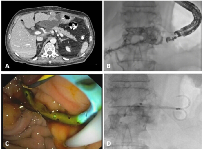
Figure 1: Multimodal imaging and EUS-guided drainage of a postoperative pancreatic fistula-related collection. (A) Pre-procedural contrast-enhanced computed tomography showing a postoperative fluid collection adjacent to the gastric wall. The collection appears non-encapsulated with ill-defined margins. (B) Fluoroscopic opacification of the collection following EUS-guided puncture. (C) Endoscopic view during transmural drainage with visualization of the guidewire and stents within the cavity. (D) Fluoroscopic view after deployment of two double-pigtail plastic stents, ensuring internal drainage. Please click here to view a larger version of this figure.
结果
A total of 20 consecutive patients underwent EUS-guided drainage for postoperative pancreatic fistula-related collections following pancreaticoduodenectomy (Table 1). Fourteen patients were treated in Toruń and six at Erasme Hospital; no patient who underwent the procedure during the study period was excluded. The mean age was 64.9 ± 9.3 years, and 15 patients (75%) were male. Nine patients (45%) had undergone pancreaticoduodenectomy with antrectomy, whereas 11 patients (55%) had undergone pancreaticoduodenectomy without antrectomy.
The median interval between surgery and EUS-guided drainage was 19.5 days (interquartile range [IQR], 12.75–26.25 days), and the median collection diameter was 70 mm (IQR, 48.75–76.25 mm). At the time of drainage, the median C-reactive protein level was 163 mg/L. Fourteen patients (70%) fulfilled the criteria for systemic inflammatory response syndrome, and eight patients (40%) had a cutaneous fistula without an indwelling percutaneous drain.
All procedures were performed using a transgastric EUS-guided approach (Figure 1). Technical success, defined as successful access to the collection or fistulous tract followed by transmural stent placement, was achieved in all 20 patients (100%). Double-pigtail plastic stents were deployed in all patients. One stent was placed in 13 patients (65%), and two stents were placed in seven patients (35%). Balloon dilation of the transmural tract was performed in seven patients (35%). Clinical success was achieved in 18 of the 20 patients (90%). Clinical success was defined as resolution of the postoperative collection or fistula, accompanied by clinical improvement, removal of the percutaneous drain when present, and avoidance of surgical intervention.
Two patients (10%) were classified as clinical failures. One patient experienced an intraprocedural perforation requiring emergency surgical intervention. Despite postoperative intensive care management, the patient died on postprocedural day 2 from multiple-organ failure in the setting of septic shock. The second patient developed intraprocedural bleeding that was successfully controlled endoscopically but subsequently underwent surgical intervention and was therefore classified as a clinical failure.
Recurrence was assessed among the 18 patients who achieved clinical success and was defined as reappearance of a POPF-related collection after initial clinical and radiological resolution. During a median follow-up of 19.5 months (IQR, 15.5–25.5 months), recurrence occurred in three patients (3/18, 16.7%).
Overall, two intraprocedural adverse events occurred among the 20 patients (10%): one perforation and one bleeding event. Both required subsequent surgery. The bleeding was controlled endoscopically during the procedure. The perforation required emergency surgery and resulted in procedure-related death on postprocedural day 2. Formal retrospective severity grading was not consistently available; therefore, adverse events are reported according to timing, management, and clinical consequences. No stent migration, stent occlusion, procedure-related infection, or other procedure-related adverse event was recorded. Clinical outcomes and adverse events are summarized in Table 1, and the complete patient flow is presented in Figure 2.
| Characteristic | Value |
| Patients | |
| Total patients, n | 20 |
| Age, years (mean ± SD) | 64.9 ± 9.3 |
| Male sex, n (%) | 15 (75) |
| Surgical data | |
| Pancreaticoduodenectomy with antrectomy, n (%) | 9 (45) |
| Pancreaticoduodenectomy without antrectomy, n (%) | 11 (55) |
| Collection characteristics | |
| Collection size, mm [IQR] | 70 [48.75–76.25] |
| Cutaneous fistula without an indwelling drain, n (%) | 8 (40) |
| Pre-drainage clinical and biological status | |
| Systemic inflammatory response syndrome (SIRS), n (%) | 14 (70) |
| C-reactive protein, mg/L [IQR] | 163 [123.75–298.75] |
| Procedural details | |
| Transgastric approach, n (%) | 20 (100) |
| Balloon dilation performed, n (%) | 7 (35) |
| One double-pigtail plastic stent, n (%) | 13 (65) |
| Two double-pigtail plastic stents, n (%) | 7 (35) |
| Clinical outcomes | |
| Technical success, n/N (%) | 20/20 (100) |
| Clinical success, n/N (%) | 18/20 (90) |
| Clinical failure, n/N (%) | 2/20 (10) |
| Need for surgical intervention, n/N (%) | 2/20 (10) |
| Recurrence among patients with clinical success, n/N (%) | 3/18 (16.7) |
| Follow-up, months [IQR] | 19.5 [15.5–25.5] |
| Procedure-related adverse events | |
| Any intraprocedural adverse event, n/N (%) | 2/20 (10) |
| Bleeding controlled endoscopically, n/N (%) | 1/20 (5) |
| Perforation, n/N (%) | 1/20 (5) |
| Stent migration, n/N (%) | 0/20 (0) |
| Stent occlusion, n/N (%) | 0/20 (0) |
| Procedure-related infection, n/N (%) | 0/20 (0) |
| Procedure-related death, n/N (%) | 1/20 (5) |
Table 1: Baseline characteristics, procedural details, and outcomes of patients undergoing EUS-guided drainage for postoperative pancreatic fistula-related collections.

Figure 2: Patient flow and clinical outcomes. The pooled cohort comprised 14 patients treated in Toruń and six treated at Erasme Hospital. All 20 consecutive patients who underwent EUS-guided transgastric drainage during the study period were included, with no exclusions. Technical success was achieved in all 20 patients. Clinical success was achieved in 18 patients, including 15 without recurrence and three with recurrence. Two patients were classified as clinical failures: one experienced a perforation requiring emergency surgery and died on postprocedural day 2, and one experienced bleeding controlled endoscopically followed by surgical intervention. Median follow-up among patients with clinical success was 19.5 months (IQR, 15.5–25.5 months). Please click here to view a larger version of this figure.
讨论
Endoscopic ultrasound (EUS)-guided drainage of postoperative pancreatic fistula (POPF)-related collections relies on a sequence of critical technical steps that must be performed in a controlled and reproducible manner18,19. The present protocol emphasizes careful collection targeting, guidewire stabilization, electrocautery-assisted tract creation, and controlled deployment of double-pigtail plastic stents. In this series, technical success was achieved in all 20 patients, whereas clinical success was achieved in 18 patients (90%). Two intraprocedural adverse events occurred, including one bleeding event and one fatal perforation. These findings demonstrate the technical feasibility of the protocol while emphasizing that successful stent deployment does not eliminate the risk of severe adverse events or subsequent clinical failure.
Accurate EUS identification of the collection and assessment of its relationship with adjacent vessels and bowel loops are essential, particularly after pancreaticoduodenectomy, when the anatomy is altered, and collections may be poorly encapsulated or have ill-defined margins18,28. Doppler imaging should be systematically used to identify intervening vessels and exclude a coexisting pseudoaneurysm before puncture10,28. If no safe access window is identified because of interposed vessels or bowel loops, the puncture site should be reassessed or the procedure postponed rather than attempted through an unsafe trajectory. After the puncture, the guidewire should be advanced sufficiently into the collection to create stable loops before device exchange. When resistance is encountered during cystotome advancement or tract dilation, forceful progression should be avoided; the guidewire position and puncture axis should be reassessed, and dilation should be limited to the diameter required for stent placement. If bleeding occurs, the procedure should be paused, and immediate endoscopic hemostasis attempted, with interventional radiology and surgery available when endoscopic control is insufficient. Although the precise mechanism of the fatal perforation could not be attributed retrospectively to a single technical step, this event reinforces the importance of maintaining guidewire access, avoiding forceful device advancement, and performing the procedure within an experienced multidisciplinary setting.
The technique can be adapted to the size, configuration, contents, and accessibility of the collection. One double-pigtail plastic stent may provide adequate drainage for a small, predominantly fluid collection, whereas two stents may be considered for larger, complex, or infected collections containing thick fluid22,29,30. Placement of a second guidewire should not be regarded as a mandatory dual-guidewire strategy but as an optional aid, at the operator’s discretion, when placement of a second stent is planned. In most patients, drainage is achieved through direct access to the collection. However, when the collection has partially resolved or is no longer clearly visible, contrast injection may delineate a persistent fistulous tract and facilitate targeted internal drainage. These adaptations allow the procedure to be tailored to the postoperative anatomy without imposing a single drainage configuration on all patients.
The choice between double-pigtail plastic stents and lumen-apposing metal stents (LAMS) remains debated. Most comparative evidence concerns pancreatic fluid collections resulting from acute pancreatitis, particularly walled-off necrosis, and should therefore be extrapolated cautiously to POPF-related collections. LAMS provide a larger drainage lumen and facilitate direct endoscopic necrosectomy, whereas plastic stents have a lower cost and may be sufficient for predominantly fluid postoperative collections with limited solid debris31,32. The PROMETHEUS randomized trial found no significant difference in short- or long-term clinical success between LAMS and plastic stents for walled-off necrosis33. Moreover, a nationwide Japanese study associated LAMS use with an increased risk of bleeding and higher healthcare costs without improvement in rescue surgery, mortality, or length of stay34. In the present cohort, double-pigtail plastic stents achieved technical success in all patients and clinical success in 90%, supporting their feasibility for this specific indication without establishing superiority over metal stents.
The median interval between surgery and drainage was 19.5 days, reflecting a relatively early intervention strategy at the cohort level. A meta-analysis of six retrospective studies reported 100% technical success and clinical success rates of 90%–97% for both early and delayed EUS-guided drainage, without a significant increase in adverse events with early intervention17. However, these studies predominantly included patients following distal pancreatectomy, and their findings cannot be directly generalized to patients with altered anatomy after pancreaticoduodenectomy. The fatal perforation in the present series further argues against interpreting early drainage as uniformly safe. Early EUS-guided drainage may instead be considered in selected symptomatic patients when a secure access route is present, and the expected benefit outweighs the procedural risk. Compared with percutaneous drainage, published meta-analyses and comparative cohort studies suggest that EUS-guided internal drainage may provide higher clinical success, lower recurrence, and fewer reinterventions while avoiding prolonged external drainage14,16,35,36. Nevertheless, these comparisons are primarily based on retrospective and heterogeneous studies and do not establish EUS-guided drainage as universally preferable.
This study has several limitations. It was retrospective, included only 20 patients from two expert European centers, and lacked a contemporaneous percutaneous or surgical control group. Although all consecutive patients selected for EUS-guided drainage were included, the cohort did not encompass all patients with POPF managed by alternative strategies at both institutions, introducing potential selection bias. Center-level denominator data regarding the total number of pancreaticoduodenectomies, clinically relevant POPF cases, and patients managed using alternative drainage strategies were not reliably available. Consequently, the proportion of potentially eligible patients undergoing EUS-guided drainage could not be determined. Time to complete resolution and formal adverse-event severity grading were not consistently documented in the retrospective records, although the timing, management, and clinical consequences of the recorded adverse events are reported. Changes in operator experience, equipment, and procedural strategy during the study period may also have influenced the outcomes. These limitations restrict the generalizability of the findings. The proposed protocol should therefore be considered a reproducible and adaptable framework for experienced interventional endoscopists working in expert multidisciplinary centers rather than a universally applicable treatment pathway. Prospective comparative studies are required to refine patient selection, determine the optimal timing of drainage, and compare plastic stents, LAMS, and percutaneous approaches in this specific postoperative population.
披露
The authors declare no conflicts of interest.
材料
| 姓名 | 公司 | 目录编号 | 评论 |
|---|---|---|---|
| Compass BDS Biliary Stent (7 Fr, 5 cm) | Cook Medical | CBSBO-7-5 / G58988 | Plastic stent used for internal drainage with radiopaque markers |
| Cystotome (10 Fr) | Cook Medical | CST-10 / G30550 | Electrocautery-enhanced device for tract creation during EUS-guided drainage |
| Electrosurgical generator | ERBE | VIO 3 | Provides cutting current for cystotome use |
| Expect Endoscopic Ultrasound Aspiration Needle (19G) | Boston Scientific | M00550006 | 19G EUS needle used for puncture and identification of the collection |
| Fluoroscopy unit (Artis zee with PURE) | Siemens Healthineers | — | Advanced angiography system providing high-resolution fluoroscopic imaging with dose optimization |
| Hurricane RX Biliary Balloon Dilation Catheter (4 mm × 4 cm) | Boston Scientific | M00545900 | Balloon dilator used to enlarge the tract between the digestive lumen and the collection |
| Hurricane RX Biliary Balloon Dilation Catheter (6 mm × 4 cm) | Boston Scientific | M00545920 | Balloon dilator used to enlarge the tract between the digestive lumen and the collection |
| Jagwire Angle Tip Guidewire (0.035″, 450 cm) | Boston Scientific | M00556590 | Hydrophilic guidewire facilitating stable access and device exchange |
| Linear echoendoscope (GF-UCT180) | Olympus | GF-UCT180 | Large-channel linear-array echoendoscope used for EUS-guided interventions |
| Syringe (10–20 mL) | — | — | Used for aspiration and contrast injection |
| Ultrasound processor (Arietta 850) | Hitachi Aloka | Arietta 850 | Ultrasound platform for real-time EUS imaging |
| Ultravist 300 (iopromide) contrast medium | Bayer | — | Non-ionic iodinated contrast agent used for fluoroscopic visualization during EUS-guided procedures |
参考文献
- Lai M, et al. Fibrin sealants for the prevention of postoperative pancreatic fistula following pancreatic surgery. Cochrane Database Syst Rev. 2023;6:CD009621.
- Roh YH, et al. Preoperative CT anthropometric measurements and pancreatic pathology increase risk for postoperative pancreatic fistula in patients following pancreaticoduodenectomy. PLoS One. 2020;15:e0243515.
- Bassi C, et al. The 2016 update of the International Study Group (ISGPS) definition and grading of postoperative pancreatic fistula: 11 years after. Surgery. 2017;161:584-591.
- Maegawa FB, et al. Postoperative pancreatic fistula after pancreaticoduodenectomy: a contemporary analysis of a large national database. Am J Surg. 2025;248:116535.
- Zhang B, et al. Risk factors of clinically relevant postoperative pancreatic fistula after pancreaticoduodenectomy: a systematic review and meta-analysis. Medicine (Baltimore). 2022;101(26):e29757.
- Hu BY, et al. Risk factors for postoperative pancreatic fistula: analysis of 539 successive cases of pancreaticoduodenectomy. World J Gastroenterol. 2016;22(34):7797-7805.
- Maeda K, et al. Optimal management of peripancreatic fluid collection with postoperative pancreatic fistula after distal pancreatectomy: significance of computed tomography values for predicting fluid infection. PLoS One. 2021;16(11):e0259701.
- Guarneri G, et al. Management of clinically relevant postoperative pancreatic fistula-related fluid collections after distal pancreatectomy. Surg Endosc. 2023;37:5623-5634.
- Trieu JA, et al. The use of endoscopic ultrasound in the management of post-surgical and pancreatic fluid collections. Best Pract Res Clin Gastroenterol. 2022;60-61:101807.
- Elmunzer BJ, et al. Endoscopic drainage of pancreatic fluid collections. Clin Gastroenterol Hepatol. 2018;16(12):1851-1863.e3.
- Baron TH, et al. American Gastroenterological Association clinical practice update: management of pancreatic necrosis. Gastroenterology. 2020;158(1):67-75.e1.
- Nealon WH, et al. Surgical management of complications associated with percutaneous and/or endoscopic management of pseudocyst of the pancreas. Ann Surg. 2005;241(6):948-957; discussion 957-960.
- Jürgensen C, et al. EUS-guided drainage in the management of postoperative pancreatic leaks and fistulas (with video). Gastrointest Endosc. 2019;89(2):311-319.e1.
- Ramouz A, et al. Systematic review and meta-analysis of endoscopic ultrasound drainage for the management of fluid collections after pancreas surgery. Surg Endosc. 2022;36(6):3708-3720.
- Fogwe DT, et al. Endoscopic ultrasound-guided versus percutaneous drainage for the management of postoperative fluid collections after distal pancreatectomy. Surg Endosc. 2023;37(9):6922-6929.
- Khizar H, et al. Efficacy and safety of endoscopic drainage versus percutaneous drainage for pancreatic fluid collection: a systematic review and meta-analysis. Ann Med. 2023;55(1):2213898.
- Mukai T, et al. Early versus delayed EUS-guided drainage for postoperative pancreatic fluid collections: a systematic review and meta-analysis. Surg Endosc. 2024;38:47-55.
- Raza MH, et al. EUS-guided drainage of symptomatic postoperative fluid collections: a retrospective cohort study evaluating timing of intervention and clinical outcomes. Surg Endosc. Published online June 2, 2026. doi:10.1007/s00464-026-12857-8.
- Terrin M, et al. First-intention EUS-guided transluminal drainage with LAMS: an effective and safe method for management of fluid collections after any kind of surgery. Surg Endosc. 2025;39(4):2415-2424.
- Oh D, et al. Effectiveness of early endoscopic ultrasound-guided drainage for postoperative fluid collection. Surg Endosc. 2022;36(1):135-142.
- Storm AC, et al. Acute and early EUS-guided transmural drainage of symptomatic postoperative fluid collections. Gastrointest Endosc. 2020;91(5):1085-1091.e1.
- Muthusamy VR, et al. The role of endoscopy in the diagnosis and treatment of inflammatory pancreatic fluid collections. Gastrointest Endosc. 2016;83(3):481-488. doi:10.1016/j.gie.2015.11.027.
- Mishra G, et al. Quality indicators for EUS. Am J Gastroenterol. 2025;120(5):973-992.
- Ramai D, et al. Early (<4 weeks) versus standard (≥4 weeks) endoscopic drainage of pancreatic walled-off fluid collections: a systematic review and meta-analysis. Gastrointest Endosc. 2023;97(3):415-421.e5.
- Calcagno P, et al. Endoscopic management of postoperative pancreatic fistula after pancreaticoduodenectomy: a single-center retrospective analysis. Surg Endosc. 2025;39(7):4186-4194.
- Seicean A, et al. Endoscopic ultrasound drainage of pancreatic fluid collections: do we know enough about the best approach? Ther Adv Gastroenterol. 2023;16:17562848231180047.
- Rahnemai-Azar AA, et al. Multidisciplinary management of complicated pancreatitis: what every interventional radiologist should know. AJR Am J Roentgenol. 2021;217(4):921-932.
- Lee HS, et al. Clinical outcomes and risk factors for adverse events associated with endoscopic ultrasound-guided drainage of postoperative pancreatic fluid collections following distal pancreatectomy. J Gastroenterol Hepatol. 2026;41(3):1021-1029.
- Vanella G, et al. EUS-guided drainage of post-surgical versus post-pancreatitis collections (the RESPELL study): a prospective comparison of clinical presentations and therapeutic outcomes. Dig Liver Dis. 2026;58(4):493-502.
- Miyagawa K, et al. EUS-TD with multiple plastic stents for postoperative pancreatic fistula: a retrospective evaluation of early postsurgery management. Surg Laparosc Endosc Percutan Tech. 2026;36(2):e1430.
- Holanda EU, et al. Metal stents versus plastic stents for drainage of pancreatic fluid collections: a systematic review and meta-analysis of randomized trials with trial sequential analysis. Surg Endosc. 2025;39(3):1449-1461.
- Boxhoorn L, et al. Acute pancreatitis. Lancet. 2020;396(10252):726-734.
- Gornals JB, et al. Plastic pigtail vs lumen-apposing metal stents for drainage of walled-off necrosis (PROMETHEUS study): an open-label, multicenter randomized trial. Surg Endosc. 2024;38(4):2148-2159.
- Hamada T, et al.; WONDERFUL Study Group in Japan and collaborators. Real-world impact of implementing lumen-apposing metal stents for pancreatic fluid collections: a nationwide Japanese study. Gut. 2025 Sep 23:gutjnl-2025-335067.
- Szakó L, et al. Endoscopic and surgical drainage for pancreatic fluid collections are better than percutaneous drainage: a meta-analysis. Pancreatology. 2020;20(1):132-141.
- Wan J, et al. Comparison of percutaneous vs endoscopic drainage in the management of pancreatic fluid collections: a prospective cohort study. J Gastroenterol Hepatol. 2020;35(12):2170-2175.
重印与许可
