本文综述了DNA甲基化在免疫调节和移植排斥反应中的作用。DNA甲基化调控免疫细胞的发育与分化,调节免疫细胞功能,并可能作为改善移植物存活率的生物标志物和治疗靶点。
综述文章
免疫调控对DNA甲基化的影响及其在同种异体移植物排斥反应中的意义
63 次观看
⸱
2026年9月11日
本文内容
摘要
摘要
移植排斥反应仍是影响移植物长期存活的主要障碍,其发生由免疫应答与分子调控机制之间的复杂相互作用所驱动。DNA甲基化是一种主要的表观遗传修饰,在调控免疫细胞的发育、分化、成熟和功能中发挥重要作用。异常的DNA甲基化可改变免疫相关基因的表达,进而影响免疫应答的性质和强度。在移植过程中,这些改变可能影响参与移植物排斥的T细胞、B细胞及固有免疫细胞。DNA甲基化还参与调控与免疫活化、免疫耐受及炎症反应相关的信号通路。因此,甲基化模式的改变可能影响排斥反应的进展以及移植物功能的维持。本文综述了当前关于DNA甲基化在免疫调控中的研究进展,并探讨其在移植排斥反应中的意义,重点包括胸腺发育、辅助性T细胞和细胞毒性T细胞、调节性T细胞、B细胞、固有免疫细胞以及相关信号通路。同时,本文还探讨了DNA甲基化作为移植领域生物标志物和治疗靶点的潜在价值。深入理解这些表观遗传机制,有助于开发更精确的策略,以评估排斥风险、调节免疫应答,并改善实体器官移植后的长期移植物预后。
引言
免疫学是一门研究免疫系统结构与功能的科学学科,包括免疫应答机制以及在识别自身与非自身成分后维持稳态的过程。该领域还涵盖由免疫功能失调引起的病理过程。免疫系统具有三种基本功能:防御、监视和稳态。这些功能共同作用,防止外来病原体和有害物质的侵入,清除体内的非自身成分,并维持稳定的内部免疫环境。根据免疫应答的特点,免疫可分为先天性免疫和适应性免疫。先天性免疫对外来病原体产生快速、非特异性的反应,而适应性免疫则针对特定抗原产生高度特异性的反应1,2。在免疫应答过程中,外部和内部因素共同调控与免疫功能相关基因的表达。这种调控涉及经典的遗传机制以及表观遗传修饰,形成一个复杂且动态的转录调控网络3。
传统遗传学关注由DNA序列改变引起的基因功能变化,而表观遗传学则研究在不改变DNA序列的情况下,基因表达发生的可逆且可遗传的变化。环境刺激(包括感染、化学物质暴露和药物)以及细胞因子和激素等内源性因素均可改变基因的表观遗传景观。这些变化调控基因表达,最终塑造细胞表型和生物学功能。调控基因表达的主要表观遗传机制包括DNA甲基化、组蛋白修饰、染色质重塑以及非编码RNA介导的调控。在早期胚胎发育过程中,DNA甲基化标记几乎被完全擦除,并在细胞分化过程中重新建立4,5。这些表观遗传修饰的精确清除与重建对于细胞正常发育、分化和成熟至关重要。相反,异常的表观遗传改变可能破坏基因功能,促进疾病的发生和发展6。在T细胞和B细胞的发育与分化过程中,DNA甲基化模式也动态变化7,8。这些发现凸显了表观遗传调控在指导骨髓造血干细胞向多种免疫细胞谱系分化中的关键作用。组蛋白修饰调控单核细胞/巨噬细胞和辅助性T细胞的分化及其功能可塑性9,10,11,12,13,14,并影响包括Toll样受体通路在内的关键信号通路15,16。染色质重塑因子Mi-2beta在胸腺细胞中高表达,可直接调控CD4基因表达和T细胞发育17。非编码RNA也在免疫应答调控中发挥重要作用。例如,干扰素-γ(IFN-γ)在宿主防御胞内细菌感染中起核心作用,而miR-21可通过直接靶向IFN-γ mRNA抑制其产生18。综上所述,这些研究结果强调了表观遗传机制在调控免疫应答中的核心地位。
免疫细胞是免疫功能的核心,也是移植排斥反应的关键驱动因素,而移植排斥反应是移植物存活的主要决定因素19,20,21。尽管已有大量免疫学研究探讨了移植排斥反应的机制,但许多方面仍知之甚少22。越来越多的证据还表明,表观遗传机制在免疫细胞的发育、分化和成熟过程中发挥着关键作用23,24。因此,从表观遗传学角度研究移植排斥反应的分子基础,可能有助于在器官移植后更早地干预和预防移植排斥反应及慢性移植肾病,从而改善移植物的长期存活。在多种表观遗传修饰中,DNA甲基化是调控哺乳动物基因组转录最稳定的机制之一25。基于此,本文综述了DNA甲基化在免疫细胞发育、分化和功能中的作用,以及其在器官移植后急性排斥反应中的参与机制。
综述与观点
1. DNA甲基化与测序技术
DNA甲基化以S-腺苷甲硫氨酸作为甲基供体,由DNA甲基转移酶催化,将甲基共价添加至CpG二核苷酸(胞嘧啶–磷酸–鸟嘌呤)中胞嘧啶的第五位碳原子上,从而将胞嘧啶转化为5-甲基胞嘧啶(5mC)。这种修饰在DNA序列本身之外,为基因表达增加了一层额外的调控机制。通常情况下,甲基化水平较低的区域允许蛋白质结合,并支持有利于基因活跃转录的染色质结构26。相反,甲基化的DNA区域可通过招募甲基化CpG结合结构域(MBD)蛋白、促进组蛋白修饰和染色质重塑复合物的形成,或直接阻断转录因子及其他DNA结合蛋白的结合,从而抑制基因的转录与表达27,28,29。然而,DNA甲基化也可能与基因激活相关,尤其是在基因体区域,其甲基化水平与基因表达呈正相关。尽管这种关联相对少见,但基因体甲基化在普遍表达的基因中较为常见30,31。
DNA甲基化的生物学功能必须在其基因组背景下进行解读。CpG二核苷酸常聚集在称为CpG岛的区域,CpG岛被定义为长度超过200个碱基对(bp)、G+C含量至少为50%、且观测值/期望值(Obs/Exp)比至少为0.6的DNA片段32。在哺乳动物基因组中,CpG二核苷酸相对稀少,仅占基因组的约1%。大约60%的人类基因启动子与CpG岛相关,这些区域在正常细胞中通常处于非甲基化状态。仅有一小部分(约6%)在早期发育或组织分化过程中发生甲基化33,34。CpG岸是指位于CpG岛上下游2,000 bp范围内的区域,其甲基化状态与转录沉默密切相关。在甲基化重编程过程中,约70%的差异甲基化区域出现在CpG岸中23,35。此外,大多数组织特异性甲基化发生在CpG岸,而非CpG岛内34,35。CpG架位于CpG岸上下游2,000 bp范围内,而“开放海域”一词则指除CpG岛、岸和架之外的所有其余基因组区域。
DNA甲基化主要通过两种过程发生:维持性甲基化和从头甲基化。维持性甲基化主要发生在半保留DNA复制过程中,此时维持性甲基转移酶将已有的甲基化模式复制到子代链上。DNMT1是主要的维持性甲基转移酶,它优先靶向半甲基化的DNA,是细胞中最丰富的甲基转移酶,并在细胞周期的S期被活跃转录36。相比之下,从头甲基化不依赖于DNA复制,通过靶向先前未甲基化的位点来建立新的甲基化模式。DNMT3A和DNMT3B作为主要的从头甲基转移酶发挥作用。它们在胚胎干细胞中高表达,在胚胎发育过程中建立甲基化模式方面起着关键作用,但随着细胞分化,其表达水平下降27。DNMT3家族还包括DNMT3L,该蛋白缺乏催化活性,但在建立母源基因组印记中至关重要37。DNMT3L能够增强DNMT3A和DNMT3B的活性,从而提高DNA甲基化的效率38。在整个基因组中,许多甲基化的胞嘧啶会经历DNA去甲基化,尤其是在基因体区域。去甲基化通过主动和被动机制发生。主动去甲基化涉及TET酶将5mC氧化为其氧化衍生物的过程39。而被动去甲基化则是在DNA复制过程中,由于缺乏维持性甲基转移酶的活性,导致甲基化胞嘧啶逐渐被稀释的结果40。
由于DNA甲基化在调控基因转录和表达中起着核心作用,过去十年中它已成为研究的主要焦点之一,部分得益于甲基化检测技术的进步。根据样本预处理方法的不同,DNA甲基化检测技术大致可分为三类41。第一种方法依赖于甲基化敏感的限制性内切酶,这些酶选择性地无法切割甲基化的DNA区域,从而根据甲基化状态产生不同大小的DNA片段。第二种方法利用能够特异性识别5-甲基胞嘧啶的甲基化结合蛋白或抗体来富集甲基化的DNA片段。第三种方法使用亚硫酸氢盐处理,将未甲基化的胞嘧啶转化为尿嘧啶,而甲基化的胞嘧啶则保持不变,从而实现甲基化与未甲基化位点的精确区分。目前,亚硫酸氢盐转化联合高通量测序是分析DNA甲基化最广泛使用的技术策略。表1总结了主要的甲基化检测方法,包括其原理、优势、局限性以及在移植研究中的潜在应用。DNA甲基化检测技术的持续进步不断拓展表观遗传学研究的技术工具箱,并推动新型表观遗传治疗策略的发展41,42,43,44,45。
早期胸腺细胞发育与分化的甲基化特征
移植排斥涉及复杂的机制,包括细胞介导和抗体介导的免疫反应。目前移植免疫学的研究证据表明,T细胞介导的免疫在移植物排斥中起核心作用46,47。胸腺是T细胞发育、分化和成熟的首要器官48。在胸腺细胞发育过程中,即T细胞的前体阶段,DNA甲基化模式经历持续而动态的重塑。这些变化包括调控T细胞转录的基因组区域发生去甲基化和甲基化,但这些过程并非完全平衡地进行。在早期发育阶段,编码关键T细胞受体(TCR)组分以及重要调控因子(包括CD3、RUNX3、RORC、RAG1和LCK)的基因主要经历去甲基化7。更广泛而言,在免疫细胞分化过程中,DNA去甲基化的发生频率高于甲基化49,50。相比之下,从头甲基化相对有限,主要发生在胸腺细胞发育的早期定型阶段7。从头甲基化对于沉默与干细胞多能性相关的基因也至关重要,从而促进细胞谱系定型和分化7,51。胸腺细胞DNA甲基化的另一个重要特征是,大多数甲基化改变一旦在分化过程中建立,便保持稳定且基本不可逆。然而,某些基因(如RAG基因)表现出动态调控:它们在免疫细胞早期发育阶段发生去甲基化,随后在后期阶段重新甲基化7。
3. 免疫细胞分化与功能中的DNA甲基化
DNA甲基化在调控多种免疫细胞群体的分化、功能和可塑性中发挥关键作用,从而影响移植免疫应答的结果。
- 适应性免疫细胞
- 辅助性T细胞(CD4+ T细胞
CD4+ T细胞在急性器官移植排斥反应中起主要效应细胞的作用52,53. 初始T细胞(Tn)分化为多种CD4+ 通过协调的遗传、表观遗传及其他调控机制,T细胞亚群在受到特定信号刺激后,其初始T细胞的表观遗传景观可发生改变,从而影响基因的转录与表达。这些变化最终可能影响移植后免疫排斥反应的类型和强度。T细胞活化后, IL2 基因经历快速去甲基化,并保持在非甲基化状态。这种表观遗传学改变对于增强支持T细胞功能的基因转录与表达至关重要54.
DNA甲基化在指导初始T细胞分化为Th1和Th2亚群的过程中也起着关键作用55,56. 在初始CD4+ T细胞, IFNG 基因高度甲基化;在Th1细胞分化过程中特异性去甲基化,而在Th2细胞中仍保持高度甲基化57相比之下, IL4 基因在初始T细胞和Th1细胞中高度甲基化,但在Th2细胞中发生部分去甲基化58外部因素可通过调节DNA甲基转移酶(DNMTs)的活性进一步调控这些甲基化模式。例如,维生素D缺乏会增加DNMT活性,并促进基因组DNA的高甲基化 IFNG 基因,而补充维生素D可逆转这些变化59生物衰老也会影响DNA甲基化模式。包括Yu在内的研究表明,DNMT3a mRNA的表达以及DNMT3a基因的甲基化状态 IL4 基因启动子的甲基化随年龄增加,而甲基化 IFNG 基因启动子活性降低60这些发现表明,与年龄相关的DNA甲基化改变可能改变Th1与Th2细胞反应之间的平衡。
初始T细胞的分化还涉及另外两种重要的CD4+ T细胞亚群:Th17细胞和调节性T(Treg)细胞61,62DNA甲基化严格调控这一过程。Th17特异性转录因子ROR的表达γt 细胞和细胞因子 IL-17 的表达依赖于其相应基因启动子区域的甲基化状态63,64,65移植组织中的 Th17 细胞可加重移植排斥反应并促进血管损伤66DNA甲基化还影响Th17细胞的功能可塑性。在慢性炎症条件下, IFNG Th17细胞中的基因保持去甲基化状态,从而使这些细胞能够产生IFN-γ因此,Th17细胞可获得Th1和Th17表型的双重特征67此外, 体外 研究表明,特定的刺激条件可诱导Th17细胞分化为具有强效免疫抑制活性的功能性Treg细胞68.
相比之下,调节性T细胞(Treg细胞)在维持免疫耐受以及保持移植物内稳态方面发挥着关键作用69这些细胞可在胸腺中产生,或在TGF-β的影响下于外周分化。β;无论其来源如何,Treg 细胞均持续表达转录因子 Foxp370表观遗传调控,尤其是DNA甲基化,在调控与Treg功能相关基因的表达中起着关键作用,包括 Foxp3 和 Ctla4从而直接调控其转录活性71,72,73,74在胸腺Treg细胞发育过程中 Foxp3 基因经历渐进性去甲基化。这种去甲基化状态在细胞迁移至外周组织后仍然持续,并需要适当强度的持续TCR信号传导。62,75,76,77在缺乏 DNMT1 的情况下,TCR 刺激能够有效诱导胸腺和外周组织中 Foxp3 阴性 T 细胞表达 Foxp3。然而,如果 TCR 刺激未能诱导充分的表观遗传重塑,细胞则无法维持稳定的 Foxp3 表达,而稳定的 Foxp3 表达对于维持 Treg 表型至关重要62,75,78Foxp3 的稳定表达主要由顺式调控元件(尤其是保守的非编码序列,CNS)上的表观遗传修饰所调控。71,79,80其中,调节性T细胞特异性去甲基化区域(TSDR)的甲基化状态可作为可靠标志,用于区分稳定的与不稳定的调节性T细胞群体71,81,82,83,84,85,86在天然存在的初始调节性T细胞(nTregs)中,TSDR保持非甲基化状态,从而允许转录因子(如Stat5、NFAT、Runx1和CREB)的募集,这些因子有助于Foxp3的稳定表达。相反,在诱导型调节性T细胞(iTregs)中,TSDR通常处于甲基化状态,导致Foxp3表达的稳定性降低。72,74,87,88,89此外,TET1或TET2等去甲基化酶的缺失会导致Foxp3基因座的高甲基化,从而损害调节性T细胞(Treg)的功能,并促进自身免疫性疾病的发生。90临床研究进一步表明,肾移植活检样本中 Foxp3 去甲基化水平较高与亚临床排斥反应患者移植物预后改善相关82,84.
调节性T细胞(Treg细胞)正越来越多地被研究用于预防同种异体移植物排斥反应及促进免疫耐受91移植物内必须有足够数量的调节性T细胞(Treg细胞)持续存在,以有效抑制驱动移植排斥反应的效应T细胞,从而维持免疫耐受。92然而,调节性T细胞(Treg细胞)表现出一定程度的表型不稳定性。在特定条件下,它们可能发生表观遗传学改变,包括FOXP3基因增强子区域的去甲基化 RORC 基因,导致促炎性细胞因子IL-17的表达93,94这种转变凸显了Treg细胞的可塑性,并对其治疗应用提出了挑战。实验研究进一步表明,表观遗传调控可增强Treg细胞功能。例如,Guo 等 在动物模型中表明,将DNA甲基化抑制剂与IL-35联合使用可增加Foxp3表达,促进调节性T细胞(Treg)分化,并延长移植物存活时间95尽管取得了这些进展,人们对调节性T细胞相关机制的理解仍不完善。未来的研究应明确促进调节性T细胞分化并稳定其表型的条件,旨在将这些发现应用于免疫介导性疾病的治疗。 - 细胞毒性T细胞(CD8+ T细胞
活化后,初始CD8+ T细胞可分化为称为细胞毒性T淋巴细胞(CTLs)的效应细胞。这些细胞通常分为两个亚群:产生IFN-γ,以及产生IL-4和IL-5的Tc2细胞96临床研究表明,CD69阳性细胞的比例+/CD8+ 心脏移植患者外周血中的T细胞与严重排斥反应相关。此外,CD8+ 心肌组织中的T细胞表达CD69+ 活化表型并表现出穿孔素介导的细胞毒性活性97,98. 实验证据进一步支持CD8在其中的作用+ 移植排斥反应中的T细胞。Jones 等 证明了在通过胸腺切除和抗CD4/CD8处理获得的T细胞耗竭小鼠中,转移TCR抗原特异性CD8+ 仅T细胞就足以诱导完全错配的同种异体心脏移植物的排斥反应99。这些发现凸显了CD8的至关重要的作用+ T细胞介导的免疫性移植物排斥反应。DNA甲基化也调控CD8+ T细胞发育与功能。在缺乏DNMT1的情况下,胸腺细胞的存活受到损害100此外,成熟CD8细胞中DNMT3a的缺失+ T细胞促进记忆细胞分化,同时减少终末效应细胞分化。这一效应部分由T细胞因子1(Tcf1)表达上调介导,因为DNMT3a靶向Tcf7基因的启动子区域,并在早期效应CD8⁺ T细胞中诱导从头甲基化。+ T细胞101此外,在急性病毒感染的小鼠模型中,T细胞内TET2的条件性缺失可增强记忆性CD8⁺ T细胞的形成。+ T细胞102这些研究结果共同表明,DNA甲基化在CD8⁺ T细胞的发育、分化及功能反应中起着关键的调控作用。+ T细胞 - B细胞
T细胞在移植排斥反应中起核心作用,但B细胞也参与细胞免疫和体液免疫排斥反应103,104体液排斥主要由B淋巴细胞介导。B细胞活化后分化为浆细胞,产生并分泌大量特异性抗体,这些抗体可引发移植物的炎症性损伤105DNA甲基化进一步调控B细胞免疫反应。Lee 等 研究表明,在B细胞早期发育过程中发生了广泛的DNA去甲基化,尤其是在基因体区域和非CpG岛区域。相比之下,DNA甲基化在B细胞成熟后期阶段的调控作用则变得较不显著。106此外,Shaknovich 等 发现生发中心B细胞与初始B细胞在DNA甲基化谱上存在显著差异107生发中心B细胞经历去甲基化,这会改变功能基因的表达并影响关键信号通路,包括NF-κB 和 MAPK 通路107,108功能研究表明,DNA甲基化可限制B细胞的活性。新甲基转移酶Dnmt3a和Dnmt3b的缺失(即Dnmt3缺陷)可促进B细胞的发育与成熟,同时增强B细胞的活化,并支持生发中心B细胞及浆细胞的扩增。这些发现表明,新生DNA甲基化通常发挥限制B细胞活化和抑制浆细胞分化的功能。109.
- 辅助性T细胞(CD4+ T细胞
- 先天免疫细胞
实体器官移植后的免疫反应涉及固有免疫与适应性免疫之间的协调相互作用。固有免疫系统包括造血和非造血细胞,如树突状细胞、巨噬细胞、自然杀伤细胞和中性粒细胞,这些细胞均可能参与移植后的免疫排斥反应110.
树突状细胞作为连接固有免疫与适应性免疫的关键调节者,具有促进免疫活化或免疫耐受的能力111,112在单核细胞向树突状细胞分化的过程中,DNA甲基化发生广泛变化,其特征是基因组多个区域的甲基化水平显著降低。这些变化主要发生在对细胞谱系特化和免疫激活至关重要的增强子区域和转录因子结合位点。113,114,115该分化过程的一个显著特征是细胞表面 CD14 的下调和 CD209 的上调。这一变化与 CD209 基因启动子区域 CpG 位点的去甲基化相关 CD209 基因116此外,使用DNA甲基化抑制剂处理可显著上调共刺激分子(如CD40和CD86)的表达,从而增强抗原呈递能力,并促进树突状细胞的功能成熟。这些结果表明,DNA甲基化直接调控树突状细胞的功能与免疫活性117.
单核细胞和巨噬细胞是免疫系统的重要组成部分,因其介导了对异常细胞和入侵病原体的吞噬与清除。大量证据表明,巨噬细胞不仅参与早期急性移植排斥反应中所观察到的炎症性损伤,还促进晚期慢性排斥反应中纤维化的进展118,119M1型巨噬细胞(经典活化)与M2型巨噬细胞(替代性活化)之间的平衡对免疫反应具有关键调控作用。M1型巨噬细胞占主导会促进炎症反应和组织损伤。相反,通过DNA甲基化抑制剂或基因敲除降低DNMT1表达,可减少过氧化物酶体增殖物激活受体位点的甲基化水平 γ1(PPARγ1)启动子。这一改变促进M2型巨噬细胞活化,有助于限制免疫介导的损伤120DNA甲基化模式的差异进一步凸显了表观遗传调控在巨噬细胞功能中的作用。研究发现,在冠状动脉疾病患者与非患者之间,外周血单核细胞/巨噬细胞中特定基因启动子区域的甲基化水平存在显著差异。121此外,单核细胞向巨噬细胞的分化过程涉及转录因子结合位点处甲基化的显著变化,表明关键调控区域的DNA甲基化在巨噬细胞分化中发挥着精确而关键的作用。122.
自然杀伤(NK)细胞在移植排斥反应中发挥效应和调节双重功能。它们既可促进移植物排斥,也可参与免疫耐受的形成。在骨髓造血干细胞向NK细胞分化的过程中,NK细胞相关基因启动子区域的甲基化水平降低 KIR 基因导致KIR表达增加,在免疫调节中发挥重要作用。此外,使用DNA甲基化抑制剂处理可诱导该基因的去甲基化 KIR 基因并促进其在NK细胞中的表达123,124这些发现表明,DNA甲基化是自然杀伤细胞功能的关键调控因子。
中性粒细胞也积极参与移植排斥反应。活化的中性粒细胞释放多种介质,募集并激活抗原呈递细胞,从而启动抗原特异性的T细胞应答,并加剧免疫排斥反应125,126,127中性粒细胞表面表达的锚定蛋白PRV-1在其启动子区域的DNA甲基化调控下,于生理和病理条件下均受到调控128.
4. 关键信号通路中的DNA甲基化
DNA甲基化不仅直接调控功能基因的转录,还可通过调节关键信号通路影响细胞行为。例如,分泌型卷曲相关蛋白(SFRP)基因启动子区域的高甲基化可导致该基因沉默。这种沉默会破坏Wnt/β-catenin信号通路的正常调控,导致其异常激活,从而促进结直肠癌的发生129。类似地,免疫细胞中mTOR信号通路上游负性调控因子的高甲基化会降低其表达水平,引起mTOR过度激活,进而导致糖尿病肾病中的炎症性损伤。采用地西他滨或RNA干扰等干预手段可通过抑制DNMT1来降低这些调控基因的甲基化水平,从而减缓糖尿病肾病的进展130。此外,DNA甲基转移酶抑制剂还可通过调控NF-κB和JAK/STAT等信号通路抑制细胞增殖,进而影响疾病的发生与发展131,132(图1)。
5. 临床意义:免疫抑制药物与DNA甲基化相互作用
标准治疗方案中的免疫抑制剂已被证实会影响免疫细胞的DNA甲基化模式,尽管不同药物和基因组背景下的效应存在差异。针对T细胞中IFNγ启动子甲基化的研究显示,霉酚酸可拮抗T细胞活化所诱导的去甲基化,而他克莫司在该特定位点则未表现出相同作用133。相比之下,西罗莫司已被证明可通过影响mTOR信号通路来调节全基因组甲基化谱型134,而DNA甲基化本身也被证实能够调控mTOR通路,从而影响肾移植后的移植物存活率及急性排斥反应135。此外,糖皮质激素(如地塞米松)可在外周免疫系统中诱导广泛的DNA甲基化重塑。中性粒细胞地塞米松甲基化指数可作为糖皮质激素暴露的敏感药效动力学标志物,并与免疫抑制性免疫特征相关136。抗胸腺细胞球蛋白(ATG)诱导治疗也被报道可改变肾移植受者的DNA甲基组,且基于甲基化的模型在预测感染风险方面优于转录组学谱型137。个体间的表观遗传变异可能影响患者对这些治疗的反应,提示甲基化生物标志物有望用于指导个体化免疫抑制方案的制定136,137。这些药物与表观基因组之间的相互作用值得进一步研究,因其可能对优化长期移植物预后具有重要意义。
6. 挑战与展望:在表观遗传调控与免疫安全性之间取得平衡
尽管DNMT抑制剂(DNMTis)如阿扎胞苷和地西他滨在移植领域具有应用前景,但其存在感染性和致癌性风险。使用这些药物的治疗已与严重的败血症并发症相关138,139,而全身性暴露于低甲基化药物还与继发性急性髓系白血病转化风险升高相关,受影响患者的中位总生存期不足5个月140。除药物相关毒性外,缺乏细胞类型特异性是另一重要挑战。全身性去甲基化可能破坏调节性T细胞(Treg)区室的稳定性,同时激活效应T细胞,从而可能加剧排斥反应141,并可能削弱CD8+记忆T细胞对巨细胞病毒(CMV)等机会性病原体的保护作用101,102。
为了克服这些局限性,新兴策略旨在实现细胞类型和基因座特异性的调控。纳米颗粒平台可将DNMT优先递送至移植物浸润细胞142,143。CRISPR/dCas9表观基因组编辑技术能够靶向去甲基化Foxp3-TSDR,从而在无全局效应的情况下稳定调节性T细胞(Tregs)144,145。基于生物标志物的监测也有助于更安全地实现临床转化146。
结论
提高移植物存活率仍是器官移植研究中的主要目标。移植排斥是一种复杂的免疫学过程,供体器官和受体的表观遗传修饰在此过程中动态相互作用,共同塑造整体的表观遗传景观。受体的免疫系统是移植物排斥的主要驱动因素。表观遗传机制在免疫细胞的发育、分化和功能调控中发挥关键作用,从而影响移植后的排斥反应和移植物存活。此外,免疫抑制药物、感染以及移植后环境等因素进一步加剧了表观遗传调控与免疫反应之间的关系复杂性。这些因素可改变表观遗传模式并调节免疫细胞行为,为移植结局增添了另一层复杂性。因此,有必要进一步研究表观遗传机制与移植排斥之间的关系,以识别新的生物标志物和治疗靶点,并改进器官移植中的诊断方法和治疗策略。
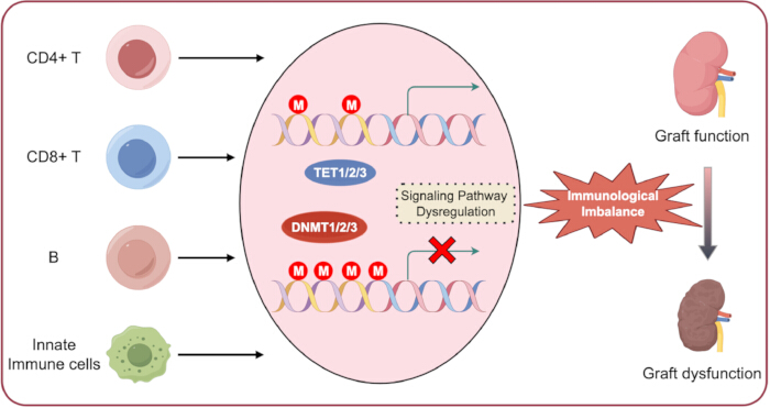
图1.移植免疫中的DNA甲基化。免疫细胞在其整个生命周期中维持着各自独特的内在DNA甲基化模式。 该生理过程的紊乱可导致异常的DNA甲基化以及下游信号通路的失调,最终引起免疫稳态失衡和同种移植物功能障碍。请点击此处查看该图的放大版本。
| 方法 | 原理 | 优点 | 局限性 | 在移植中的应用 |
| MeDIP-seq⁴¹ | 抗-5mC 抗体富集 + NGS | 全基因组分析成本较低;适用于大样本队列 | 分辨率低(~100–300 bp);无法区分 5mC 与 5hmC | 筛查移植受者外周血单个核细胞(PBMCs)中的全基因组甲基化变化 |
| WGBS⁴² | 亚硫酸氢盐转化 + NGS | 单碱基分辨率;全基因组覆盖 | 成本高;需要大量数据量 | 移植物活检样本的全面甲基化组作图;鉴定新的与排斥反应相关的差异甲基化区域(DMRs) |
| RRBS⁴³ | 限制性内切酶富集 + 亚硫酸氢盐转化 + NGS | 成本效益高;对富含 CpG 的区域具有高覆盖率 | 对非 CpG 区域及 CpG 稀疏区域的覆盖有限 | 在移植物组织中进行靶向甲基化组分析,降低测序成本 |
| 甲基化特异性 PCR⁴⁴ | 亚硫酸氢盐转化 + 使用甲基化特异性引物进行 PCR | 高灵敏度;操作简单、快速且成本低 | 仅针对特定基因座;定性或有限的定量分析 | 验证发现队列中的候选 DMRs;临床生物标志物开发 |
| 焦磷酸测序⁴⁵ | 亚硫酸氢盐转化 + 合成测序法 | 可定量;在靶向位点实现单碱基分辨率 | 通量中等;需要预先确定目标区域 | 精确量化特定 CpG 位点的甲基化水平,例如 Treg 细胞中 FOXP3 TSDR 区域 |
表1:主要DNA甲基化检测方法的概述。 本表总结了常用的DNA甲基化检测方法,包括其原理、优势、局限性以及在移植研究中的潜在应用。
披露
作者声明不存在竞争性利益或潜在的利益冲突。
致谢
作者感谢 Figdraw 在图表制备方面提供的帮助。
参考文献
- Güell-Alonso R, Cabrera-Pérez R, Vives J. Immunocompatibility in transplantation: adapting to a changing therapeutic landscape. Front Immunol. 2025;16:1648534.
- Li XQ, Ding RD, Cai JZ. Organ transplantation: current status, challenges, and future prospects. MedComm. 2026;7(1):e70567.
- Medzhitov R, Horng T. Transcriptional control of the inflammatory response. Nat Rev Immunol. 2009;9:692–703.
- Bagci H, Fisher AG. DNA demethylation in pluripotency and reprogramming: the role of tet proteins and cell division. Cell Stem Cell. 2013;13:265–269.
- Gu TP, et al. The role of Tet3 DNA dioxygenase in epigenetic reprogramming by oocytes. Nature. 2011;477:606–610.
- Cedar H, Bergman Y. Programming of DNA methylation patterns. Annu Rev Biochem. 2012;81:97–117.
- Rodriguez RM, et al. Regulation of the transcriptional program by DNA methylation during human αβ T-cell development. Nucleic Acids Res. 2015;43:760–774.
- Rauch TA, et al. A human B cell methylome at 100-base pair resolution. Proc Natl Acad Sci U S A. 2009;106:671–678.
- Hardbower DM, et al. Ornithine decarboxylase regulates M1 macrophage activation and mucosal inflammation via histone modifications. Proc Natl Acad Sci U S A. 2017;114:E751–E760.
- De Santa F, et al. The histone H3 lysine-27 demethylase Jmjd3 links inflammation to inhibition of polycomb-mediated gene silencing. Cell. 2007;130:1083–1094.
- Antignano F, Zaph C. Regulation of CD4 T-cell differentiation and inflammation by repressive histone methylation. Immunol Cell Biol. 2015;93:245–252.
- Wei G, et al. Global mapping of H3K4me3 and H3K27me3 reveals specificity and plasticity in lineage fate determination of differentiating CD4+ T cells. Immunity. 2009;30:155–167.
- Liu Z, et al. The histone H3 lysine-27 demethylase Jmjd3 plays a critical role in specific regulation of Th17 cell differentiation. J Mol Cell Biol. 2015;7:505–516.
- He H, et al. Histone methylation mediates plasticity of human FOXP3(+) regulatory T cells by modulating signature gene expressions. Immunology. 2014;141:362–376.
- Menden H, et al. Histone deacetylase 6 regulates endothelial MyD88-dependent canonical TLR signaling, lung inflammation, and alveolar remodeling in the developing lung. Am J Physiol Lung Cell Mol Physiol. 2019;317:L332–L346.
- Irizarry-Caro RA, et al. TLR signaling adapter BCAP regulates inflammatory to reparatory macrophage transition by promoting histone lactylation. Proc Natl Acad Sci U S A. 2020;117:30628–30638.
- Williams CJ, et al. The chromatin remodeler Mi-2beta is required for CD4 expression and T cell development. Immunity. 2004;20:719–733.
- Yang F, et al. MiR-21 is remotely governed by the commensal bacteria and impairs anti-TB immunity by down-regulating IFN-γ. Front Microbiol. 2020;11:512581.
- Nankivell BJ, Kuypers DR. Diagnosis and prevention of chronic kidney allograft loss. Lancet. 2011;378:1428–1437.
- Zununi Vahed S, et al. Genetics and epigenetics of chronic allograft dysfunction in kidney transplants. Iran J Kidney Dis. 2016;10:1–9.
- Goldberg RJ, Weng FL, Kandula P. Acute and chronic allograft dysfunction in kidney transplant recipients. Med Clin North Am. 2016;100:487–503.
- Wood KJ, Bushell A, Hester J. Regulatory immune cells in transplantation. Nat Rev Immunol. 2012;12:417–430.
- Ji H, et al. Comprehensive methylome map of lineage commitment from haematopoietic progenitors. Nature. 2010;467:338–342.
- Suarez-Alvarez B, Rodriguez RM, Fraga MF, Lopez-Larrea C. DNA methylation: a promising landscape for immune system-related diseases. Trends Genet. 2012;28:506–514.
- Eckhardt F, et al. DNA methylation profiling of human chromosomes 6, 20 and 22. Nat Genet. 2006;38:1378–1385.
- Thomson JP, et al. CpG islands influence chromatin structure via the CpG-binding protein Cfp1. Nature. 2010;464:1082–1086.
- Esteller M. Epigenetic gene silencing in cancer: the DNA hypermethylome. Hum Mol Genet. 2007;16(Spec No 1):R50–R59.
- Lopez-Serra L, Esteller M. Proteins that bind methylated DNA and human cancer: reading the wrong words. Br J Cancer. 2008;98:1881–1885.
- Kuroda A, et al. Insulin gene expression is regulated by DNA methylation. PLoS One. 2009;4:e6953.
- Hellman A, Chess A. Gene body-specific methylation on the active X chromosome. Science. 2007;315:1141–1143.
- Jones PA. Functions of DNA methylation: islands, start sites, gene bodies and beyond. Nat Rev Genet. 2012;13:484–492.
- Esteller M. Epigenetics in evolution and disease. Lancet. 2008;372:S90–S96.
- Straussman R, et al. Developmental programming of CpG island methylation profiles in the human genome. Nat Struct Mol Biol. 2009;16:564–571.
- Irizarry RA, et al. The human colon cancer methylome shows similar hypo- and hypermethylation at conserved tissue-specific CpG island shores. Nat Genet. 2009;41:178–186.
- Doi A, et al. Differential methylation of tissue- and cancer-specific CpG island shores distinguishes human induced pluripotent stem cells, embryonic stem cells and fibroblasts. Nat Genet. 2009;41:1350–1353.
- Portela A, Esteller M. Epigenetic modifications and human disease. Nat Biotechnol. 2010;28:1057–1068.
- Bourc'his D, et al. Dnmt3L and the establishment of maternal genomic imprints. Science. 2001;294:2536–2539.
- Gowher H, et al. Mechanism of stimulation of catalytic activity of Dnmt3A and Dnmt3B DNA-(cytosine-C5)-methyltransferases by Dnmt3L. J Biol Chem. 2005;280:13341–13348.
- Wu X, Zhang Y. TET-mediated active DNA demethylation: mechanism, function and beyond. Nat Rev Genet. 2017;18:517–534.
- Bochtler M, Kolano A, Xu GL. DNA demethylation pathways: additional players and regulators. Bioessays. 2017;39:1–13.
- Laird PW. Principles and challenges of genomewide DNA methylation analysis. Nat Rev Genet. 2010;11:191–203.
- Lister R, et al. Human DNA methylomes at base resolution show widespread epigenomic differences. Nature. 2009;462:315–322.
- Meissner A, et al. Reduced representation bisulfite sequencing for comparative high-resolution DNA methylation analysis. Nucleic Acids Res. 2005;33:5868–5877.
- Herman JG, et al. Methylation-specific PCR: a novel PCR assay for methylation status of CpG islands. Proc Natl Acad Sci U S A. 1996;93:9821–9826.
- Colella S, et al. Sensitive and quantitative universal pyrosequencing methylation analysis of CpG sites. Biotechniques. 2003;35:114–120.
- Nankivell BJ, Alexander SI. Rejection of the kidney allograft. N Engl J Med. 2010;363:1451–1462.
- Gelman AE, et al. CD4+ T lymphocytes are not necessary for the acute rejection of vascularized mouse lung transplants. J Immunol. 2008;180:4754–4762.
- Seo W, Taniuchi I. Transcriptional regulation of early T-cell development in the thymus. Eur J Immunol. 2016;46:531–538.
- Calvanese V, et al. A promoter DNA demethylation landscape of human hematopoietic differentiation. Nucleic Acids Res. 2012;40:116–131.
- Lee ST, et al. A global DNA methylation and gene expression analysis of early human B-cell development reveals a demethylation signature and transcription factor network. Nucleic Acids Res. 2012;40:11339–11351.
- Challen GA, et al. Dnmt3a is essential for hematopoietic stem cell differentiation. Nat Genet. 2011;44:23–31.
- Chitilian HV, et al. The strength of cell-mediated xenograft rejection in the mouse is due to the CD4+ indirect response. Xenotransplantation. 1998;5:93–98.
- Pierson RN, et al. Xenogeneic skin graft rejection is especially dependent on CD4+ T cells. J Exp Med. 1989;170:991–996.
- Bruniquel D, Schwartz RH. Selective, stable demethylation of the interleukin-2 gene enhances transcription by an active process. Nat Immunol. 2003;4:235–240.
- Sanders VM. Epigenetic regulation of Th1 and Th2 cell development. Brain Behav Immun. 2006;20:317–324.
- Wilson CB, Rowell E, Sekimata M. Epigenetic control of T-helper-cell differentiation. Nat Rev Immunol. 2009;9:91–105.
- Schoenborn JR, et al. Comprehensive epigenetic profiling identifies multiple distal regulatory elements directing transcription of the gene encoding interferon-gamma. Nat Immunol. 2007;8:732–742.
- Santangelo S, et al. Chromatin structure and DNA methylation of the IL-4 gene in human T(H)2 cells. Chromosome Res. 2009;17:485–496.
- Jiao X, et al. Vitamin D deficiency during pregnancy affects the function of Th1/Th2 cells and methylation of IFN-γ gene in offspring rats. Immunol Lett. 2019;212:98–105.
- Xia Y, et al. Age-related changes in DNA methylation associated with shifting Th1/Th2 balance. Inflammation. 2016;39:1892–1903.
- Muranski P, Restifo NP. Essentials of Th17 cell commitment and plasticity. Blood. 2013;121:2402–2414.
- Ohkura N, et al. T cell receptor stimulation-induced epigenetic changes and Foxp3 expression are independent and complementary events required for Treg cell development. Immunity. 2012;37:785–799.
- Janson PCJ, et al. Profiling of CD4+ T cells with epigenetic immune lineage analysis. J Immunol. 2011;186:92–102.
- Thomas RM, Sai H, Wells AD. Conserved intergenic elements and DNA methylation cooperate to regulate transcription at the il17 locus. J Biol Chem. 2012;287:25049–25059.
- Huang Y, et al. Decipherer manifestations and Treg/Th17 imbalance in multi-staging rheumatoid arthritis and correlation with TSDR/RORC methylation. Mol Immunol. 2020;127:1–11.
- Yuan X, et al. A novel role of CD4 Th17 cells in mediating cardiac allograft rejection and vasculopathy. J Exp Med. 2008;205:3133–3144.
- Cohen CJ, et al. Human Th1 and Th17 cells exhibit epigenetic stability at signature cytokine and transcription factor loci. J Immunol. 2011;187:5615–5626.
- Ye J, et al. Human tumor-infiltrating Th17 cells have the capacity to differentiate into IFN-γ+ and FOXP3+ T cells with potent suppressive function. Eur J Immunol. 2011;41:936–951.
- Issa F, Chandrasekharan D, Wood KJ. Regulatory T cells as modulators of chronic allograft dysfunction. Curr Opin Immunol. 2011;23:648–654.
- Josefowicz SZ, Rudensky A. Control of regulatory T cell lineage commitment and maintenance. Immunity. 2009;30:616–625.
- Baron U, et al. DNA demethylation in the human FOXP3 locus discriminates regulatory T cells from activated FOXP3(+) conventional T cells. Eur J Immunol. 2007;37:2378–2389.
- Kim HP, Leonard WJ. CREB/ATF-dependent T cell receptor-induced FoxP3 gene expression: a role for DNA methylation. J Exp Med. 2007;204:1543–1551.
- Morikawa H, et al. Differential roles of epigenetic changes and Foxp3 expression in regulatory T cell-specific transcriptional regulation. Proc Natl Acad Sci U S A. 2014;111:5289–5294.
- Polansky JK, et al. Methylation matters: binding of Ets-1 to the demethylated Foxp3 gene contributes to the stabilizationization of Foxp3 expression in regulatory T cells. J Mol Med (Berl). 2010;88:1029–1040.
- Toker A, et al. Active demethylation of the Foxp3 locus leads to the generation of stable regulatory T cells within the thymus. J Immunol. 2013;190:3180–3188.
- Jordan MS, et al. Thymic selection of CD4+CD25+ regulatory T cells induced by an agonist self-peptide. Nat Immunol. 2001;2:301–306.
- Kawahata K, et al. Generation of CD4(+)CD25(+) regulatory T cells from autoreactive T cells simultaneously with their negative selection in the thymus and from nonautoreactive T cells by endogenous TCR expression. J Immunol. 2002;168:4399–4405.
- Josefowicz SZ, Wilson CB, Rudensky AY. Cutting edge: TCR stimulation is sufficient for induction of Foxp3 expression in the absence of DNA methyltransferase 1. J Immunol. 2009;182:6648–6652.
- Floess S, et al. Epigenetic control of the foxp3 locus in regulatory T cells. PLoS Biol. 2007;5:e38.
- Polansky JK, et al. DNA methylation controls Foxp3 gene expression. Eur J Immunol. 2008;38:1654–1663.
- Barzaghi F, et al. Demethylation analysis of the FOXP3 locus shows quantitative defects of regulatory T cells in IPEX-like syndrome. J Autoimmun. 2012;38:49–58.
- Bestard O, et al. Intragraft regulatory T cells in protocol biopsies retain foxp3 demethylation and are protective biomarkers for kidney graft outcome. Am J Transplant. 2011;11:2162–2172.
- Liu J, et al. T regulatory cells in cord blood—FOXP3 demethylation as reliable quantitative marker. PLoS One. 2010;5:e13267.
- Wieczorek G, et al. Quantitative DNA methylation analysis of FOXP3 as a new method for counting regulatory T cells in peripheral blood and solid tissue. Cancer Res. 2009;69:599–608.
- Zhuo C, et al. Higher FOXP3-TSDR demethylation rates in adjacent normal tissues in patients with colon cancer were associated with worse survival. Mol Cancer. 2014;13:153.
- Lal G, et al. Epigenetic regulation of Foxp3 expression in regulatory T cells by DNA methylation. J Immunol. 2009;182:259–273.
- Zhou L, Chong MM, Littman DR. Plasticity of CD4+ T cell lineage differentiation. Immunity. 2009;30:646–655.
- Burchill MA, et al. IL-2 receptor beta-dependent STAT5 activation is required for the development of Foxp3+ regulatory T cells. J Immunol. 2007;178:280–290.
- Kanamori M, et al. Induced regulatory T cells: their development, stability, and applications. Trends Immunol. 2016;37:803–811.
- Yang R, et al. Hydrogen sulfide promotes Tet1- and Tet2-mediated Foxp3 demethylation to drive regulatory T cell differentiation and maintain immune homeostasis. Immunity. 2015;43:251–263.
- Issa F, Robb RJ, Wood KJ. The where and when of T cell regulation in transplantation. Trends Immunol. 2013;34:107–113.
- Issa F, Wood KJ. CD4+ regulatory T cells in solid organ transplantation. Curr Opin Organ Transplant. 2010;15:757–764.
- Suarez-Alvarez B, Baragano Raneros A, Ortega F, Lopez-Larrea C. Epigenetic modulation of the immune function: a potential target for tolerance. Epigenetics. 2013;8:694–702.
- Schmidl C, et al. Epigenetic reprogramming of the RORC locus during in vitro expansion is a distinctive feature of human memory but not naive Treg. Eur J Immunol. 2011;41:1491–1498.
- Guo H, et al. Inhibiting cardiac allograft rejection with interleukin-35 therapy combined with decitabine treatment in mice. Transpl Immunol. 2013;29:99–104.
- Betts RJ, Kemeny DM. CD8+ T cells in asthma: friend or foe? Pharmacol Ther. 2009;121:123–131.
- Schwengerdt KO, Fricker FJ, Bahjat KS, Kuntz ST. Increased expression of the lymphocyte early activation marker CD69 in peripheral blood correlates with histologic evidence of cardiac allograft rejection. Transplantation. 2000;69:2102–2107.
- Rutella S, et al. Induction of CD69 antigen on normal CD4+ and CD8+ lymphocyte subsets and its relationship with the phenotype of responding T-cells. Cytometry. 1999;38:95–101.
- Jones ND, et al. T-cell activation, proliferation, and memory after cardiac transplantation in vivo. Ann Surg. 1999;229:570–578.
- Lee PP, et al. A critical role for Dnmt1 and DNA methylation in T cell development, function, and survival. Immunity. 2001;15:763–774.
- Ladle BH, et al. De novo DNA methylation by DNA methyltransferase 3a controls early effector CD8+ T-cell fate decisions following activation. Proc Natl Acad Sci U S A. 2016;113:10631–10636.
- Carty SA, et al. The loss of TET2 promotes CD8(+) T cell memory differentiation. J Immunol. 2018;200:82–91.
- Schiffer L, et al. Chemokine CXCL13 as a new systemic biomarker for B-cell involvement in acute T cell-mediated kidney allograft rejection. Int J Mol Sci. 2019;20(10):2552. doi:10.3390/ijms20102552.
- Xu H, He X, Xu R. B cell activating factor, renal allograft antibody-mediated rejection, and long-term outcome. J Immunol Res. 2018;2018:5251801.
- Schmitz R, et al. B cells in transplant tolerance and rejection: friends or foes? Transpl Int. 2020;33:30–40.
- Lee ST, et al. A global DNA methylation and gene expression analysis of early human B-cell development reveals a demethylation signature and transcription factor network. Nucleic Acids Res. 2012;40:11339–11351.
- Shaknovich R, et al. DNA methyltransferase 1 and DNA methylation patterning contribute to germinal center B-cell differentiation. Blood. 2011;118:3559–3569.
- Caron G, et al. Cell-cycle-dependent reconfiguration of the DNA methylome during terminal differentiation of human B cells into plasma cells. Cell Rep. 2015;13:1059–1071.
- Barwick BG, et al. B cell activation and plasma cell differentiation are inhibited by de novo DNA methylation. Nat Commun. 2018;9:1900.
- Kaur BP, Secord E. Innate immunity. Pediatr Clin North Am. 2019;66:905–911.
- Steinman RM, Banchereau J. Taking dendritic cells into medicine. Nature. 2007;449:419–426.
- Merad M, Collin M, Bromberg J. Dendritic cell homeostasis and trafficking in transplantation. Trends Immunol. 2007;28:353–359.
- Zhang X, et al. DNA methylation dynamics during ex vivo differentiation and maturation of human dendritic cells. Epigenetics Chromatin. 2014;7:21.
- Pacis A, et al. Bacterial infection remodels the DNA methylation landscape of human dendritic cells. Genome Res. 2015;25:1801–1811.
- Klug M, et al. Active DNA demethylation in human postmitotic cells correlates with activating histone modifications, but not transcription levels. Genome Biol. 2010;11:R63.
- Bullwinkel J, Lüdemann A, Deberry J, Singh PB. Epigenotype switching at the CD14 and CD209 genes during differentiation of human monocytes to dendritic cells. Epigenetics. 2011;6:45–51.
- Frikeche J, et al. Impact of the hypomethylating agent 5-azacytidine on dendritic cells function. Exp Hematol. 2011;39:1056–1063.
- van den Bosch TP, et al. CD16+ monocytes and skewed macrophage polarization toward M2 type hallmark heart transplant acute cellular rejection. Front Immunol. 2017;8:346.
- Wang YY, et al. Macrophage-to-myofibroblast transition contributes to interstitial fibrosis in chronic renal allograft injury. J Am Soc Nephrol. 2017;28:2053–2067.
- Wang X, et al. Epigenetic regulation of macrophage polarization and inflammation by DNA methylation in obesity. JCI Insight. 2016;1:e87748.
- Bakshi C, Vijayvergiya R, Dhawan V. Aberrant DNA methylation of M1-macrophage genes in coronary artery disease. Sci Rep. 2019;9:1429.
- Dekkers KF, et al. Human monocyte-to-macrophage differentiation involves highly localized gain and loss of DNA methylation at transcription factor binding sites. Epigenetics Chromatin. 2019;12:34.
- Li G, Yu M, Weyand CM, Goronzy JJ. Epigenetic regulation of killer immunoglobulin-like receptor expression in T cells. Blood. 2009;114:3422–3430.
- Schenk A, Bloch W, Zimmer P. Natural killer cells—an epigenetic perspective of development and regulation. Int J Mol Sci. 2016;17:326.
- Scapini P, et al. The neutrophil as a cellular source of chemokines. Immunol Rev. 2000;177:195–203.
- Chertov O, Yang D, Howard OM, Oppenheim JJ. Leukocyte granule proteins mobilize innate host defenses and adaptive immune responses. Immunol Rev. 2000;177:68–78.
- Hirayama S, et al. Prevention of neutrophil migration ameliorates rat lung allograft rejection. Mol Med. 2006;12:208–213.
- Jelinek J, et al. Epigenetic control of PRV-1 expression on neutrophils. Exp Hematol. 2007;35:1677–1683.
- Suzuki H, et al. Epigenetic inactivation of SFRP genes allows constitutive WNT signaling in colorectal cancer. Nat Genet. 2004;36:417–422.
- Chen G, et al. Aberrant DNA methylation of mTOR pathway genes promotes inflammatory activation of immune cells in diabetic kidney disease. Kidney Int. 2019;96:409–420.
- Shang D, Han T, Xu X, Liu Y. Decitabine induces G2/M cell cycle arrest by suppressing p38/NF-κB signaling in human renal clear cell carcinoma. Int J Clin Exp Pathol. 2015;8:11140–11148.
- Zhu Z, et al. STAT3 signaling pathway is involved in decitabine induced biological phenotype regulation of acute myeloid leukemia cells. Am J Transl Res. 2015;7:1896–1907.
- Peters F, et al. Interferon-gamma DNA methylation is affected by mycophenolic acid but not by tacrolimus after T-cell activation. Front Immunol. 2017;8:822.
- Kraig E, et al. A randomized control trial to establish the feasibility and safety of rapamycin treatment in an older human cohort: immunological, physical performance, and cognitive effects. Exp Gerontol. 2018;105:53–69.
- Zhu C, et al. DNA methylation modulates allograft survival and acute rejection after renal transplantation by regulating the mTOR pathway. Am J Transplant. 2021;21(2):567–581.
- Wiencke JK, et al. DNA methylation as a pharmacodynamic marker of glucocorticoid response and glioma survival. Nat Commun. 2022;13(1):5537.
- Hsu FM, et al. DNA methylation predicts infection risk in kidney transplant recipients. Life Sci Alliance. 2025;8(7):e202403124. doi:10.26508/lsa.202403124.
- Bai JF, et al. Efficacy and safety of venetoclax and azacitidine for acute myeloid leukemia in China: a real-world single-center study. BMC Cancer. 2025;25(1):990.
- Arora S, et al. Tumor lysis syndrome and infectious complications during treatment with venetoclax combined with azacitidine or decitabine in patients with acute myeloid leukemia. Leuk Res. 2022;117:106844.
- Batra A, et al. Characteristics and survival of secondary acute myeloid leukemia from myelodysplastic syndromes in older adults: a population analysis. Clin Lymphoma Myeloma Leuk. 2025;25(6):e417–e423.
- Bai L, Hao X, Keith J, Feng Y. DNA methylation in regulatory T cell differentiation and function: challenges and opportunities. Biomolecules. 2022;12(9):1282.
- Trinh A, Huang Y, Shao H, et al. Targeting the ADPKD methylome using nanoparticle-mediated combination therapy. APL Bioeng. 2023;7(2):026111.
- Yuan N, Ma Z, Xue Z, et al. DNMT1-targeted macrophages delivering drug-loaded nanoparticles attenuate heart allograft fibrosis via PTPRD-STAT3/6 axis. Cell Death Discov. 2026. doi:10.1038/s41420-026-03295-5.
- Cameron J, Martino P, Nguyen L, Li X. Cutting edge: CRISPR-based transcriptional regulators reveal transcription-dependent establishment of epigenetic memory of Foxp3 in regulatory T cells. J Immunol. 2020;205(11):2953–2958.
- Liu XS, et al. Rescue of fragile X syndrome neurons by DNA methylation editing of the FMR1 gene. Cell. 2018;172:979–992.e6.
- Velut Y, Poulet G, Bersez T, et al. Epigenetic signatures on plasma cell-free DNA to detect kidney allograft rejection in a non-invasive way: development of a 10-plex digital PCR assay. Biomark Res. 2025;13:118.