本文综述了颅内脊索瘤病理分类及治疗领域近期进展的临床与转化医学证据,包括手术入路、放射治疗和靶向治疗。
本文内容
摘要
摘要
脊索瘤是一种罕见的恶性骨肿瘤,根据监测、流行病学和最终结果(SEER)数据库的数据,其年发病率约为每10万人0.08例,其中颅内脊索瘤约占所有脊索瘤病例的三分之一。由于该病罕见,人们对它的认识仍有限,尤其是对不常见的病理亚型了解更少,目前在病理分类和最佳治疗策略方面尚未形成普遍共识。颅内脊索瘤还具有难以实现肉眼全切、对常规放疗和化疗不敏感以及高复发率等特点,使临床管理变得复杂。本篇叙述性综述整合了有关颅内脊索瘤病理分类和治疗的临床研究及转化研究,重点讨论手术入路、放疗和靶向治疗。随着对病理亚型更详细的表征,以及神经内镜手术、质子束治疗、碳离子放疗和分子靶向治疗的发展,疾病分类与管理的可用手段已得到扩展。对这些进展的评估为颅内脊索瘤当前诊断与治疗策略及尚存挑战提供了更新的概述。
引言
脊索瘤是一种罕见的、局部侵袭性的恶性骨肿瘤,起源于脊索组织的残余。根据监测、流行病学和最终结果(SEER)数据库的数据,其年发病率约为每10万人中0.08例,占原发性恶性骨肿瘤的1%–4%1,2。脊索瘤主要发生在中轴骨骼,包括斜坡、脊柱和骶尾部区域。颅内脊索瘤约占所有脊索瘤的27%–42%,通常为生长缓慢的肿瘤,转移较少见1,3,4,5,6,7。然而,由于对常规放疗和化疗具有耐药性,且难以实现完全的手术切除,导致其复发率高达约30%–51%8,9,10,11,12,13,14,15。
脊索瘤在病理学上分为三种亚型:普通型或经典型脊索瘤、低分化脊索瘤和去分化脊索瘤。普通型脊索瘤预后相对较好,主要通过完全手术切除进行治疗(可行时)。相比之下,低分化和去分化脊索瘤更具侵袭性,临床预后较差,可能需要包括手术、放疗和靶向治疗在内的多模式治疗。因此,病理学分类在治疗选择和临床管理中具有重要作用。本文综述了颅内脊索瘤病理特征及治疗的最新进展,重点探讨病理亚型与治疗策略之间的关系。
综述与观点
1. 分类
- 传统型/经典脊索瘤
传统型脊索瘤是最常见的病理亚型16,17显微镜下,该肿瘤主要由上皮样细胞构成,这些细胞嵌于黏液样间质中,并排列成小叶状结构,小叶之间由纤维性间隔分隔。11这些上皮样细胞的细胞质通常含有空泡,被称为 physaliphorous 细胞或空泡细胞,这一特征最早由德国病理学家 Rudolf Virchow 于 1857 年描述。18某些脊索瘤表现出空泡状的细胞形态和透明软骨样基质,其组织学特征与软骨肉瘤相似;这类肿瘤被称为软骨样脊索瘤6,19软骨样脊索瘤既往被归类为一种独立的病理亚型,但在最新的世界卫生组织(WHO)骨与软组织肿瘤分类中已被纳入传统脊索瘤范畴。尽管早期研究提示软骨样脊索瘤的预后较传统脊索瘤更为良好,但近期的研究表明两者之间的预后差异甚微。20,21,22,23分化良好的经典型脊索瘤在组织学上可能与良性脊索细胞肿瘤(BNCT)相似,后者预后较好,通常无需手术干预,因此需仔细鉴别。在三种脊索瘤亚型中,经典型脊索瘤的预后最佳,中位总生存期为4–7年,10年生存率约为40%–60%12. - 低分化脊索瘤
低分化脊索瘤是一种极为罕见的亚型。在20世纪80年代和90年代,Sibley、Coffin及其同事报道了儿童中存在恶性颅底肿瘤,这些肿瘤缺乏脊索瘤典型的空泡状细胞,但表现出与脊索瘤一致的免疫组织化学特征,包括细胞角蛋白、波形蛋白和S-100蛋白呈阳性24,25。因此,该类型是否应归类为一种独立的病理亚型长期存在争议,文献中曾使用多种异质性术语,如细胞性脊索瘤和非典型脊索瘤24,25,26,27,28,29低分化脊索瘤在2020年《世界卫生组织软组织和骨肿瘤分类》中被正式认定为一种独特的亚型。显微镜下,肿瘤细胞呈现横纹肌样和印戒样特征,缺乏经典脊索瘤所特有的空泡化细胞,且很少显示黏液样基质。30,31,32该亚型的预后明显差于传统型脊索瘤。Hoch 等人的研究显示,6 例低分化脊索瘤患者中有 5 例死亡,死亡率达 83%;其中 2 例出现肺转移,唯一存活的患者发生了颈淋巴结转移。33平均总生存期报道为大约 9–53 个月32,34,35. - 去分化型脊索瘤
去分化型脊索瘤是另一种罕见的病理亚型,主要影响成年人。组织病理学上,这类肿瘤具有双相形态学特征,即经典的空泡状脊索瘤细胞与高级别肉瘤成分并存,后者由梭形或异型细胞构成。两种成分之间边界清晰,但亦可见过渡区域,显示出从脊索瘤样向肉瘤样分化演变的过程36免疫组织化学上,经典型成分类似于传统脊索瘤,而肉瘤样成分则表现为Brachyury表达缺失,且细胞角蛋白呈阴性。37. Heffelfinger 等 称之为肉瘤样转化“去分化”,后续研究普遍采用了“去分化型脊索瘤”这一术语6,38,39,40术语如“肉瘤样脊索瘤”和“间变性脊索瘤”也曾被使用。由于在一些研究中“间变性脊索瘤”指代的是低分化脊索瘤,因此不建议继续使用这些含义重叠的术语。41去分化型脊索瘤在脊索瘤亚型中表现出最侵袭性的临床行为和最差的预后,诊断后的中位总生存期通常不足24个月38,42.
2. 免疫组织化学与分子生物学
Brachyury 基因属于 T-box(TBX)基因家族,其编码的蛋白在脊索发育过程中具有重要作用43,44,45。Brachyury 的表达高度局限于胚胎脊索和脊索瘤,因此作为诊断标志物具有较高的敏感性和特异性22,43,45,46。因此,Brachyury 是用于区分脊索瘤与其他肿瘤的重要免疫组织化学标志物,其表达也为脊索瘤起源于脊索提供了分子层面的证据。一些研究提示,Brachyury 阴性的脊索瘤可能比 Brachyury 阳性的肿瘤预后更差46。然而,这一关联仅在少数研究中被报道,尚需进一步验证。
SWI/SNF相关、基质相关、肌动蛋白依赖性染色质调节因子亚家族B成员1(SMARCB1)是SWI/SNF(switch/sucrose-non-fermentable)染色质重塑复合体的核心组分。该肿瘤抑制基因的缺失与非典型畸胎样/横纹肌样瘤的发生密切相关34,47。除低分化型脊索瘤外,其他各亚型脊索瘤通常保留SMARCB1表达,因此SMARCB1有助于将低分化型脊索瘤与其他亚型相鉴别34,48。脊索瘤还特征性地表达细胞角蛋白,且大多数病例表达上皮膜抗原(EMA)和S100蛋白21,46,49,50。这些免疫组织化学标志物有助于将脊索瘤与其他肿瘤进行鉴别诊断。脊索瘤的病理学亚型及免疫组织化学特征详见表1。
| 类型 | 高危人群 | 病理特征 | 免疫组织化学/分子生物学 | 预后 |
| 经典型脊索瘤 | 成人 | 存在空泡样细胞 | Brachyury、细胞角蛋白、S100蛋白和SMARCB1呈阳性 | 三种亚型中预后最佳 |
| 低分化型脊索瘤 | 儿童及青少年 | 缺乏特征性空泡样细胞 | SMARCB1缺失 | 中等 |
| 去分化型脊索瘤 | 成人 | 同时含有空泡样脊索瘤细胞和高级别肉瘤样成分 | 经典型成分与经典型脊索瘤相似;去分化成分表现为Brachyury缺失且细胞角蛋白阴性 | 差 |
表1:脊索瘤的病理亚型及免疫组织化学特征。 脊索瘤病理亚型及其主要免疫组织化学特征的总结。SMARCB1,SWI/SNF相关、基质相关、肌动蛋白依赖性染色质调节因子,亚家族B,成员1。
3. 治疗
- 外科治疗
手术切除仍是颅内脊索瘤的主要治疗方式。颅内脊索瘤常起源于斜坡,该区域存在重要的神经和血管结构,使得肿瘤的完全切除在技术上具有挑战性。切除范围是重要的预后因素。51因此,最大程度的安全切除仍是外科治疗的主要目标。
手术入路 broadly 可分为前侧、外侧和后侧52,53,54北京天坛医院的数据显示,约60%的颅内脊索瘤发生于斜坡上段的三分之二区域55因此,许多颅内脊索瘤可通过经鼻经蝶窦的腹侧入路进行手术,从而在切除肿瘤的同时保护周围解剖结构。经蝶窦手术可采用内镜辅助或显微镜辅助技术。Komotar 的一项系统性综述 等 分析了766例颅内脊索瘤患者的治疗结果,报道显示内镜经鼻手术(EES)的全切除率高于传统开放手术(61.0%) 与 48.1%)56据报道,EES 术后神经损伤、脑脊液(CSF)漏出和脑膜炎的发生率分别为 1.3%、5.0% 和 0.9%,而开放手术后分别为 24.2%、10.7% 和 5.9%56死亡率和复发率在EES队列中也低于开放手术队列(4.7% 与 21.6% 和 16.8% 与 40%)56肺炎、败血症、尿崩症、伤口感染或脑积水方面均未报告具有统计学显著性的差异。56游离脂肪移植物可能有助于修复硬脑膜缺损,减少脑脊液漏,并在硬脑膜缺损处提供物理屏障55.
EES 的局限性主要与肿瘤位置、浸润范围和肿瘤大小有关。尽管脊索瘤更常位于斜坡上部和中部,但约有三分之一发生在斜坡下部9,55斜坡下部位于经蝶入路的远端,使得完全切除更为困难。据报道,斜坡下部脊索瘤术后的残留肿瘤及复发率高于斜坡上部和中部的肿瘤。8在此类情况下,可考虑采用远外侧入路。肿瘤的浸润范围也会影响手术入路的选择。当肿瘤向两侧超出斜坡时,内镜入路所能提供的手术空间和视野可能受限,因此已有其他替代入路的描述。对于向视神经外侧延伸的肿瘤,推荐采用眶额开颅术;而对于向颈内动脉外侧延伸的肿瘤,则可考虑采用远外侧或经岩骨后方入路。57肿瘤大小是另一个重要因素。直径大于 4 cm 或体积超过 80 cm³ 的脊索瘤3 仅使用内镜经鼻手术(EES)可能难以完全切除,可能需要联合外科手术入路58.
因此,内镜扩大经鼻入路(EES)是上部和中部斜坡脊索瘤的重要手术选择。对于位于下斜坡、体积过大或伴有广泛双侧侵犯的肿瘤,应根据其解剖学与生物学特征个体化地制定手术方案。由于脊索瘤罕见且缺乏大规模随机对照试验,目前比较不同手术入路的证据仍十分有限。除少数几项荟萃分析外,迄今最大规模的单中心病例系列由北京天坛医院报道。各研究在肿瘤切除范围和残留肿瘤定义上的差异,进一步限制了治疗结果的直接比较。因此,亟需采用标准化结局定义的多中心大样本研究,以明确不同手术入路的相对优势与局限性。 - 放射治疗
传统放射治疗在颅内脊索瘤中的疗效有限。质子束治疗(PBT)和碳离子放射治疗(CIRT)与较低的复发率和更优的生存结局相关。23,32这些基于粒子的方法会在辐射路径末端附近产生集中的能量沉积,即布拉格峰,从而可在向肿瘤递送治疗剂量的同时,减少对邻近正常组织的辐射暴露。59,60,61PBT 和 CIRT 已在脊柱、骶骨及颅内脊索瘤人群中得到评估62,63,64,65,66,67. Rodrigues 的一项系统评价与荟萃分析 等 报道的质子束治疗(PBT)和碳离子放射治疗(CIRT)的5年总生存率(OS)分别为83%和89%,5年无进展生存率(PFS)分别为84%和81%67质子束治疗和碳离子放射治疗需要专门的设施和大型粒子加速器,这限制了其可及性。包括治疗方式选择和剂量处方在内的标准化放射治疗方案尚未建立。早期研究多评估颅外脊索瘤患者群体,而针对颅内脊索瘤的证据则相对有限67,68,69这些因素继续限制了粒子放射治疗技术的广泛应用及其直接比较。 - 药物治疗
- 靶向治疗
脊索瘤中已评估的靶向治疗包括血小板衍生生长因子受体(PDGFR)、表皮生长因子受体(EGFR)和血管内皮生长因子受体(VEGFR)的抑制剂。
甲磺酸伊马替尼是一种靶向血小板源性生长因子受体(PDGFR)的酪氨酸激酶抑制剂(TKI),在脊索瘤中已得到广泛研究70,71报道的部分缓解(PR)率为 0%–5%,疾病稳定(SD)率为 69%–74%,疾病进展(PD)率为 26%–28%;中位无进展生存期(PFS)约为 9 个月,总生存期(OS)约为 30–35 个月72,73,74伊马替尼还曾与哺乳动物雷帕霉素靶蛋白(mTOR)抑制剂(如依维莫司或西罗莫司)以及环磷酰胺联合应用进行评估。75,76,77达沙替尼是另一种PDGFR抑制剂,也已在脊索瘤中进行评估。一项纳入32例患者的Ⅱ期临床试验显示,中位无进展生存期为6.3个月,2年和5年总生存率分别为43%和18%。78.
在脊索瘤中评估的EGFR抑制剂包括厄洛替尼、拉帕替尼和西妥昔单抗。厄洛替尼的临床证据仍较有限。一项针对五名接受厄洛替尼单药治疗并根据实体瘤疗效评价标准(RECIST)进行评估的研究显示,一名患者达到部分缓解(PR),四名患者达到疾病稳定(SD)。74厄洛替尼还与其他药物(如西妥昔单抗、林西替尼及其他药物)联合进行了评估。79,80,81,82,83,84Stacchiotti 进行的一项 II 期临床试验 等 评估了拉帕替尼单药治疗,报告的PR、SD和PD率分别为40%、50%和10%,中位PFS为8个月,中位OS为25个月85. 已有文献报道西妥昔单抗、吉非替尼及其他EGFR抑制剂在脊索瘤中的应用,但现有证据仍有限,尚需进一步研究86,87.
索拉非尼是一种靶向 VEGFR 和 PDGFR 的酪氨酸激酶抑制剂88,89在一项纳入27例患者的2期临床试验中,总体缓解率(PR)为4%,6个月无进展生存率(PFS)为85.35%,6个月总生存率(OS)为100%,9个月无进展生存率约为73%,12个月总生存率约为86.5%89另一项临床研究报道的PR、SD和PD率分别为9%、82%和9%74舒尼替尼和帕唑帕尼是另外两种具有相似药理机制的多靶点酪氨酸激酶抑制剂90,91,92. 舒尼替尼的报道中,SD 和 PD 率分别为 44% 和 56%;而帕唑帕尼的 SD 和 PD 率均为 50%;部分患者的 PFS 长达 15 个月93,94.
总体而言,现有研究表明,靶向 PDGFR、EGFR 和 VEGFR 的疗法可能具有疾病控制作用。然而,现有证据基础仍受限于样本量小、个案报告和回顾性分析。迄今为止规模最大的 2 期试验仅纳入 56 例患者,凸显了大规模系统性临床研究的匮乏。大多数研究在评估晚期脊索瘤时,未按病理亚型进行分层。因此,这些药物在不同病理亚型中的治疗活性尚不明确。部分非个案报告的研究总结如下 表2. 在脊索瘤中评估的靶向治疗包括血小板衍生生长因子受体(PDGFR)、表皮生长因子受体(EGFR)和血管内皮生长因子受体(VEGFR)的抑制剂(图1). - 细胞毒性药物
基于喜树碱的药物和烷化剂也已在脊索瘤中进行过评估。一项关于口服9-硝基喜树碱治疗晚期或转移性脊索瘤患者的II期临床试验报道,中位无进展生存期为9.9周,3个月和6个月无进展生存率分别为47%和33%。95在一项小型回顾性系列研究中,Dhall 等 报道了三例接受术后环磷酰胺和依托泊苷治疗的患者;在长期随访期间均未出现复发,中位随访时间为9年,随访范围为6–13年96另一份报告描述了一名接受长春新碱、环磷酰胺和依托泊苷治疗的儿科患者达到完全缓解;磁共振成像显示,治疗3个月时肿瘤缩小,随访12个月和30个月时均未检测到肿瘤。97蒽环类药物和顺铂也被报道在部分患者中产生部分缓解。然而,这些报道主要涉及低分化或去分化型脊索瘤,且现有证据仍然有限。 - 其他药物
Palbociclib 是一种细胞周期蛋白依赖性激酶(CDK)抑制剂。CDK 属于丝氨酸/苏氨酸激酶,参与细胞周期调控和转录过程。在部分脊索瘤中已报道存在 CDKN2A 和 CDKN2B 的纯合性缺失,该缺失与 CDK 活性改变及肿瘤增殖相关98帕博西尼已在脊索瘤细胞系和患者来源的异种移植模型中进行了评估,且已启动一项2期临床试验。99,100,101,102.
正在研究的免疫治疗策略包括免疫检查点抑制剂、T细胞疗法、单克隆抗体以及靶向Brachyury的疫苗疗法。多数研究仍处于早期阶段,目前临床获益的证据尚有限103,104,105,106其潜在的调控机制仍需进一步阐明。因此,这些方法仍是脊索瘤的探索性治疗策略。
- 靶向治疗
| 靶点 | 药物 | 研究类型 | 样本量 | PR (%) | SD (%) | PD (%) | PFS | OS |
| PDGFR | Imatinib | 2 期临床试验 | 56 | 2 | 70 | 28 | 9.2 个月 | 35 个月 |
| Imatinib | 回顾性病例系列研究 | 46 | 0 | 74 | 26 | 9.9 个月 | 30 个月 | |
| Imatinib | 回顾性研究 | 62 | 5 | 69 | 26 | NR | NR | |
| Dasatinib | 2 期临床试验 | 32 | NR | NR | NR | 6.3 个月 | NR | |
| EGFR | Erlotinib | 回顾性研究 | 5 | 20 | 80 | NR | 4 个月 | NR |
| Lapatinib | 2 期临床试验 | 18 | 40 | 50 | 10 | 8 个月 | 25 个月 | |
| VEGFR | Sorafenib | 2 期临床试验 | 27 | 4 | NR* | NR* | 6 个月无进展生存率:85.35%;9 个月无进展生存率:73.0% | 6 个月总生存率:100%;12 个月总生存率:86.5% |
| Sorafenib | 回顾性研究 | 11 | 9 | 82 | 9 | NR | NR | |
| Sunitinib | 2 期临床试验 | 9 | NR | 44 | 56 | NR | NR | |
| Pazopanib | 病例系列研究 | 4 | NR | 50 | 50 | 8.5 个月 | NR | |
| 缩写:PR,部分缓解;SD,疾病稳定;PD,疾病进展;PFS,无进展生存期;OS,总生存期 | ||||||||
| 注:在 Bompas 等人89的研究中,未报告中位 PFS 和 OS,此处数据为 6/9 个月和 12 个月的 PFS 率 | ||||||||
表2:脊索瘤中的靶向治疗研究(部分)。 部分评估脊索瘤靶向治疗的研究总结,包括治疗反应和生存结局。PR,部分缓解;SD,疾病稳定;PD,疾病进展;PFS,无进展生存期;OS,总生存期。
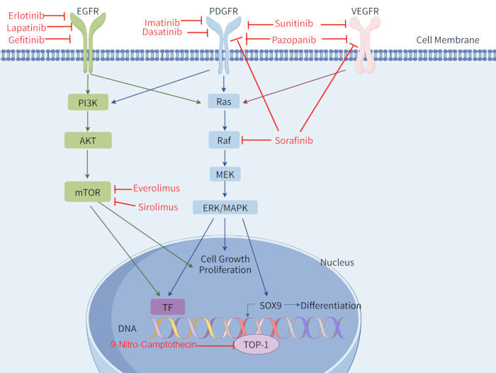
图1:脊索瘤相关治疗靶点及信号转导通路。 脊索瘤相关治疗靶点及其相关的信号转导通路概览。EGFR,表皮生长因子受体;PDGFR,血小板源性生长因子受体;VEGFR,血管内皮生长因子受体;PI3K,磷脂酰肌醇3-激酶;AKT,蛋白激酶B;mTOR,雷帕霉素靶蛋白;Ras,大鼠肉瘤病毒癌基因同源物;Raf,迅速加速成纤维细胞肉瘤激酶;MEK,丝裂原活化蛋白激酶激酶;ERK,细胞外信号调节激酶;MAPK,丝裂原活化蛋白激酶;TF,转录因子;SOX9,SRY-box转录因子9;TOP-1,拓扑异构酶I。 使用FigDraw软件绘制。请点击此处查看该图的高清版本。
结论
病理分类和治疗的进展已提高了对脊索瘤的理解和管理水平。2020年世界卫生组织(WHO)分类通过将低分化型脊索瘤确认为一种独立的病理类型,进一步完善了脊索瘤的分类体系。手术切除仍是颅内脊索瘤的主要治疗方法。内镜经鼻手术在累及颅底上部和中部的肿瘤治疗中已显示出良好的疗效,包括观察性研究中更高的肉眼全切除率和更低的复发率。然而,目前的证据主要基于回顾性研究和汇总的临床病例系列,因此手术入路的选择应根据肿瘤的位置、累及范围和大小而定。
对于不可切除、残留或复发性疾病,质子束治疗和碳离子放射治疗是重要的治疗选择,已显示出良好的无进展生存和总生存结果。靶向治疗,包括PDGFR抑制剂及其他分子靶向药物,在晚期脊索瘤中也显示出一定疗效,但现有支持证据仍有限。治疗方案应考虑病理亚型。经典型脊索瘤通常预后较好,主要采用手术治疗,是否加用放射治疗需根据具体临床情况决定。低分化型和去分化型脊索瘤临床行为更具侵袭性,常需采用包括手术、放疗和系统性治疗在内的多模式综合治疗。
目前仍存在一些局限性。脊索瘤较为罕见,许多现有研究纳入的患者队列较小。标准化的化疗方案(包括剂量和治疗周期)尚未建立。质子束治疗与碳离子放射治疗之间的直接比较仍然有限,且这两种方法均需要专门的设施。放射治疗和系统性治疗的研究也主要集中于晚期疾病,针对不同病理亚型之间治疗效果的比较证据有限。未来还需开展更多临床研究,以明确这些治疗方法在不同脊索瘤亚型中的作用,并优化治疗方案的选择。
披露
未披露任何利益冲突。
致谢
未从公共、商业或非营利机构等资助组织获得特定资助。
参考文献
- McMaster ML et al. Chordoma: incidence and survival patterns in the United States, 1973–1995. Cancer Causes Control. 2001;12(1):1-11. doi:10.1023/A:1008947301735.
- Healey JH, Lane JM. Chordoma: a critical review of diagnosis and treatment. Orthop Clin North Am. 1989;20(3):417-426.
- Eriksson B, Gunterberg B, Kindblom LG. Chordoma: a clinicopathologic and prognostic study of a Swedish national series. Acta Orthop Scand. 1981;52(1):49-58. doi:10.3109/17453678108991758.
- Higinbotham NL, Phillips RF, Farr HW, Hustu HO. Chordoma: thirty-five-year study at Memorial Hospital. Cancer. 1967;20(11):1841-1850. doi:10.1002/1097-0142(196711)20:11<1841::AID-CNCR2820201107>3.0.CO;2-2.
- Chambers KJ et al. Incidence and survival patterns of cranial chordoma in the United States. Laryngoscope. 2014;124(5):1097-1102. doi:10.1002/lary.24420.
- Heffelfinger MJ, Dahlin DC, MacCarty CS, Beabout JW. Chordomas and cartilaginous tumors at the skull base. Cancer. 1973;32(2):410-420. doi:10.1002/1097-0142(197308)32:2<410::AID-CNCR2820320219>3.0.CO;2-S.
- Soo MY. Chordoma: review of clinicoradiological features and factors affecting survival. Australas Radiol. 2001;45(4):427-434. doi:10.1046/j.1440-1673.2001.00950.x.
- Jahangiri A et al. Factors predicting recurrence after resection of clival chordoma using variable surgical approaches and radiation modalities. Neurosurgery. 2015;76(2):179–186. doi:10.1227/NEU.0000000000000611.
- Sen C et al. Clival chordomas: clinical management, results, and complications in 71 patients. J Neurosurg. 2010;113(5):1059-1071. doi:10.3171/2009.9.JNS08596.
- Di Maio S, Temkin N, Ramanathan D, Sekhar LN. Current comprehensive management of cranial base chordomas: 10-year meta-analysis of observational studies. J Neurosurg. 2011;115(6):1094-1105. doi:10.3171/2011.7.JNS11355.
- Wasserman JK, Gravel D, Purgina B. Chordoma of the head and neck: a review. Head Neck Pathol. 2018;12(2):261–268. doi:10.1007/s12105-017-0860-8.
- Yamaguchi T, Imada H, Iida S, Szuhai K. Notochordal tumors. Surg Pathol Clin. 2017;10(3):637-656. doi:10.1016/j.path.2017.04.008.
- Colia V, Stacchiotti S. Medical treatment of advanced chordomas. Eur J Cancer. 2017;83:220-228. doi:10.1016/j.ejca.2017.06.038.
- Radaelli S et al. Sacral chordoma: long-term outcome of a large series of patients surgically treated at two reference centers. Spine. 2016;41(12):1049-1057. doi:10.1097/BRS.0000000000001604.
- Fischbein NJ, Kaplan MJ, Holliday RA, Dillon WP. Recurrence of clival chordoma along the surgical pathway. AJNR Am J Neuroradiol. 2000;21(3):578-583.
- Lee IJ, Lee RJ, Fahim DK. Prognostic factors and survival outcome in patients with chordoma in the United States: a population-based analysis. World Neurosurg. 2017;104:346-355. doi:10.1016/j.wneu.2017.04.118.
- Tauziède-Espariat A et al. Prognostic and therapeutic markers in chordomas: a study of 287 tumors. J Neuropathol Exp Neurol. 2016;75(2):111-120. doi:10.1093/jnen/nlv010.
- Sahyouni R, Goshtasbi K, Mahmoodi A, Chen JW. A historical recount of chordoma. J Neurosurg Spine. 2018;28(4):422-428. doi:10.3171/2017.7.SPINE17668.
- Rosenberg AE, Brown GA, Bhan AK, Lee JM. Chondroid chordoma—a variant of chordoma: a morphologic and immunohistochemical study. Am J Clin Pathol. 1994;101(1):36-41. doi:10.1093/ajcp/101.1.36.
- Forsyth PA et al. Intracranial chordomas: a clinicopathological and prognostic study of 51 cases. J Neurosurg. 1993;78(5):741-747. doi:10.3171/jns.1993.78.5.0741.
- Mitchell A et al. Chordoma and chondroid neoplasms of the spheno-occiput: an immunohistochemical study of 41 cases with prognostic and nosologic implications. Cancer. 1993;72(10):2943-2949. doi:10.1002/1097-0142(19931115)72:10<2943::AID-CNCR2820721014>3.0.CO;2-6.
- Vujovic S et al. Brachyury, a crucial regulator of notochordal development, is a novel biomarker for chordomas. J Pathol. 2006;209(2):157-165. doi:10.1002/path.1969.
- Jian BJ et al. Adjuvant radiation therapy and chondroid chordoma subtype are associated with a lower tumor recurrence rate of cranial chordoma. J Neurooncol. 2010;98(1):101-108. doi:10.1007/s11060-009-0068-1.
- Sibley RK, Day DL, Dehner LP, Trueworthy RC. Metastasizing chordoma in early childhood: a pathological and immunohistochemical study with review of the literature. Pediatr Pathol. 1987;7(3):287-301. doi:10.1080/15513818709177131.
- Coffin CM, Swanson PE, Wick MR, Dehner LP. Chordoma in childhood and adolescence: a clinicopathologic analysis of 12 cases. Arch Pathol Lab Med. 1993;117(9):927-933.
- Becker LE, Yates AJ, Hoffman HJ, Norman MG. Intracranial chordoma in infancy: case report. J Neurosurg. 1975;42(3):349-352. doi:10.3171/jns.1975.42.3.0349.
- Wold LE, Laws ER. Cranial chordomas in children and young adults. J Neurosurg. 1983;59(6):1043-1047. doi:10.3171/jns.1983.59.6.1043.
- Borba LA, Al-Mefty O, Mrak RE, Suen J. Cranial chordomas in children and adolescents. J Neurosurg. 1996;84(4):584-591. doi:10.3171/jns.1996.84.4.0584.
- Sassin JF, Chutorian AM. Intracranial chordoma in children. Arch Neurol. 1967;17(1):89-93. doi:10.1001/archneur.1967.00470250093010.
- Tirabosco R, O'Donnell P, Flanagan AM. Notochordal tumors. Surg Pathol Clin. 2021;14(4):619-643. doi:10.1016/j.path.2021.06.006.
- Karpathiou G et al. Chordomas: a review with emphasis on their pathophysiology, pathology, molecular biology, and genetics. Pathol Res Pract. 2020;216(9):153089. doi:10.1016/j.prp.2020.153089.
- Yeter HG, Kosemehmetoglu K, Soylemezoglu F. Poorly differentiated chordoma: review of 53 cases. APMIS. 2019;127(9):607–615. doi:10.1111/apm.12978.
- Hoch BL, Nielsen GP, Liebsch NJ, Rosenberg AE. Base of skull chordomas in children and adolescents: a clinicopathologic study of 73 cases. Am J Surg Pathol. 2006;30(7):811-818. doi:10.1097/01.pas.0000209828.39477.ab.
- Hasselblatt M et al. Poorly differentiated chordoma with SMARCB1/INI1 loss: a distinct molecular entity with dismal prognosis. Acta Neuropathol. 2016;132(1):149-151. doi:10.1007/s00401-016-1574-9.
- Shih AR et al. Clinicopathologic characteristics of poorly differentiated chordoma. Mod Pathol. 2018;31(8):1237-1245. doi:10.1038/s41379-018-0002-1.
- Morimitsu Y, Aoki T, Yokoyama K, Hashimoto H. Sarcomatoid chordoma: chordoma with a massive malignant spindle-cell component. Skeletal Radiol. 2000;29(12):721-725. doi:10.1007/s002560000264.
- Hung YP et al. Dedifferentiated chordoma: clinicopathologic and molecular characteristics with integrative analysis. Am J Surg Pathol. 2020;44(9):1213-1223. doi:10.1097/PAS.0000000000001501.
- Meis JM et al. “Dedifferentiated” chordoma: a clinicopathologic and immunohistochemical study of three cases. Am J Surg Pathol. 1987;11(7):516-525. doi:10.1097/00000478-198707000-00003.
- Smith J, Reuter V, Demas B. Case report 576. Anaplastic sacrococcygeal chordoma (dedifferentiated chordoma). Skeletal Radiol. 1989;18(7):561-564. doi:10.1007/BF00351762.
- Ng WK, Tang V. Crush preparation findings of “sarcomatoid” chordoma of the sacrum: report of a case with histologic, immunohistochemical, and ultrastructural correlation. Diagn Cytopathol. 2001;25(6):406-410. doi:10.1002/dc.10024.
- Chavez JA, Nasir Ud Din, Memon A, Perry A. Anaplastic chordoma with loss of INI1 and brachyury expression in a 2-year-old girl. Clin Neuropathol. 2014;33(6):418-420. doi:10.5414/NP300724.
- Fernandez-Miranda JC et al. Clival chordomas: a pathological, surgical, and radiotherapeutic review. Head Neck. 2014;36(6):892-906. doi:10.1002/hed.23415.
- Miettinen M et al. Nuclear Brachyury expression is consistent in chordoma, common in germ cell tumors and small cell carcinomas, and rare in other carcinomas and sarcomas: an immunohistochemical study of 5229 cases. Am J Surg Pathol. 2015;39(10):1305-1312. doi:10.1097/PAS.0000000000000462.
- Showell C, Binder O, Conlon FL. T-box genes in early embryogenesis. Dev Dyn. 2004;229(1):201-218. doi:10.1002/dvdy.10480.
- Jambhekar NA et al. Revisiting chordoma with brachyury, a “new age” marker: analysis of a validation study on 51 cases. Arch Pathol Lab Med. 2010;134(8):1181-1187. doi:10.5858/2009-0476-OA.1.
- Oakley GJ, Fuhrer K, Seethala RR. Brachyury, SOX-9, and podoplanin, new markers in the skull base chordoma vs chondrosarcoma differential: a tissue microarray-based comparative analysis. Mod Pathol. 2008;21(12):1461-1469. doi:10.1038/modpathol.2008.144.
- Roberts CWM, Leroux MM, Fleming MD, Orkin SH. Highly penetrant, rapid tumorigenesis through conditional inversion of the tumor suppressor gene Snf5. Cancer Cell. 2002;2(5):415-425. doi:10.1016/S1535-6108(02)00185-X.
- Mobley BC et al. Loss of SMARCB1/INI1 expression in poorly differentiated chordomas. Acta Neuropathol. 2010;120(6):745-753. doi:10.1007/s00401-010-0767-x.
- Tirabosco R et al. Brachyury expression in extra-axial skeletal and soft tissue chordomas: a marker that distinguishes chordoma from mixed tumor/myoepithelioma/parachordoma in soft tissue. Am J Surg Pathol. 2008;32(4):572-580. doi:10.1097/PAS.0b013e31815b693a.
- Al-Adnani M, Cannon SR, Flanagan AM. Chordomas do not express CD10 and renal cell carcinoma antigen: an immunohistochemical study. Histopathology. 2005;47(5):535-537. doi:10.1111/j.1365-2559.2005.02131.x.
- Barber SM et al. Chordoma—current understanding and modern treatment paradigms. J Clin Med. 2021;10(5):1054. doi:10.3390/jcm10051054.
- Crumley RL, Gutin PH. Surgical access for clivus chordoma: the University of California, San Francisco, experience. Arch Otolaryngol Head Neck Surg. 1989;115(3):295-300. doi:10.1001/archotol.1989.01860270037011.
- Blevins NH, Jackler RK, Kaplan MJ, Gutin PH. Combined transpetrosal-subtemporal craniotomy for clival tumors with extension into the posterior fossa. Laryngoscope. 1995;105(9):975-982. doi:10.1288/00005537-199509000-00018.
- Bertalanffy H, Seeger W. The dorsolateral, suboccipital, transcondylar approach to the lower clivus and anterior portion of the craniocervical junction. Neurosurgery. 1991;29(6):815-821. doi:10.1097/00006123-199112000-00002.
- Wang L et al. Clinical features and surgical outcomes of patients with skull base chordoma: a retrospective analysis of 238 patients. J Neurosurg. 2017;127(6):1257-1267. doi:10.3171/2016.9.JNS16559.
- Komotar RJ et al. The endoscope-assisted ventral approach compared with open microscope-assisted surgery for clival chordomas. World Neurosurg. 2011;76(3-4):318-327. doi:10.1016/j.wneu.2011.02.026.
- Koutourousiou M et al. Endoscopic endonasal approach for resection of cranial base chordomas: outcomes and learning curve. Neurosurgery. 2012;71(3):614-625. doi:10.1227/NEU.0b013e31825ea3e0.
- Fraser JF et al. Endoscopic endonasal transclival resection of chordomas: operative technique, clinical outcome, and review of the literature. J Neurosurg. 2010;112(5):1061-1069. doi:10.3171/2009.7.JNS081504.
- Yaniv D et al. Skull base chordomas: review of current treatment paradigms. World J Otorhinolaryngol Head Neck Surg. 2020;6(2):125-131. doi:10.1016/j.wjorl.2020.01.008.
- Konieczkowski DJ, DeLaney TF, Yamada Y. Radiation strategies for spine chordoma: proton beam, carbon ions, and stereotactic body radiation therapy. Neurosurg Clin N Am. 2020;31(2):263-288. doi:10.1016/j.nec.2019.12.002.
- Wu S et al. Carbon ion radiotherapy for patients with extracranial chordoma or chondrosarcoma: initial experience from Shanghai Proton and Heavy Ion Center. J Cancer. 2019;10(15):3315-3322. doi:10.7150/jca.29667.
- Mima M et al. Particle therapy using carbon ions or protons as a definitive therapy for patients with primary sacral chordoma. Br J Radiol. 2014;87(1033):20130512. doi:10.1259/bjr.20130512.
- Uhl M et al. Carbon ion beam treatment in patients with primary and recurrent sacrococcygeal chordoma. Strahlenther Onkol. 2015;191(7):597-603. doi:10.1007/s00066-015-0825-3.
- Imai R et al. Carbon ion radiation therapy for unresectable sacral chordoma: an analysis of 188 cases. Int J Radiat Oncol Biol Phys. 2016;95(1):322-327. doi:10.1016/j.ijrobp.2016.02.012.
- Fuchs B et al. Operative management of sacral chordoma. J Bone Joint Surg Am. 2005;87(10):2211-2216. doi:10.2106/JBJS.D.02693.
- Stacchiotti S et al. Chordoma of the mobile spine and sacrum: a retrospective analysis of a series of patients surgically treated at two referral centers. Ann Surg Oncol. 2010;17(1):211-219. doi:10.1245/s10434-009-0740-x.
- Rodrigues ACLF et al. Proton beam and carbon ion radiotherapy in skull base chordoma: a systematic review, meta-analysis and meta-regression with trial sequential analysis. Neurosurg Rev. 2024;47(1):893. doi:10.1007/s10143-024-03117-1.
- Potter GM et al. Skull base chordoma and chondrosarcoma: neuroradiologist's guide to diagnosis, surgical management, and proton beam therapy. Radiographics. 2024;44(10):e240036. doi:10.1148/rg.240036.
- Gersey ZC et al. A refined treatment strategy for skull base chordoma: a protocol and management algorithm. Neurosurg Rev. 2025;48(1):729. doi:10.1007/s10143-025-03869-4.
- Tamborini E et al. Molecular and biochemical analyses of platelet-derived growth factor receptor B, PDGFRA, and KIT receptors in chordomas. Clin Cancer Res. 2006;12(23):6920-6928. doi:10.1158/1078-0432.CCR-06-1584.
- Zeinali M et al. Systematic review of novel target therapies and clinical trials in chordoma. Clin Neurol Neurosurg. 2025;259:109222. doi:10.1016/j.clineuro.2025.109222.
- Hindi N et al. Imatinib in advanced chordoma: a retrospective case series analysis. Eur J Cancer. 2015;51(17):2609-2614. doi:10.1016/j.ejca.2015.07.038.
- Stacchiotti S et al. Phase II study of imatinib in advanced chordoma. J Clin Oncol. 2012;30(9):914-920. doi:10.1200/JCO.2011.35.3656.
- Lebellec L et al. Advanced chordoma treated by first-line molecular targeted therapies: outcomes and prognostic factors. A retrospective study of the French Sarcoma Group (GSF/GETO) and the Association des Neuro-Oncologues d'Expression Française (ANOCEF). Eur J Cancer. 2017;79:119-128. doi:10.1016/j.ejca.2017.03.037.
- Stacchiotti S et al. Imatinib and everolimus in patients with progressing advanced chordoma: a phase 2 clinical study. Cancer. 2018;124(20):4056-4063. doi:10.1002/cncr.31685.
- Stacchiotti S et al. Response to imatinib plus sirolimus in advanced chordoma. Ann Oncol. 2009;20(11):1886-1894. doi:10.1093/annonc/mdp210.
- Adenis A et al. A dose-escalating phase I of imatinib mesylate with fixed dose of metronomic cyclophosphamide in targeted solid tumours. Br J Cancer. 2013;109(10):2574-2578. doi:10.1038/bjc.2013.648.
- Schuetze SM et al. Phase 2 study of dasatinib in patients with alveolar soft part sarcoma, chondrosarcoma, chordoma, epithelioid sarcoma, or solitary fibrous tumor. Cancer. 2017;123(1):90-97. doi:10.1002/cncr.30379.
- Singhal N, Kotasek D, Parnis FX. Response to erlotinib in a patient with treatment refractory chordoma. Anticancer Drugs. 2009;20(10):953-955. doi:10.1097/CAD.0b013e328330c7f0.
- Launay SG et al. Efficacy of epidermal growth factor receptor targeting in advanced chordoma: case report and literature review. BMC Cancer. 2011;11:423. doi:10.1186/1471-2407-11-423.
- Houessinon A et al. Sustained response of a clivus chordoma to erlotinib after imatinib failure. Case Rep Oncol. 2015;8(1):25-29. doi:10.1159/000371843.
- Macaulay VM et al. Phase I dose-escalation study of linsitinib (OSI-906) and erlotinib in patients with advanced solid tumors. Clin Cancer Res. 2016;22(12):2897-2907. doi:10.1158/1078-0432.CCR-15-2218.
- Migliorini D et al. First report of clinical responses to immunotherapy in 3 relapsing cases of chordoma after failure of standard therapies. Oncoimmunology. 2017;6(8):e1338235. doi:10.1080/2162402X.2017.1338235.
- Asklund T et al. Durable stabilization of three chordoma cases by bevacizumab and erlotinib. Acta Oncol. 2014;53(7):980-984. doi:10.3109/0284186X.2013.878472.
- Stacchiotti S et al. Phase II study on lapatinib in advanced EGFR-positive chordoma. Ann Oncol. 2013;24(7):1931-1936. doi:10.1093/annonc/mdt117.
- Hof H, Welzel T, Debus J. Effectiveness of cetuximab/gefitinib in the therapy of a sacral chordoma. Onkologie. 2006;29(12):572-574. doi:10.1159/000096283.
- Lindèn O, Stenberg L, Kjellén E. Regression of cervical spinal cord compression in a patient with chordoma following treatment with cetuximab and gefitinib. Acta Oncol. 2009;48(1):158-159. doi:10.1080/02841860802266672.
- Wilhelm SM et al. BAY 43-9006 exhibits broad spectrum oral antitumor activity and targets the RAF/MEK/ERK pathway and receptor tyrosine kinases involved in tumor progression and angiogenesis. Cancer Res. 2004;64(19):7099-7109. doi:10.1158/0008-5472.CAN-04-1443.
- Bompas E et al. Sorafenib in patients with locally advanced and metastatic chordomas: a phase II trial of the French Sarcoma Group (GSF/GETO). Ann Oncol. 2015;26(10):2168-2173. doi:10.1093/annonc/mdv300.
- Chow LQM, Eckhardt SG. Sunitinib: from rational design to clinical efficacy. J Clin Oncol. 2007;25(7):884–896. doi:10.1200/JCO.2006.06.3602.
- Chellappan DK et al. The role of pazopanib on tumour angiogenesis and in the management of cancers: a review. Biomed Pharmacother. 2017;96:768-781. doi:10.1016/j.biopha.2017.10.058.
- Meng T et al. Molecular targeted therapy in the treatment of chordoma: a systematic review. Front Oncol. 2019;9:30. doi:10.3389/fonc.2019.00030.
- George S et al. Multicenter phase II trial of sunitinib in the treatment of nongastrointestinal stromal tumor sarcomas. J Clin Oncol. 2009;27(19):3154-3160. doi:10.1200/JCO.2008.20.9890.
- Lipplaa A, Dijkstra S, Gelderblom H. Efficacy of pazopanib and sunitinib in advanced axial chordoma: a single reference centre case series. Clin Sarcoma Res. 2016;6:19. doi:10.1186/s13569-016-0059-x.
- Chugh R et al. Phase II study of 9-nitro-camptothecin in patients with advanced chordoma or soft tissue sarcoma. J Clin Oncol. 2005;23(15):3597-3604. doi:10.1200/JCO.2005.02.170.
- Dhall G et al. The role of chemotherapy in pediatric clival chordomas. J Neurooncol. 2011;103(3):657-662. doi:10.1007/s11060-010-0441-0.
- Al-Rahawan MM, Siebert JD, Mitchell CS, Smith SD. Durable complete response to chemotherapy in an infant with a clival chordoma. Pediatr Blood Cancer. 2012;59(2):323-325. doi:10.1002/pbc.23297.
- Martinez Moreno M et al. Shedding light on emerging therapeutic targets for chordoma. Expert Opin Ther Targets. 2023;27(8):705-713. doi:10.1080/14728222.2023.2248382.
- Von Witzleben A et al. Preclinical characterization of novel chordoma cell systems and their targeting by pharmacological inhibitors of the CDK4/6 cell-cycle pathway. Cancer Res. 2015;75(18):3823-3831. doi:10.1158/0008-5472.CAN-14-3270.
- Liu T et al. CDK4 expression in chordoma: a potential therapeutic target. J Orthop Res. 2018;36(6):1581-1589. doi:10.1002/jor.23819.
- Passeri T et al. In vivo efficacy assessment of the CDK4/6 inhibitor palbociclib and the PLK1 inhibitor volasertib in human chordoma xenografts. Front Oncol. 2022;12:960720. doi:10.3389/fonc.2022.960720.
- Sharifnia T et al. Small-molecule targeting of brachyury transcription factor addiction in chordoma. Nat Med. 2019;25(2):292-300. doi:10.1038/s41591-018-0312-3.
- Godinez C et al. Combinatorial therapies for epigenetic, immunotherapeutic, and genetic targeting of chordoma. J Neurooncol. 2025;172(2):307-315. doi:10.1007/s11060-024-04920-y.
- Heery CR et al. Phase I trial of a yeast-based therapeutic cancer vaccine (GI-6301) targeting the transcription factor Brachyury. Cancer Immunol Res. 2015;3(11):1248-1256. doi:10.1158/2326-6066.CIR-15-0119.
- Heery CR et al. Phase I study of a poxviral TRICOM-based vaccine directed against the transcription factor Brachyury. Clin Cancer Res. 2017;23(22):6833-6845. doi:10.1158/1078-0432.CCR-17-1087.
- DeMaria PJ et al. Randomized, double-blind, placebo-controlled phase II study of yeast-Brachyury vaccine (GI-6301) in combination with standard-of-care radiotherapy in locally advanced, unresectable chordoma. Oncologist. 2021;26(5):e847-e858. doi:10.1002/onco.13720.