Acupuncture at Baihui and Yintang reduces the required sedative dosage of dexmedetomidine, alleviates mechanical ventilation- and dexmedetomidine-induced diaphragm dysfunction, and suppresses excessive autophagy in mechanically ventilated rats.
研究文章
Analgesic and Sedative Effect of Acupuncture Combined with Medicine on Patients Undergoing Cardiac Surgery
本文内容
摘要
摘要
Acupuncture (AP) has been reported to modulate inflammation and oxidative stress, but whether it can reduce dexmedetomidine (Dex) requirements or protect against Dex-induced diaphragm toxicity during mechanical ventilation (MV) remains unknown. This study investigated whether AP could alleviate Dex-aggravated diaphragmatic dysfunction and explored its underlying mechanisms, focusing on autophagy. Female Sprague-Dawley rats were subjected to MV and treated with Dex, AP (Baihui and Yintang), and the autophagy inducer rapamycin (RAPA). Arterial blood gas parameters, vital signs, blood cell counts, and sedative dosage were recorded. Diaphragm function was evaluated by measuring contractile force, muscle fiber cross-sectional area, apoptosis, and oxidative stress markers, including superoxide dismutase (SOD), catalase (CAT), malondialdehyde (MDA), and angiotensin II (ANG II). Autophagy levels were assessed using the LC3-II/LC3-I ratio, p62 expression, and autophagosome accumulation. AP reduced the total Dex dosage by 54.7% (0.102 ± 0.005 mg vs. 0.225 ± 0.009 mg in the MV + Dex group, p < 0.001) while maintaining stable physiological parameters. MV decreased diaphragm contractility and myofiber cross-sectional area and increased MAFbx expression, apoptosis, and oxidative stress markers. Dex further worsened these parameters, whereas AP improved diaphragm function and attenuated atrophy, apoptosis, and oxidative stress. MV and MV + Dex groups exhibited excessive autophagy, as shown by autophagic vacuole accumulation, an increased LC3-II/LC3-I ratio, and decreased p62 expression. AP inhibited these autophagic changes, while RAPA partially reversed its protective effects. AP at Baihui and Yintang reduced Dex requirements and alleviated MV- and Dex-induced diaphragm dysfunction in rats by inhibiting excessive autophagy.
引言
MV is a life-saving intervention for patients with respiratory failure1. However, prolonged MV is associated with diaphragm dysfunction, a condition known as ventilator-induced diaphragmatic dysfunction (VIDD)2. VIDD is a key risk factor for several poor clinical outcomes, including difficult weaning, longer durations of ventilation and intensive care unit (ICU) stays, and complications related to acute respiratory failure3. Therefore, strategies to protect diaphragm function during MV are of clinical importance.
Sedatives are commonly used during MV to reduce patient-ventilator asynchrony and improve tolerance. Dex, a selective alpha-2 adrenergic receptor agonist, is widely used in intensive care units because of its sedative, analgesic, and sympatholytic properties and relatively low risk of respiratory depression4,5,6. However, recent studies have shown that Dex may paradoxically exacerbate MV-induced diaphragm dysfunction, an effect linked to excessive autophagy and oxidative stress7. Moreover, high-dose Dex may cause bradycardia, hypotension, and even sinus arrest8. This has raised concerns about its safety profile, despite its sedative advantages. AP, a key component of traditional Chinese medicine, has been reported to modulate inflammation, oxidative stress, and autophagy in various disease models9,10,11,12. In particular, AP at the Baihui and Yintang points is known to exert neuroprotective and anti-inflammatory effects13,14,15. However, whether AP can reduce the required sedative dosage of Dex or protect against Dex-induced diaphragm toxicity during MV remains unclear.
The pathogenesis of VIDD is multifactorial, driven by a complex interplay of oxidative stress, proteolysis, apoptosis, and dysregulated autophagy16. Both MV and Dex have been reported to promote autophagy and exacerbate diaphragmatic dysfunction7,17, yet the modulatory role of AP in this process remains poorly understood. This study hypothesized that AP combined with Dex would reduce the required sedative dosage of Dex and attenuate MV- and Dex-induced diaphragm dysfunction in rats through inhibition of excessive autophagy. Accordingly, the Dex-sparing effect of AP under MV conditions was evaluated, followed by assessment of diaphragm contractility, structural integrity, oxidative stress, and autophagy. Finally, RAPA, an autophagy inducer, was used to examine the role of autophagy in the protective effect of AP.
方案
Ethics statement:
This study was approved by the Animal Ethical and Welfare Committee of Jiangsu Huachuang Yutong Medical Laboratory Co., Ltd. (approval no. HC25041-P001-01; November 14, 2025) and was conducted in accordance with the U.K. Animals (Scientific Procedures) Act 1986 and associated guidelines and EU Directive 2010/63/EU on the protection of animals used for scientific purposes.
Details of all materials and equipment used in this study are provided in the Table of Materials.
Animal experiments
Healthy female Sprague-Dawley rats (3 months old, 300 g) were housed under a 12 h light/dark cycle with free access to food and water for 1 week before the experiments. In the first part of the study, rats were randomly assigned into five groups (n = 5): Control (sham surgery), MV (mechanical ventilation, model group), MV + Dex, MV + AP (acupuncture), and MV + Dex + AP. In the second part of the study, four groups were used (n = 5): MV, MV + Dex, MV + Dex + AP, and MV + Dex + AP + RAPA. Five animals per group were included in the general phenotypic assessments. For histological staining, TUNEL assay, and western blotting, tissues from three randomly selected animals per group were analyzed for these ex vivo experiments.
For MV model construction, all surgical procedures were performed under aseptic conditions. Rats were anesthetized with a combination of Zoletil (30 mg/kg) and Xylazine (7 mg/kg), and subsequently stabilized on a circulating heating blanket. After tracheotomy, rats were connected to a volume-controlled small animal ventilator for 24 h with the following settings: respiratory rate, 60 breaths/min; tidal volume, 6–7 mL/kg; inspiratory/expiratory ratio, 1:1. The inspiratory gas was enriched with oxygen and humidified with sterile saline. To prevent systemic hypoxia or hypercapnia during ventilation, arterial blood was sampled from the carotid artery every 3 h for blood gas analysis, and ventilator settings were adjusted to maintain PaCO2 at 35–45 mmHg and PaO2 at 80–100 mmHg. Control animals underwent sham surgery without MV.
For Dex treatment, Dex was continuously infused via the jugular vein during MV to maintain stable sedation, with the dosage adjusted based on tail-flick and pedal reflex tests. For AP treatment, needles (0.3 mm diameter, 25 mm length) were inserted at Baihui acupuncture point (GV20) and Yintang acupuncture point (GV29) at a 15° angle to a depth of 5 mm. Treatment lasted 20 min every 12 h. For RAPA treatment, RAPA (20 mg/kg) was administered intraperitoneally for two consecutive days, including 3 h before MV on the day of ventilation, to induce autophagy.
Animal care and physiological monitoring
Throughout the 24 h experimental period, all animals received continuous infusion of 0.9% saline at 2 mL/h via a central venous catheter to maintain normovolemia. To prevent bladder distension and discomfort, the bladder was manually compressed every hour to promote voiding. After 24 h, terminal anesthesia was induced with a combination of Zoletil (50–60 mg/kg) and Xylazine (10–12 mg/kg), followed by removal of the diaphragm under maintained respiratory and circulatory conditions. All animals were then euthanized by intraperitoneal injection of pentobarbital (100 mg/kg).
All rats underwent continuous electrocardiographic monitoring and monitoring of invasive mean arterial pressure (MAP) and rectal temperature (maintained at 37 °C). Simultaneously, noninvasive tail artery blood pressure (BP) and heart rate (HR) were measured and recorded in real time using tail-cuff plethysmography. In the MV + Dex and MV + Dex + AP groups, the total sedative dosage of Dex was recorded at the end of the experiment.
Diaphragm contractility measurement
After MV, the diaphragm was rapidly excised and placed in Krebs-Henseleit solution equilibrated with 95% O2 and 5% CO2 at 25 °C. Muscle strips were mounted with one end connected to a force transducer and the other end fixed. Optimal contraction length (Lo) was determined using a maximum stimulation voltage (15 V). Muscles were then stimulated at 20, 40, 60, 80, 100, and 120 Hz for 600 ms with 1-min intervals between stimuli. The force-frequency relationship was determined and contractile force was normalized to physiological cross-sectional area.
Hematoxylin and eosin (H&E) staining
H&E staining was performed to evaluate muscle fiber cross-sectional area (CSA). Diaphragm tissues were fixed in 4% formaldehyde for 24 h, dehydrated through graded ethanol (50% to absolute), cleared in xylene, and embedded in paraffin. Sections (4–7 µm) were deparaffinized, rehydrated, stained with hematoxylin and eosin, dehydrated, cleared, and mounted with neutral balsam. Sections were examined under a light microscope, and myofiber CSA was quantified from the acquired images.
Biochemical assays
Commercial assay kits were performed to detect the superoxide dismutase (SOD), CAT, and MDA in diaphragm tissues and ANG II in serum. Diaphragm tissues were homogenized in pre-chilled phosphate-buffered saline (PBS) (1:9 w/v) and centrifuged. The corresponding kits were: ANG II-related ELISA kit, SOD-related kit, CAT-related kit, and MDA-related assay kit.
Western blotting
Expression of MAFbx and the autophagy markers LC3-I, LC3-II, and p62 in diaphragm tissue was quantified by western blotting. Briefly, tissues were lysed in radioimmunoprecipitation assay (RIPA) buffer containing phenylmethylsulfonyl fluoride (PMSF). Protein concentrations were determined using a bicinchoninic acid (BCA) assay kit. Equal amounts of protein (25 µg) were separated by SDS-PAGE and transferred onto polyvinylidene fluoride (PVDF) membranes. Membranes were blocked with 5% non-fat milk and incubated overnight at 4 °C with primary antibodies: rabbit anti-MAFbx (1:1000), rabbit anti-LC3A/LC3B (1:1000), rabbit anti-p62 (1:1000), and rabbit anti-β-actin (1:2500). After washing, membranes were incubated with horseradish peroxidase (HRP)-conjugated goat anti-rabbit immunoglobulin G (IgG) (1:2000). Signals were detected using enhanced chemiluminescence. Band intensities were quantified by densitometry and normalized to β-actin.
TUNEL staining
A TUNEL assay was performed to detect apoptotic cells in diaphragm tissue. Diaphragm sections were deparaffinized, rehydrated, and digested with proteinase K (37 °C, 30 min). TUNEL reaction mixture (5 µL terminal deoxynucleotidyl transferase (TdT) enzyme and 45 µL fluorescence labeling solution) was applied for 60 min at 37 °C in the dark. Nuclei were counterstained with 4′,6-diamidino-2-phenylindole (DAPI). Images were captured under a fluorescence microscope.
Transmission electron microscopy
TEM was used to examine autophagic structures in diaphragm tissue. Diaphragm tissue (≈1 mm3) was fixed in 2.5% glutaraldehyde for more than 24 h, postfixed in 1% osmium tetroxide, dehydrated in acetone, and embedded in EPON812. Ultrathin sections (50–70 nm) were stained with uranyl acetate and lead citrate, and examined using a transmission electron microscope at 80 kV.
Statistical analysis
All analyses were conducted using GraphPad Prism. Data are presented as mean ± standard deviation (SD). Comparisons among multiple groups were performed using one-way or repeated-measures two-way analysis of variance (ANOVA) followed by Tukey’s post hoc tests. A p-value < 0.05 was considered statistically significant.
结果
Acupuncture reduces the dexmedetomidine requirement in mechanically ventilated rats
Arterial blood gas parameters and vital signs remained stable and did not differ significantly among the Dex, AP, and Dex + AP intervention groups (p > 0.05; Table 1). However, the total sedative dosage of Dex was significantly lower in the MV + Dex + AP group (0.102 ± 0.005 mg) compared with the MV + Dex group (0.225 ± 0.009 mg, p < 0.001), representing a 54.7% reduction (Table 1). These findings indicate that AP treatment was associated with a substantial reduction in the required Dex dosage.
Acupuncture mitigates MV-induced diaphragm dysfunction and oxidative stress
The effects of MV, Dex, AP, and combined Dex + AP treatment on diaphragm contractility and structural integrity were assessed. Diaphragm force did not differ significantly among groups at 20 Hz, increased sharply from 20 to 60 Hz, and increased more gradually from 60 to 120 Hz (Figure 1A). At 120 Hz, MV reduced diaphragm force; under MV conditions, the application of Dex further decreased diaphragm force, while AP alleviated this reduction (Figure 1A). H&E staining revealed that MV and Dex led to a decrease in the CSA of diaphragmatic fibers and widened interstitial spaces; AP treatment significantly increased fiber CSA and improved muscle morphology (Figure 1B). Simultaneously, western blotting and TUNEL assays demonstrated that AP significantly downregulated the expression of the atrophy marker MAFbx and reduced the rate of cell apoptosis in the diaphragm (Figure 1C,D). Moreover, the oxidative stress levels were investigated. AP intervention reduced the MV-induced elevation in MDA levels and increased SOD and CAT activities (Figure 1E). AP also reduced the elevated ANG II levels in the MV group (Figure 1E), consistent with the reported role of ANG II in oxidative stress and skeletal muscle atrophy18. Collectively, acupuncture treatment significantly alleviated MV-induced diaphragm dysfunction, as evidenced by improved contractility, preserved myofiber structure, reduced atrophy and apoptosis, and attenuated oxidative stress.
Acupuncture reduces the side effects of dexmedetomidine on diaphragm function by inhibiting excessive autophagy
To investigate the underlying mechanism, we focused on the autophagic pathway. TEM observations confirmed that the control group showed intact sarcomeres with no obvious ultrastructural abnormalities, while the MV and MV + Dex groups exhibited accumulation of autophagic vacuoles, with the highest accumulation in the MV + Dex group (Figure 2A). Similarly, western blotting presented a significant increase in LC3-II/LC3-I ratio and a reduction in p62 expression in the MV and MV + Dex groups (Figure 2B), indicating excessive autophagy. AP intervention effectively inhibited these autophagic markers, bringing the LC3-II/LC3-I ratio and p62 levels closer to those in the control group (Figure 2B).
To confirm the role of autophagy, RAPA, an autophagy inducer, was used to further examine its involvement. RAPA treatment reversed the decrease of LC3-II/LC3-I ratio and the increase of p62 observed in the MV + Dex + AP group (Figure 3A). Notably, the administration of RAPA (MV + Dex + AP + RAPA) partially blocked the protective effects of AP, leading to decreased diaphragm force (Figure 3B), reduced fiber CSA (Figure 3C), and elevated ANG II levels (Figure 3D) compared with the MV + Dex + AP group. These results demonstrate that AP mitigates the diaphragm toxicity associated with Dex by suppressing excessive MV-induced autophagy.
DATA AVAILABILITY:
All raw data have been provided in Supplementary File.
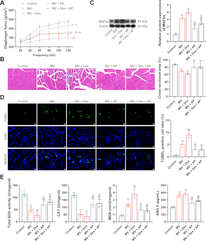
Figure 1: Acupuncture alleviates mechanical ventilation-induced diaphragm dysfunction and oxidative stress. (A) Diaphragm contractile force at stimulation frequencies of 20–120 Hz (n = 5). (B) H&E staining and myofiber CSA (n = 3). Scale bar = 20 µm. (C) MAFbx protein expression (n = 3). (D) TUNEL staining and quantification of TUNEL-positive cells (n = 3). Scale bar = 20 µm. (E) MDA, SOD, CAT, and serum ANG II levels (n = 5). Data are presented as mean ± SD. For (A), repeated-measures two-way ANOVA followed by Tukey’s post hoc test was used; for (B–E), one-way ANOVA followed by Tukey’s post hoc test was used. *p < 0.05, **p < 0.01 vs. Control; #p < 0.05, ##p < 0.01 vs. MV; $p < 0.05, $$p < 0.01 vs. MV + Dex; &p < 0.05, &&p < 0.01 vs. MV + AP. Please click here to view a larger version of this figure.
Figure 2: Acupuncture inhibits mechanical ventilation- and dexmedetomidine-induced excessive autophagy in the diaphragm. (A) Transmission electron microscopy images of diaphragm tissue (n = 3). Scale bar = 2 µm. (B) LC3-I, LC3-II, LC3-II/LC3-I ratio, and p62 protein expression (n = 3). Data are presented as mean ± SD. One-way ANOVA followed by Tukey’s post hoc test was used for (B). *p < 0.05, **p < 0.01 vs. Control; #p < 0.05, ##p < 0.01 vs. MV; $p < 0.05, $$p < 0.01 vs. MV + Dex; &p < 0.05, &&p < 0.01 vs. MV + AP. Please click here to view a larger version of this figure.
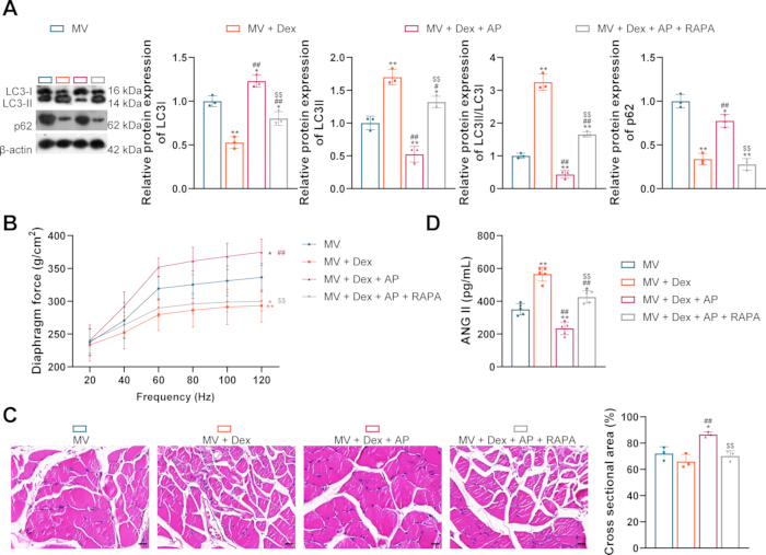
Figure 3: Rapamycin reverses the protective effects of acupuncture against dexmedetomidine-induced diaphragm dysfunction. (A) LC3-I, LC3-II, LC3-II/LC3-I ratio, and p62 protein expression (n = 3). (B) Diaphragm contractile force at stimulation frequencies of 20–120 Hz (n = 5). (C) H&E staining and myofiber CSA (n = 3). Scale bar = 20 µm. (D) Serum ANG II levels (n = 5). Data are presented as mean ± SD. For (A, C, D), one-way ANOVA followed by Tukey’s post hoc test was used; for (B), repeated-measures two-way ANOVA followed by Tukey’s post hoc test was used. *p < 0.05, **p < 0.01 vs. MV; #p < 0.05, ##p < 0.01 vs. MV + Dex; $p < 0.05, $$p < 0.01 vs. MV + Dex + AP. Abbreviations: MV = mechanical ventilation; Dex = dexmedetomidine; AP = acupuncture. Please click here to view a larger version of this figure.
| Group | MV + Dex | MV + AP | MV + Dex + AP |
| Arterial blood gas | |||
| pH | 7.44 ± 0.05 | 7.42 ± 0.04 | 7.44 ± 0.07 ns |
| PaCO₂ (mmHg) | 33 ± 4 | 33 ± 5 | 34 ± 4 ns |
| PaO₂ (mmHg) | 107 ± 9 | 109 ± 7 | 110 ± 10 ns |
| Lactate (mmol/L) | 1.1 ± 0.3 | 1.3 ± 0.4 | 1.4 ± 0.2 ns |
| HCO₃⁻ (mmol/L) | 25.8 ± 2.2 | 26.7 ± 2.8 | 26.2 ± 2.3 ns |
| BEecf (mmol/L) | 3 ± 1 | 3 ± 1 | 4 ± 2 ns |
| Vital signs | |||
| Heart rate (beats/min) | 393 ± 17 | 387 ± 15 | 388 ± 12 ns |
| Rectal temperature (°C) | 37.3 ± 0.1 | 36.9 ± 0.3 | 37.0 ± 0.3 ns |
| MAP (mmHg) | 102 ± 5 | 104 ± 4 | 106 ± 4 ns |
| Glucose (mmol/L) | 5.2 ± 0.6 | 5.5 ± 0.8 | 5.1 ± 0.5 ns |
| Blood cell counts | |||
| Erythrocytes (× 10¹²/L) | 6.9 ± 0.6 | 7.2 ± 0.7 | 6.6 ± 0.4 ns |
| Hemoglobin (g/L) | 166 ± 7 | 162 ± 7 | 159 ± 9 ns |
| Neutrophils (%) | 45.5 ± 2.6 | 45.9 ± 3.0 | 44.3 ± 1.6 ns |
| Lymphocytes (%) | 37.8 ± 3.6 | 36.9 ± 3.3 | 36.5 ± 4.5 ns |
| Sedative dosages (mg) | 0.225 ± 0.009 | - | 0.102 ± 0.005*** |
Table 1: Arterial blood gas parameters, vital signs, blood cell counts, and total sedative dosages at the end of the experiment in mechanically ventilated groups. The table summarizes physiological parameters across the experimental groups and compares the total dexmedetomidine dosage between the MV + Dex and MV + Dex + AP groups. Data are presented as mean ± SD. ***p < 0.001; ns = not significant. Comparisons among three groups were performed using one-way analysis of variance (ANOVA) followed by Tukey’s post hoc tests. Comparisons between two groups were performed using unpaired t-test.
讨论
AP at Baihui and Yintang significantly reduced the required Dex dosage in mechanically ventilated rats by more than 50%. In addition, acupuncture alleviated MV-induced diaphragm dysfunction, as evidenced by improved contractile force, preserved myofiber structure, reduced expression of the atrophy marker MAFbx, and decreased apoptosis. Furthermore, acupuncture mitigated oxidative stress by reducing MDA and ANG II levels and modulating SOD and CAT activities. Mechanistically, acupuncture inhibited excessive autophagy in the diaphragm, as shown by decreased LC3-II/LC3-I ratio and increased p62 expression. The protective effect of acupuncture was partially reversed by the autophagy inducer RAPA, confirming that the beneficial effects are mediated, at least in part, by suppression of excessive autophagy.
AP has been investigated as a nonpharmacological complementary therapy in critical care11,19. In mechanically ventilated patients, previous studies have reported that AP, compared with conventional ICU treatment, may reduce ICU stay and the duration of arrhythmia, and also lower the overall mortality rate of patients20. Previous studies have also reported that AP can significantly reduce the demand for sedative drugs in ICU patients21,22,23. In canine studies, subcutaneous administration of sedatives at specific acupuncture points, including Baihui, produced more stable and longer-lasting sedation than conventional intramuscular administration24,25,26. These findings further support a potential role of acupuncture in pain relief. The present findings are consistent with these reports; AP at Baihui and Yintang maintained stable physiological parameters while reducing the required Dex dosage during MV by 54.7%. These two acupoints, both located on the Governor Vessel, are traditionally used to calm the mind and promote sleep23. This finding suggests that AP exerts a sedative-sparing effect. While the exact mechanism of this synergy is not fully understood, one possible explanation is that AP may reduce central excitation, alleviate brain injury, and regulate pain-related neurotransmission21,27.
The beneficial effects of acupuncture on diaphragm function have also been reported in a previous meta-analysis28. In the present study, AP treatment significantly improved diaphragm function in MV rats and in MV rats treated with Dex. MV alone significantly reduced diaphragm contractility, decreased myofiber cross-sectional area, increased MAFbx expression and apoptosis, and elevated oxidative stress markers. The addition of Dex (MV + Dex group) further worsened these parameters compared with MV alone. This is in line with a previous report that Dex aggravates VIDD through metabolic and oxidative pathways7. AP treatment (MV + AP and MV + Dex + AP groups) significantly improved diaphragm function, reduced atrophy and apoptosis, and lowered oxidative stress. Notably, in the MV + Dex + AP group, AP partially restored diaphragm contractility and structure that had been worsened by Dex. These results indicate that acupuncture not only protects against MV-induced damage but also counteracts the additional diaphragm toxicity induced by Dex.
The potential involvement of autophagy in AP-mediated protection against Dex-aggravated diaphragm dysfunction was also investigated. The MV and MV + Dex groups showed accumulation of autophagic vacuoles by TEM, increased LC3-II/LC3-I ratio, and decreased p62 expression, indicative of excessive autophagy. Acupuncture reversed these changes, suggesting inhibition of autophagic flux. To confirm that the protective effect of acupuncture is mediated via autophagy inhibition, RAPA was used as an autophagy inducer. RAPA treatment reversed the effects of acupuncture, leading to decreased diaphragm force, reduced fiber CSA, and elevated ANG II levels compared with the MV + Dex + AP group. These findings support suppression of excessive autophagy as a mechanism contributing to the protective effect of AP against Dex-induced diaphragm toxicity.
This study has several notable features. AP at Baihui and Yintang demonstrated a Dex-sparing effect during MV, reducing the required sedative dosage by more than half. In addition to its effects on MV-induced diaphragm dysfunction, AP attenuated the additional diaphragm impairment observed with Dex treatment. The RAPA experiments further support the involvement of excessive autophagy in this protective effect. Several limitations should be acknowledged. First, only female rats were used, so the findings may not be directly generalizable to male animals. Second, the sedative depth was not quantitatively scored (e.g., using the Ramsay scale), which would have provided a more objective assessment of the sedative-sparing effect. Third, the exact molecular pathway linking acupuncture to autophagy regulation—such as whether it acts via AMPK, mTOR, or other signaling pathways—was not investigated. Finally, the effects of AP alone without Dex under MV conditions on autophagy markers were not evaluated in detail across all time points. Future studies should address these gaps.
In summary, AP at Baihui and Yintang reduced the required Dex dosage and attenuated MV- and Dex-associated diaphragm dysfunction in mechanically ventilated rats. Suppression of excessive autophagy appears to contribute to these protective effects. These findings support further investigation of AP as an adjunctive approach for reducing sedative requirements and preserving diaphragm function during mechanical ventilation.
披露
The authors have no relevant financial or non-financial interests to disclose.
AUTHOR CONTRIBUTIONS:
Bixia Huang contributed to conceptualization, methodology, formal analysis, funding acquisition, and writing the original draft. Hailong Ding contributed to data curation, funding acquisition, and visualization. Wei Liang contributed to formal analysis, investigation, and software. Lingen Xu contributed to conceptualization, project administration, formal analysis, supervision, funding acquisition, and review and editing of the manuscript. All authors read and approved the final manuscript
致谢
The authors thank Xinchang County Hospital of Traditional Chinese Medicine and Jiangsu Huachuang Yutong Medical Laboratory Co., Ltd. for providing the research facilities used in this study. This study was supported by the Shaoxing Municipal Science and Technology Bureau (Nos. 2020A13100 and 2025SKY137).
材料
| 姓名 | 公司 | 目录编号 | 评论 |
|---|---|---|---|
| Acetone | China National Pharmaceutical Group Co., Ltd. (Sinopharm) | 10000418 | Transmission electron microscopy |
| Acupuncture needles | Zhongyan Taihe | 20222200133 | Animal experiments/drug |
| Angiotensin II (ANG II) ELISA kit | Mlbio | ml106670 | Biochemical assays |
| Bicinchoninic acid (BCA) assay kit | Bio-rad | 1610374 | Western blotting |
| Blood-gas analyzer | Wuhan EasyDiagnosis | PT1000 | Equipment |
| Catalase (CAT) assay kit | Beyotime | S0051 | Biochemical assays |
| 4′,6-Diamidino-2-phenylindole (DAPI) | Beyotime | C1005 | TUNEL staining |
| Dexmedetomidine | Macklin | D833149 | Drug |
| Enhanced chemiluminescence (ECL) reagent | Applygen | P1000 | Western blotting |
| EPON 812 | MERCK | 45345 | Transmission electron microscopy |
| Ethanol | China National Pharmaceutical Group Co., Ltd. (Sinopharm) | 10009218 | Hematoxylin and eosin (H&E) staining |
| Female Sprague-Dawley rats | Beijing Huafukang Bioscience Co., Ltd. | 22001A | Animal |
| Force transducer | Aurora Scientific | 305C-LR-FP-R | Diaphragm contractility measurement |
| Formaldehyde | China National Pharmaceutical Group Co., Ltd. (Sinopharm) | 10010018 | Hematoxylin and eosin (H&E) staining |
| Glutaraldehyde | China National Pharmaceutical Group Co., Ltd. (Sinopharm) | 30092436 | Transmission electron microscopy |
| Hematoxylin and eosin (H&E) staining kit | Beyotime | C0105S | Hematoxylin and eosin (H&E) staining |
| HRP-conjugated goat anti-rabbit IgG | CST | 7074 | Western blotting |
| Krebs–Henseleit solution | Thermo | J67820.AP | Diaphragm contractility measurement |
| Lead citrate | Macklin | L885990 | Transmission Electron Microscopy |
| Light microscope | Leica | DM3000 | Hematoxylin and eosin (H&E) staining |
| Malondialdehyde (MDA) assay kit | Nanjing Jiancheng | A003-1-2 | Biochemical assays |
| Neutral balsam | Solarbio | G8590 | Hematoxylin and eosin (H&E) staining |
| Osmium tetroxide | China National Pharmaceutical Group Co., Ltd. (Sinopharm) | L01210302 | Transmission Electron Microscopy |
| Paraffin | China National Pharmaceutical Group Co., Ltd. (Sinopharm) | 69019061 | Hematoxylin and eosin (H&E) staining |
| Phosphate-buffered saline (PBS) | Beyotime | C0221A | Biochemical assays |
| Physiological monitoring equipment | Techman | BL-420 F | Equipment |
| Phenylmethylsulfonyl fluoride (PMSF) | Beyotime | ST506 | Western blotting |
| Proteinase K | Thermo | 4333793 | TUNEL staining |
| Polyvinylidene fluoride (PVDF) membranes | Beyotime | FFP24 | Western blotting |
| Rabbit anti-LC3A/LC3B | Thermo | PA1-16931 | Western blotting |
| Rabbit anti-MAFbx | Thermo | PA5-76680 | Western blotting |
| Rabbit anti-p62 | Thermo | PA5-20839 | Western blotting |
| Rabbit anti-β-actin | Thermo | PA1-16889 | Western blotting |
| Rapamycin | Thermo | J62473.MF | Animal experiments/drug |
| Radioimmunoprecipitation assay (RIPA) buffer | Beyotime | P0013B | Western blotting |
| SDS-PAGE gel/reagent kit | Beyotime | P0015A | Western blotting |
| Superoxide dismutase (SOD) assay kit | Nanjing Jiancheng | A001-3 | Biochemical assays |
| Statistical software | GraphPad Software, LLC | Version 7.0 | Data analysis |
| Transmission electron microscope | HITACHI | HT7800 | Equipment |
| TUNEL assay kit | Beyotime | C1086 | TUNEL staining |
| Uranyl acetate | Yaji Biotechnology | YS25690U | Transmission electron microscopy |
| Volume-controlled small animal ventilator | Shanghai Alcott Biotech | ALC-V8A | Animal experiments |
| Xylazine | Dunhua Shengda Animal Medicine Co., Ltd. | BN 20260114 | Animal experiments/drug |
| Xylene | China National Pharmaceutical Group Co., Ltd. (Sinopharm) | 10023418 | Hematoxylin and eosin (H&E) staining |
| Zoletil | Virbac | BN 9KWE | Animal experiments/drug |
参考文献
- Demoule A et al. Prevalence, Intensity, and Clinical Impact of Dyspnea in Critically Ill Patients Receiving Invasive Ventilation. American Journal of Respiratory and Critical Care Medicine. 2022;205(8):917-26.
- Xie D et al. Mechanosensitive Piezo1 Channels Mediate Diaphragm Fibrosis Induced by Prolonged Mechanical Ventilation. Journal of Cachexia, Sarcopenia and Muscle. 2025;16(6):e70136.
- Fu W et al. Ventilator-induced diaphragmatic dysfunction: pathophysiology, monitoring and advances in potential treatment and prevention. European Respiratory Review. 2025;34(178):250069.
- Kawazoe Y et al. Effect of Dexmedetomidine on Mortality and Ventilator-Free Days in Patients Requiring Mechanical Ventilation With Sepsis: A Randomized Clinical Trial. JAMA. 2017;317(13):1321-28.
- Lim JY et al. Dexmedetomidine for analgesia and sedation in newborn infants receiving mechanical ventilation. Cochrane Database of Systematic Reviews. 2024;5(5):CD012361.
- Chen L et al. The effect of dexmedetomidine in mechanically ventilated patients with sepsis and septic shock: a meta-analysis of randomized controlled trials. Annals of Medicine. 2026;58(1):2643971.
- Breuer T et al. Dexmedetomidine Impairs Diaphragm Function and Increases Oxidative Stress but Does Not Aggravate Diaphragmatic Atrophy in Mechanically Ventilated Rats. Anesthesiology. 2018;128(4):784-95.
- Shehabi Y et al. Early Sedation with Dexmedetomidine in Critically Ill Patients. New England Journal of Medicine. 2019;380(26):2506-17.
- Wang M, Liu W, Ge J, Liu S. The immunomodulatory mechanisms for acupuncture practice. Frontiers in Immunology. 2023;14:1147718.
- Bao QN et al. The effect of acupuncture on oxidative stress in animal models of vascular dementia: a systematic review and meta-analysis. Systematic Reviews. 2024;13(1):59.
- Liu T et al. Acupuncture for Rheumatoid Arthritis Treatment: Biological Mechanisms of Anti-Inflammation and Analgesia. American Journal of Chinese Medicine. 2026;54(2):349-74.
- He J et al. The Mechanism of Acupuncture Regulating Autophagy: Progress and Prospect. Biomolecules. 2025;15(2):263.
- Jiang H et al. Effects of acupuncture on regulating the hippocampal inflammatory response in rats exposed to post-traumatic stress disorder. Neuroscience Letters. 2023;796:137056.
- Wang W et al. Effect of acupuncture Baihui acupoint-Yintang acupoint-Neiguan acupoint combined with conventional Western medicine treatment on cerebral blood flow in insomnia patients: a 3D-ASL functional magnetic resonance imaging study. Frontiers in Neurology. 2026;17:1679885.
- Wu BX et al. Acupuncture as A Potential Therapeutic Approach for Tourette Syndrome: Modulation of Neurotransmitter Levels and Gut Microbiota. Chinese Journal of Integrative Medicine. 2025;31(8):735-42.
- Powers SK. Ventilator-induced diaphragm dysfunction: phenomenology and mechanism(s) of pathogenesis. Journal of Physiology. 2024;602(19):4729-52.
- Smuder AJ et al. Crosstalk between autophagy and oxidative stress regulates proteolysis in the diaphragm during mechanical ventilation. Free Radical Biology and Medicine. 2018;115:179-90.
- Kwon OS et al. AT1 receptor blocker losartan protects against mechanical ventilation-induced diaphragmatic dysfunction. Journal of Applied Physiology. 2015;119(10):1033-41.
- Choi Y et al. Acupuncture as adjunctive treatment for schizophrenia: a systematic review and meta-analysis. Asian Journal of Psychiatry. 2026;117:104844.
- Ben-Arie E et al. Acupuncture reduces mechanical ventilation time in critically ill patients: A systematic review and meta-analysis of randomized control trials. Explore. 2024;20(4):477-92.
- Xie C et al. [Observation on the clinical efficacy of acupuncture combined with conventional Western medicine treatment in reducing ICU postoperative delirium]. Journal of Hunan University of Chinese Medicine. 2024;44(12):2239-44.
- Yu HJ et al. [Analgesic and Sedative Effect of Acupuncture Combined with Medicine on Patients Undergoing Cardiac Surgery]. Zhongguo Zhong Xi Yi Jie He Za Zhi. 2016;36(3):289-93.
- Yuan SC et al. [Effect of acupuncture on analgesia and sedation in elderly patients with severe pneumonia during invasive mechanical ventilation]. Zhongguo Zhen Jiu. 2021;41(9):971-78.
- Leriquier C et al. Comparison of sedation with dexmedetomidine/atipamezole administered subcutaneously at GV20 acupuncture point with usual routes of administration in dogs presented for orthopaedic radiographs. Journal of Small Animal Practice. 2023;64(12):759-68.
- Llido M et al. Comparison of sedation with dexmedetomidine administered subcutaneously at 2 different locations on the head in dogs. Canadian Veterinary Journal. 2024;65(4):351-58.
- Pons A et al. Effects of dexmedetomidine administered at acupuncture point GV20 compared to intramuscular route in dogs. Journal of Small Animal Practice. 2017;58(1):23-28.
- Li P et al. Acupuncture for ICU patients: evidence, mechanisms, and implementation challenges. Frontiers in Neurology. 2025;16:1711600.
- Liu R et al. Rehabilitation effects of acupuncture on the diaphragmatic dysfunction in respiratory insufficiency: A systematic review and meta-analysis. Complementary Therapies in Medicine. 2024;87:103105.