This article presents concentrated purified fraction (CPF) as a proposed concentrated growth factor-derived preparation and outlines its rationale, provisional workflow, analytical requirements, and potential relevance to hair regeneration.
综述文章
Concentrated Purified Fraction Derived from Concentrated Growth Factor: Rationale, Proposed Preparation, and Potential Applications
本文内容
摘要
摘要
Platelet concentrates have become an important class of autologous biologics for regenerative medicine and are increasingly applied in dermatology, wound healing, and hair restoration. However, different generational classification frameworks for autologous platelet concentrates coexist in the literature. Based on an evolutionary framework that distinguishes platelet-rich plasma (PRP), platelet-rich fibrin (PRF), and concentrated growth factor (CGF) according to their preparation principles and technological progression, this review proposes the concentrated purified fraction (CPF), a novel CGF-derived preparation generated through sequential physical fractionation. The proposed strategy is intended to produce a resuspendable extracellular-particle-associated fraction while reducing fibrin fragments, platelet aggregates, and cellular components. Existing platelet concentrates remain limited by compositional heterogeneity, retention of larger particulate components, and incomplete characterization of extracellular vesicle (EV)-associated fractions, which together may contribute to variability in biological activity and clinical outcomes. This review summarizes the biological rationale for CPF, outlines its proposed preparation workflow and characterization strategy, evaluates current evidence relevant to hair regeneration, and presents the proposed EV gradient hypothesis, in which differences in EV-associated components may contribute to the biological performance of successive platelet concentrate preparations. CPF should currently be regarded as a conceptual and methodological framework that requires direct compositional, biological, mechanistic, and clinical validation before its therapeutic value and potential advantages can be determined.
引言
Platelet concentrates have evolved over the past two decades and are now widely used in dentistry, orthopedics, dermatology, aesthetic medicine, and other regenerative applications. The first generation is represented by platelet-rich plasma (PRP), a liquid platelet concentrate generally prepared by centrifugation of anticoagulated blood. Depending on the preparation protocol, PRP may be administered with or without exogenous activation, and the absence of a stable fibrin scaffold results in a relatively rapid release of platelet-derived mediators1,2,3. The second generation, platelet-rich fibrin (PRF), was introduced by Choukroun in 2001 and relies on anticoagulant-free preparation and natural coagulation to form a fibrin network that retains platelets and supports the gradual release of bioactive mediators4. Subsequent variants, including injectable platelet-rich fibrin (i-PRF) and extended platelet-rich fibrin (e-PRF), were developed to broaden the handling characteristics and potential applications of fibrin-based preparations5,6. The third generation, concentrated growth factor (CGF), employs variable-speed centrifugation to produce a denser fibrin matrix containing platelet-derived factors and CD34-positive cellular components7,8. However, generational nomenclature for autologous platelet concentrates remains non-uniform: recent articles in Periodontology 2000 include CGF among modifications of the anticoagulant-free leukocyte- and platelet-rich fibrin (L-PRF) family within their broader second-generation category9,10, whereas other evolutionary frameworks and a recent expert consensus document continue to describe CGF as a third-generation preparation11,12,13. In parallel with this technological progression, clinical research in androgenetic alopecia (AGA) has expanded from evaluating the overall efficacy of platelet-rich plasma (PRP) to examining how preparation protocols, patient characteristics, adjunctive treatments, and administration techniques influence hair-regrowth outcomes. Meta-analytic evidence suggests that PRP may improve several hair-growth measures, although substantial heterogeneity in product preparation and treatment schedules continues to limit direct comparisons across studies14. Comparative studies of activated and non-activated PRP have reported inconsistent findings, and no activation strategy has been established as universally superior15,16. Although PRP has been investigated in both male AGA and female pattern hair loss, limited sex-stratified evidence prevents reliable conclusions regarding differential responses between men and women14,17. Microneedling, low-level light/laser therapy (LLLT), and multimodal treatment strategies have also been explored, while manual and device-assisted delivery represent additional sources of procedural variability18,19,20. Collectively, these findings indicate that clinical outcomes depend not only on the type of platelet concentrate but also on product preparation, patient selection, adjunctive treatment, and delivery technique; however, methodological heterogeneity continues to constrain the interpretation and reproducibility of treatment outcomes.
Despite continued advances in platelet concentrate technologies, currently available preparations remain subject to three interrelated limitations. First, different preparation methods retain substantially different amounts of cells, platelet aggregates, fibrin, and other large particulate components, which may affect product homogeneity, injectability, and tissue distribution21. Second, current research and clinical applications have predominantly focused on the release of growth factors, whereas the potential contributions of platelet-derived extracellular vesicles (EVs) and their molecular cargoes to therapeutic activity have received comparatively limited attention22. Third, variations in donor characteristics, centrifugation parameters, platelet activation methods, and final product composition may contribute to both inter-product and inter-batch variability, thereby affecting the consistency of clinical outcomes23. Accumulating evidence indicates that platelet activation can induce the release of EVs carrying functional microRNAs (miRNAs) and protein cargoes, which may participate in tissue repair and regeneration by mediating intercellular communication. Studies of hair regeneration further support this biological rationale. Exosomal miR-218-5p derived from dermal papilla cell spheroids has been shown to promote the transition of hair follicles from the telogen to the anagen phase by suppressing secreted frizzled-related protein 2 (SFRP2) and activating the Wnt/β-catenin signaling pathway24. More recent studies have also suggested that PRP-derived exosomes may promote hair follicle growth through Wnt/β-catenin- and sirtuin 1 (SIRT1) / FoxO3a-related pathways. Although these findings do not directly establish the therapeutic efficacy of CPF, they provide a biological basis for further investigation of platelet-derived particulate components and their EV-associated cargoes in regenerative applications (Table 1).
| Generation | Representative product | Core feature | Primary mechanism | Relative EV abundance |
| 1st generation | PRP, PRGF | Double centrifugation + anticoagulant | Burst growth factor release | Low |
| 2nd generation | PRF, A-PRF | Natural coagulation | Sustained growth factor release | Moderate |
| 3rd generation | CGF | Variable-speed centrifugation | Dense fibrin matrix + CD34⁺ cell retention | Moderate–High |
| Proposed CPF strategy | CPF (CGF-PF) | CGF + sequential purification | Extracellular vesicle (EV)-mediated signaling | High (≈2–5 × CGF) |
Table 1: Comparative features of platelet concentrate generations and the proposed CPF strategy.
In light of these limitations and the emerging biological rationale, this review proposes concentrated purified fraction (CPF) as a CGF-derived preparation strategy. Using CGF as the starting material, CPF is obtained through mechanical dissociation followed by sequential differential centrifugation to generate a resuspendable, particle-enriched fraction. This approach is intended to reduce larger cellular components, platelet aggregates, and fibrin-associated material while retaining extracellular particles with potential biological activity. The proposed novelty of CPF does not lie in defining it directly as a new generation of platelet concentrate, but rather in integrating the physical fractionation and purification of CGF with the systematic characterization of EV-associated components. This shifts the focus from the conventional application of platelet concentrates as whole preparations toward the isolation and investigation of their potentially bioactive particulate fractions. Although the composition, reproducibility, and biological activity of CPF require further clarification through direct comparative studies, this strategy provides a testable methodological framework for developing CGF-derived injectable fractions that are more compositionally uniform, readily characterizable, and potentially relevant to regenerative applications.
综述与观点
Rationale and scientific positioning of CPF
Recent literature illustrates that generational labels for autologous platelet concentrates are framework-dependent rather than standardized. Quirynen et al. described CGF as a later modification of L-PRF and included it within the broader second-generation L-PRF family, reflecting a preparation-based classification centered on anticoagulant-free processing and natural fibrin formation9,10. Li et al. further highlighted the coexistence of classifications that group CGF with PRF in the second generation and those that distinguish CGF as the third generation, before proposing a function-oriented framework of their own12. By contrast, Jia et al. explicitly classified CGF as a third-generation platelet-rich derivative and distinguished it from earlier preparations by its programmed acceleration–deceleration centrifugation protocol, while a 2026 expert consensus document also referred to CGF as a “third-generation platelet concentrate"11,13. These usages reflect different organizing criteria rather than a single settled taxonomy. The present review favors the PRP-PRF-CGF evolutionary sequence because it captures the later introduction of programmed variable-speed centrifugation and the characteristic fibrin, growth-factor, and cellular composition reported for CGF, rather than because this sequence represents a universally accepted taxonomy7,11. Within this explicitly defined working framework, the present review proposes CPF as a putative fourth-generation preparation generated through further physical fractionation of CGF; this designation remains conceptual pending direct compositional, biological, and clinical validation.
Platelet-derived extracellular vesicles constitute a substantial proportion of circulating EVs and carry growth factors, functional miRNAs, messenger RNAs (mRNAs), and signaling lipids22,25. Current-generation products have three key limitations that CPF is intended to address: (1) a large particle burden, as CGF contains fibrin fragments and platelet aggregates, and also cellular debris, that limits tissue penetration and causes post-injection discomfort21 (2) an underutilized EV compartment, because EV-associated components within CGF may be obscured by the bulk fibrin matrix and soluble proteins, thereby limiting their therapeutic potential usage22,25 and (3) a mechanistic blind spot. The reported clinical advantages of CGF over PRP and PRF have been attributed in part to its higher growth factor content; however, PRP can also be prepared to achieve high peak growth factor levels, yet this does not necessarily translate into superior clinical outcomes26.
CPF is designed to address these limitations. We centrifuge and filter CGF sequentially, so that CPF retains the nanometer-sized bioactive fraction (50–200 nm, corresponding to small EVs and exosomes), while larger particulates and soluble proteins are removed. Importantly, CPF is not conceived merely as CGF subjected to an additional purification step. Rather, sequential physical fractionation is intended to reduce larger cellular, platelet-associated, and fibrin-related components while retaining a potentially bioactive extracellular particle fraction. In this context, CPF may represent a methodological shift from the use of platelet concentrates as bulk cellular or fibrin-rich preparations toward the investigation of more compositionally defined, cell-reduced, and EV-associated fractions27 (Figure 1).
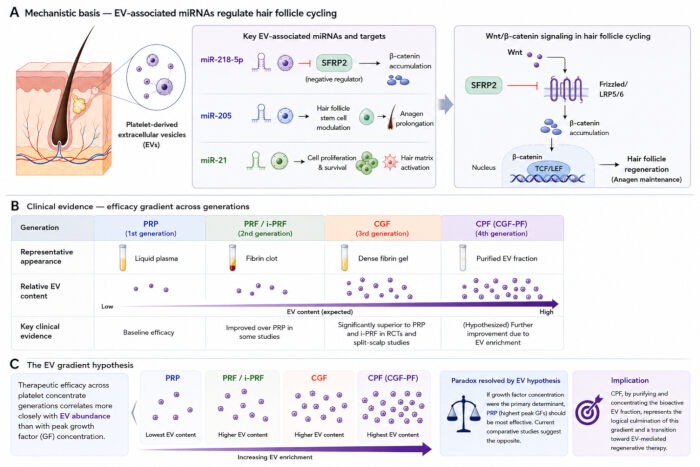
Figure 1: Clinical evidence and the proposed extracellular vesicle (EV) gradient hypothesis across platelet concentrate preparations. (A) Proposed mechanism for EV-mediated hair regeneration. Platelet-derived EVs serve as carriers for bioactive microRNAs. These microRNAs may regulate multiple stages of hair follicle cycle. Indeed, miR-218-5p is a crucial microRNA in this list, which suppresses SFRP2 to activate the Wnt/β-catenin pathway, which is anticipated to promote hair regeneration. We believe this mechanism is vital in the regeneration process. Also, there are other EV-associated microRNAs such as miR-205 and miR-21. They engage in hair follicle stem cell regulation and cell proliferation, which contributes to the maintenance of anagen and hair growth, thus enhancing the overall effect of EV treatment. (B) We analyze the clinical evidence regarding the efficacy gradient. Relative EV abundance should increase from platelet-rich plasma (PRP) to platelet-rich fibrin (PRF/i-PRF), concentrated growth factor (CGF), and concentrated purified fraction (CPF). It's important to note that published studies indicate that CGF performs better than PRP and i-PRF in androgenetic alopecia. Also, CPF is a biologic that is EV-enriched and represents the next generation of platelet concentrates. (C) Visual representation of the EV gradient hypothesis. Efficacy trends across various generations of platelet concentrates show a more aligned relationship with the EV abundance. It is not related to peak growth factor release. In reality, EV enrichment increases from PRP to PRF/i-PRF, then CGF, and finally CPF, this pattern can explain the differences in therapeutic performance reported. Moreover, this can account for the fact that PRP has not always achieved superior clinical results. Although PRP achieves the highest peak growth factor levels, the results are not consistently superior. By focusing on the EV fraction and eliminating larger particulate components, CPF may represent a potential future strategy in a standardized regenerative therapy based on mechanism. Please click here to view a larger version of this figure.
Preparation principles and characterization of the proposed CPF
Current approaches to generating liquid derivatives from concentrated growth factor (CGF) can be broadly divided into rapid physical extraction and prolonged release from the fibrin matrix. Freeze–thaw-based protocols disrupt the CGF clot and recover released components through low-speed centrifugation and extraction into culture medium. Using this strategy, Xin et al. obtained a rat-derived CGF extract that increased proliferation-related absorbance, alkaline phosphatase activity, and osteogenic gene expression in MC3T3-E1 cells cultured on titanium surfaces28. Dong et al. subsequently generated CGF extract through repeated freeze–thaw cycles and low-speed centrifugation; the preparation enhanced osteogenic readouts in vitro and, when combined with Bio-Oss Collagen, was associated with increased new-bone formation in a rat calvarial-defect model29. By contrast, Calabriso et al. cultured intact human CGF clots for 14 days and examined both the proangiogenic activity of the resulting conditioned medium and the phenotypic characteristics of CGF-released cells30. Together, these studies establish that bioactive CGF-derived preparations can be generated either by disrupting the fibrin network or by allowing its contents to be released over time. However, these approaches were designed principally to investigate soluble mediators, crude releases, or CGF-associated cellular components and did not incorporate a dedicated procedure for separating an extracellular-particle-enriched fraction from the bulk fibrin matrix, residual cells, platelet aggregates, and plasma proteins.
Methodological guidance for further fractionation is provided by studies of platelet- and platelet-rich plasma-derived extracellular vesicles (EVs), although these preparations should not be regarded as direct equivalents of CGF-derived fractions. Differential centrifugation is attractive because staged increases in centrifugal force progressively reduce cells and coarse debris while concentrating denser extracellular particles. Nevertheless, the resulting fractions are operational rather than biologically discrete. Aatonen et al. demonstrated substantial size overlap among platelet-derived EV populations recovered at different centrifugal forces31, whereas Kowal et al. showed that intermediate- and high-speed pellets contain compositionally heterogeneous and overlapping extracellular-particle populations32. More intensive separation workflows can increase stringency but introduce different trade-offs. Saumell-Esnaola et al. isolated a 100,000 × g EV-enriched fraction released by platelets obtained from PRP and combined ultracentrifugation with transmission electron microscopy and molecular-marker analysis; this fraction was termed platelet-derived exosomes by the original authors33. Size-exclusion chromatography can reduce soluble-protein and lipoprotein contamination, although vesicle recovery remains incomplete34, whereas CD61-based immunoaffinity separation improves platelet-source specificity but, by design, enriches only antigen-positive particles35. Collectively, these studies indicate that no single method simultaneously maximizes recovery, purity, source specificity, and procedural accessibility. They also underscore that centrifugal force alone cannot establish vesicle identity and that any CGF-derived fraction must be defined by its experimentally measured composition.
To our knowledge, no previous study has specifically described the fractionation of preformed CGF into a resuspendable extracellular-particle-enriched preparation. Based on previously reported approaches for CGF preparation/extraction and platelet-derived EV fractionation28,29,30,31,32,33, we propose a provisional workflow for generating concentrated purified fraction (CPF). The following conditions represent a proposed methodological framework and require experimental optimization and validation rather than an established or validated CPF protocol. In the proposed workflow, 18 mL of venous blood is collected into two 9 mL plain glass vacuum tubes without anticoagulant and with a silica clot activator on the inner tube wall. CGF is prepared using a programmed variable-speed centrifugation sequence consisting of 400 × g for 2 min, 720 × g for 4 min, 400 × g for 2 min, and 720 × g for 4 min, followed by natural deceleration without braking. The isolated middle CGF layer is transferred into 5 mL of sterile PBS pre-warmed to 37 °C, mechanically fragmented into approximately 1 mm3 pieces. The suspension is then vortexed at approximately 800 × g for 30 s and incubated at 37 °C for 10 min. Sequential centrifugation at 500 × g for 10 min and 2,000 × g for 20 min at 4 °C is proposed to reduce intact cells, coarse fibrin fragments, residual cellular material, and larger platelet-associated aggregates. A subsequent centrifugation step at 10,000 × g for 30 min at 4 °C is proposed to recover an operationally defined particle-enriched pellet. This final centrifugation step is intended to provide a relatively simple particle-enrichment approach without requiring ultracentrifugation.
For the proposed CPF preparation framework, we recommend physicochemical and molecular characterization using orthogonal approaches in accordance with MISEV202336. Nanoparticle tracking analysis may be used to assess particle concentration and size distribution. Based on our practical experience, a provisional internal criterion of > 70% of particles within the 50–200 nm range may be considered for CPF. This size range is consistent with the approximate 50–200 nm range proposed for small extracellular vesicles in a harmonized four-society position statement37. Ráez-Meseguer et al. reported mean particle diameters of 116–127 nm across three independent batches of platelet lysate-derived EVs isolated by size-exclusion chromatography, which is comparable to the particle-size range observed in our experience with CPF38. The pellet is resuspended in 500 µL of sterile PBS or physiological saline; based on our experience, the total protein concentration of CPF typically ranges from 150 to 450 µg/mL, which is of a comparable order of magnitude to the 119–295 µg/mL reported by Ráez-Meseguer et al. for SEC-purified platelet-derived EV preparations38.
Optional terminal filtration through a 0.22 µm membrane should be considered a supplementary translational processing step rather than an intrinsic component of CPF production, because it may alter particle recovery and does not independently establish sterility. In this context, “purified” refers to the physical reduction of bulk fibrin, cellular material, and larger aggregates relative to the starting CGF and does not imply biochemical isolation of a homogeneous EV population. Accordingly, we recommend a complementary assessment of particle yield and size distribution, single-particle morphology, EV-associated proteins, platelet-origin markers, and co-isolated contaminants in accordance with MISEV202336. For molecular characterization, we recommend CD63 and CD81 as EV-associated positive markers and Calnexin as an endoplasmic-reticulum-associated negative marker. CD63 and CD81 are commonly used EV-associated tetraspanins, whereas Calnexin can be used to assess residual endoplasmic-reticulum-associated cellular material. This marker panel is consistent with the multiparametric characterization principles emphasized in MISEV202336. In addition, miR-218-5p, miR-205, and miR-21 may be assessed by qRT-PCR, with cel-miR-39 used as an exogenous spike-in control; these miRNAs are more appropriately regarded as exploratory functional cargo markers rather than criteria for EV identity. Robust analytical criteria for CPF should ultimately integrate particle enrichment, depletion of fibrin- and cell-associated material, batch consistency, and biologically relevant activity (Figure 2).
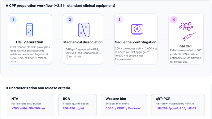
Figure 2: Preparation and quality-control workflow for concentrated purified fraction (CPF). (A) Schematic representation of the four-step CPF preparation workflow. First, venous blood is processed to produce concentrated growth factor (CGF), followed by mechanical dissociation of the CGF gel and sequential differential centrifugation to remove debris, platelet aggregates, and other larger particulate components while collecting a small extracellular particle fraction. Finally, the pellet is resuspended in sterile phosphate-buffered saline (PBS) or saline to obtain CPF. The workflow can be completed in approximately 2.5 h using standard clinical laboratory equipment, supporting its potential compatibility with a point-of-care preparation workflow. (B) CPF is characterized with reference to the MISEV guidelines. Nanoparticle tracking analysis (NTA) is used to determine particle size distribution, with more than 70% of detected particles falling within the range of 50–200 nm. In our hands, the protein concentration of CPF typically ranges from 150 to 450 µg/mL, as determined using the bicinchoninic acid (BCA) assay. Western blot analysis is performed to assess the presence of EV-associated markers, including CD63 and CD81, together with the absence of the negative marker calnexin. Quantitative reverse-transcription polymerase chain reaction (qRT-PCR) is used to quantify selected hair-regeneration-associated microRNAs, including miR-218-5p, miR-205, and miR-21. Please click here to view a larger version of this figure.
Clinical potential and future research directions
For AGA treatment, the potential advantages of CPF can be considered at both the formulation and biological levels. From a formulation perspective, converting fibrin-rich CGF into a resuspendable particle-enriched fraction is expected to reduce larger fibrin fragments, platelet aggregates, and cellular components. Compared with bulk CGF, this liquid formulation may facilitate more uniform dose dispersion and standardized intradermal administration. These anticipated handling advantages should ultimately be evaluated using objective parameters such as injection force, needle obstruction, particle recovery, and dose reproducibility. Their potential clinical relevance is supported by evidence showing that the outcomes of platelet-based treatments are influenced by both product preparation and delivery technique. Gentile and Garcovich reported greater hair-density gains with non-activated PRP than with activated PRP in a retrospective case-series comparison15, whereas Singh and Singh found in a randomized split-scalp study that PRP activation and platelet concentration significantly affected hair density and thickness16. Ozcan et al. showed that manual point-by-point injection and dermapen-mediated PRP delivery both improved hair count, hair density, terminal hair count, and terminal hair density; dermapen-mediated delivery showed additional advantages only in selected parameters, including anagen hair, telogen hair, and average hair length20. Gentile et al. also reported increased hair density following the combined use of non-activated PRP, microneedling, and low-level light/laser therapy, although the multimodal design did not allow the contribution of each component to be distinguished18. These findings provide a rationale for evaluating CPF using controlled intradermal or microneedle-assisted delivery, but they do not establish the superiority of CPF or any specific administration method. The biological potential of CPF may extend beyond its handling characteristics. Yang et al. demonstrated that PRP-derived exosomes promoted hair follicle stem cell proliferation and migration, inhibited apoptosis, and reduced oxidative-stress-related injury in vitro. Subcutaneous administration also increased anagen-phase follicle numbers and dermal thickness in a murine hair-removal model, with the observed effects associated with SIRT1 upregulation and FoxO3a deacetylation39. These findings support investigating whether CPF retains functionally active platelet-derived EV cargo, although equivalent activity cannot be assumed without direct characterization. Potential applications may also extend beyond alopecia. Johnson et al. reported that purified allogeneic platelet-derived EVs promoted dermal fibroblast proliferation and migration and enhanced the angiogenic activity of dermal endothelial cells in vitro. In their first-in-human phase I study, EV injection was safe and well tolerated, but the single dose tested did not reduce wound-closure time in healthy participants40. In skin rejuvenation, Proffer et al. reported improvements in several clinical and imaging measures of facial photodamage and cutaneous aging after six weeks of a topical platelet-derived exosome product in a prospective, single-arm study41. Collectively, these studies provide a biological rationale for investigating CPF in hair regeneration, wound repair, and skin rejuvenation. Its application in scar remodeling and the proposed combination of CGF with CPF remain testable clinical hypotheses rather than established indications.
Some challenges still remain. Actually, the purification efficiency is affected by donor-related variables like age, platelet count, and metabolic status, so we need standard quality control procedures. Also, the optimal CPF dosage for various clinical indications is not yet determined, and we need to study this via dose-escalation and dose-finding studies21. Direct head-to-head trials comparing CPF and conventional CGF remain indispensable to validate its superior efficacy. For the scaled manufacturing process, additional steps like tangential flow filtration might be required. We propose to conduct prospective clinical trials to test the EV gradient hypothesis and determine the position of CPF in therapeutic armamentarium.
结论
Concentrated purified fraction (CPF) provides a conceptual and methodological extension of concentrated growth factor (CGF) technology by shifting the focus from the use of a bulk fibrin-rich preparation toward the isolation and investigation of a resuspendable, particle-enriched fraction. Through mechanical dissociation and sequential differential centrifugation, the proposed workflow is intended to reduce larger fibrin fragments, platelet aggregates, and cellular components while retaining extracellular particles with potential biological activity. These formulation characteristics may facilitate more uniform dose dispersion, standardized intradermal administration, and compatibility with controlled injection or microneedle-assisted delivery. Evidence from studies of platelet-rich plasma and platelet-derived extracellular vesicles further provides a biological rationale for investigating CPF in hair regeneration, wound repair, and skin rejuvenation. The proposed extracellular vesicle gradient hypothesis and the potential combination of CGF with CPF also provide testable frameworks for integrating the growth-factor-rich environment of CGF with EV-associated intercellular signaling.
CPF should nevertheless currently be regarded as a proposed CGF-derived preparation strategy rather than an established clinical product. Its composition, EV-associated cargo, batch-to-batch reproducibility, biological activity, and comparative performance against CGF and conventional platelet concentrates require direct experimental and clinical validation. Future studies should establish standardized preparation, characterization, and quality-control criteria; determine appropriate doses and administration protocols; and evaluate the influence of patient characteristics, adjunctive treatments, and delivery techniques on clinical outcomes. Well-designed comparative studies and long-term safety assessments will ultimately determine whether CPF can translate the physical fractionation of CGF into a more readily characterizable, reproducible, and clinically useful platelet-derived regenerative platform.
Abbreviations: AGA, androgenetic alopecia; CGF, concentrated growth factor; CPF, concentrated purified fraction; EV, extracellular vesicle; PRF, platelet-rich fibrin; PRP, platelet-rich plasma; SFRP2, secreted frizzled-related protein 2; PRGF, plasma rich in growth factors; A-PRF, advanced platelet-rich fibrin
披露
The authors have no conflicts of interest.
致谢
This work was supported by the Ministry of Education of China (No. 231103242232720 and 2408271251), the Clinical Research Plan of Shanghai East Hospital (No. SEHHH-2021(KJ)-0669-KJB-485 and No. SEHHH-2023(KJ)-0143-KYB-111), the Featured Clinical Discipline Project of Shanghai Pudong Fund (No. PWYts2021-07), and the East Hospital Affiliated to Tongji University Introduced Talent Research Startup Fund (No. DFRC2019008).
参考文献
- Dohan Ehrenfest DM, Rasmusson L, Albrektsson T. Classification of platelet concentrates: from pure platelet-rich plasma (P-PRP) to leucocyte- and platelet-rich fibrin (L-PRF). Trends Biotechnol. 2009;27:158-167.
- Marx RE. Platelet-rich plasma: evidence to support its use. J Oral Maxillofac Surg. 2004;62:489-496.
- Gentile P, Garcovich S. Systematic review of platelet-rich plasma use in androgenetic alopecia compared with minoxidil®, finasteride®, and adult stem cell-based therapy. Int J Mol Sci. 2020;21:2702.
- Dohan DM, et al. Platelet-rich fibrin (PRF): a second-generation platelet concentrate. Part II: platelet-related biologic features. Oral Surg Oral Med Oral Pathol Oral Radiol Endod. 2006;101:e45-e50.
- Miron RJ, et al. Injectable platelet rich fibrin (i-PRF): opportunities in regenerative dentistry Clin Oral Investig. 2017;21:2619-2627.
- Miron RJ, et al. Extended platelet-rich fibrin. Periodontol 2000. 2024;94:114-130.
- Rodella LF, et al. Growth factors, CD34 positive cells, and fibrin network analysis in concentrated growth factors fraction. Microsc Res Tech. 2011;74:772-777.
- Bonazza V, et al. How the different material and shape of the blood collection tube influences the concentrated growth factors production. Microsc Res Tech. 2016;79:1173-1178.
- Quirynen M, et al. Instructions for the use of L-PRF in different clinical indications. Periodontol 2000. 2025;97:421-432.
- Quirynen M, et al. Introduction and overview on autogenous platelet concentrates. Periodontol 2000. 2025;97:7-15.
- Jia K, et al. Platelet-rich fibrin as an autologous biomaterial for bone regeneration: mechanisms, applications, optimization. Front Bioeng Biotechnol. 2024;12:1286035.
- Li Y, et al. The evolution of three generations of platelet concentrates products: a leap from classical formulations to the era of extracellular vesicles. Front Bioeng Biotechnol. 2025;13:1628565.
- Sun R, et al. Expert consensus on the management of third molar health. Int J Oral Sci. 2026;18:36-49.
- Papakonstantinou M, et al. Autologous platelet-rich plasma treatment for androgenic alopecia: a systematic review and meta-analysis of clinical trials. Plast Reconstr Surg. 2023;151:739e-747e.
- Gentile P, Garcovich S. Autologous activated platelet-rich plasma (AA-PRP) and non-activated (a-PRP) in hair growth: a retrospective, blinded, randomized evaluation in androgenetic alopecia. Expert Opin Biol Ther. 2020;20:327-337.
- Singh SK, Singh S. Effect of platelet counts and activator in platelet-rich plasma on the treatment of androgenetic alopecia, split-head comparison: a randomised, double-blind study. Indian J Dermatol Venereol Leprol. 2023;89:647-655.
- Gentile P, Garcovich S. Systematic review: the platelet-rich plasma use in female androgenetic alopecia as effective autologous treatment of regenerative plastic surgery. J Plast Reconstr Aesthet Surg. 2022;75:851-859.
- Gentile P, et al. A randomized blinded retrospective study: the combined use of micro-needling technique, low-level laser therapy and autologous non-activated platelet-rich plasma improves hair re-growth in patients with androgenic alopecia. Expert Opin Biol Ther. 2020;20:1099-1109.
- Gentile P, Garcovich S. The effectiveness of low-level light/laser therapy on hair loss. Facial Plast Surg Aesthet Med. 2024;26:228-235.
- Ozcan KN, Sener S, Altunisik N, Turkmen D. Platelet rich plasma application by dermapen microneedling and intradermal point-by-point injection methods, and their comparison with clinical findings and trichoscan in patients with androgenetic alopecia. Dermatol Ther. 2022;35:e15182.
- Everts P, et al. Platelet-rich plasma: new performance understandings and therapeutic considerations in 2020. Int J Mol Sci. 2020;21:7794.
- Johnson J, et al. Prospective therapeutic applications of platelet extracellular vesicles. Trends Biotechnol. 2021;39:598-612.
- Cruciani M, et al. Platelet-rich plasma for the treatment of alopecia: a systematic review and meta-analysis: PRP for treatment of alopecia. Blood Transfus. 2021;21:24-36.
- Hu S, et al. Dermal exosomes containing miR-218-5p promote hair regeneration by regulating β-catenin signaling. Sci Adv. 2020;6:eaba1685.
- Antich-Rosselló M, Forteza-Genestra MA, Monjo M, Ramis JM. Platelet-derived extracellular vesicles for regenerative medicine. Int J Mol Sci. 2021;22:8580.
- Li M, et al. Comparative efficacy and safety of platelet-rich plasma (PRP), injectable platelet-rich fibrin (i-PRF) and concentrated growth factors (CGF) for female pattern hair loss (FPHL): a prospective multicenter randomized clinical trial. J Dermatol Treat. 2026;37:2659498.
- Fan Z, Gan Y, Hu Y. The potential utilization of platelet-derived extracellular vesicles in clinical treatment. Platelets. 2024;35:2397592.
- Xin L, Zhihong J, Zhonghao L. Effects of concentrated growth factor extract on the proliferation and differentiation of MC3T3-E1 cells attached to titanium surfaces. West China J Stomatol. 2015;33:84-87.
- Dong K, Zhou W-J, Liu Z-H, Hao P-J. The extract of concentrated growth factor enhances osteogenic activity of osteoblast through PI3K/AKT pathway and promotes bone regeneration in vivo. Int J Implant Dent. 2021;7:70.
- Calabriso N, et al. Angiogenic properties of concentrated growth factors (CGFs): the role of soluble factors and cellular components. Pharmaceutics. 2021;13:635.
- Aatonen MT, et al. Isolation and characterization of platelet-derived extracellular vesicles. J Extracell Vesicles. 2014;3:24692.
- Kowal J, et al. Proteomic comparison defines novel markers to characterize heterogeneous populations of extracellular vesicle subtypes. Proc Natl Acad Sci U S A. 2016;113:E968-E977.
- Saumell-Esnaola M, et al. Isolation of platelet-derived exosomes from human platelet-rich plasma: biochemical and morphological characterization. Int J Mol Sci. 2022;23:2861.
- Böing AN, et al. Single-step isolation of extracellular vesicles by size-exclusion chromatography. J Extracell Vesicles. 2014;3:23430.
- Multia E, et al. Fast isolation of highly specific population of platelet-derived extracellular vesicles from blood plasma by affinity monolithic column, immobilized with anti-human CD61 antibody. Anal Chim Acta. 2019;1091:161-168.
- Welsh JA, et al. Minimal information for studies of extracellular vesicles (MISEV2023): from basic to advanced approaches. J Extracell Vesicles. 2024;13:e12404.
- Witwer KW, et al. Defining mesenchymal stromal cell (MSC)-derived small extracellular vesicles for therapeutic applications. J Extracell Vesicles. 2019;8:1609206.
- Ráez-Meseguer C, et al. Comparative analysis of platelet-derived extracellular vesicle protein extraction methodologies for mass spectrometry. J Proteome Res. 2025;24:3931-3942.
- Yang X, et al. Exosomes derived from platelet-rich plasma promote hair regeneration by regulating the SIRT1/FoxO3a pathway to alleviate oxidative stress. Regen Ther. 2025;30:711-718.
- Johnson J, et al. First-in-human clinical trial of allogeneic, platelet-derived extracellular vesicles as a potential therapeutic for delayed wound healing. J Extracell Vesicles. 2023;12:12332.
- Proffer SL, et al. Efficacy and tolerability of topical platelet exosomes for skin rejuvenation: six-week results. Aesthetic Surg J. 2022;42:1185-1193.