This protocol provides a reproducible workflow for processing human stool samples for activity-based protein profiling, enabling functional characterization of active bacterial enzymes in the human gut microbiome.
方法文章
Stool Processing for Functional Characterization by Activity-Based Protein Profiling of Bacterial Enzymes in Complex Human Gut Microbiome Systems
* These authors contributed equally
本文内容
摘要
摘要
The human gut microbiome is a complex ecosystem composed of trillions of microorganisms whose metabolic activities influence human health, disease progression, and therapeutic outcomes. While sequencing-based approaches provide valuable information regarding microbial composition and genetic potential, they do not directly measure enzyme activity within microbial communities. Activity-based protein profiling (ABPP) enables the selective labeling, enrichment, and identification of active enzymes, providing a functional readout of microbiome activity. Successful application of ABPP to human stool samples requires robust sample processing methods that efficiently isolate bacterial cells while minimizing interference from dietary material, host-derived components, and other contaminants present within the fecal matrix. The goal of this protocol is to provide a reproducible workflow for processing human stool samples for functional characterization of active bacterial enzymes using ABPP and liquid chromatography-tandem mass spectrometry (LC-MS/MS)-based proteomics. This method describes stool homogenization, bacterial cell isolation, lysate preparation, probe labeling, enrichment of labeled proteins, and downstream LC-MS/MS measurement. The protocol is designed to generate high-quality bacterial protein extracts suitable for profiling active enzyme populations within complex gut microbiome samples.
引言
The human gut microbiome is a densely populated microbial ecosystem that plays a central role in host metabolism, immune regulation, and resistance to pathogen colonization1,2. Disruptions in microbiome composition and function have been associated with a wide range of diseases, including inflammatory bowel diseases (IBD), colorectal cancer, metabolic disorders, and neurological conditions. As a result, substantial efforts have been devoted to characterizing microbial communities using sequencing-based approaches such as 16S rRNA gene sequencing, metagenomics, metatranscriptomics, and metaproteomics3,4,5,6. While these methods provide valuable information regarding community composition, genetic potential, and protein abundance, they generally do not directly measure enzyme activity. Consequently, the presence of a gene, transcript, or protein does not necessarily indicate that the corresponding enzyme is catalytically active within the microbiome environment3,7. Because microbial metabolism is ultimately governed by active enzymes rather than genetic potential alone, techniques capable of directly measuring enzyme activity are needed to better understand microbiome function.
ABPP is a chemoproteomic strategy that enables the selective labeling, enrichment, and identification of active enzymes within complex biological systems8,9. Activity-based probes (ABPs) typically contain a reactive group that covalently labels active enzymes, a reporter handle for enrichment or visualization, and a linker connecting these functional elements10. Following labeling, probe-bound proteins can be enriched and identified by LC-MS/MS, enabling direct characterization of active enzyme populations. ABPP has been successfully applied to numerous enzyme classes, including hydrolases, proteases, glycosidases, and oxidoreductases, and has emerged as a powerful tool for studying enzyme function in mammalian and microbial systems11,12. More recently, ABPs have been adapted for studying bacterial metabolism within complex microbial systems, enabling functional investigations that extend beyond traditional abundance-based measurements13,14.
Despite advances in microbiome-focused chemical biology approaches, human stool remains a particularly challenging sample type and matrix for functional proteomic analyses. Human fecal material contains a heterogeneous mixture of bacterial cells, dietary debris, host-derived proteins, insoluble particulates, and metabolites that can interfere with probe labeling, protein extraction, and downstream mass spectrometry analyses. Furthermore, variability in sample handling and processing can significantly impact protein recovery and reproducibility. Therefore, robust sample preparation procedures are essential for obtaining bacterial protein extracts that accurately represent microbial enzyme activity15,16. The method described here was developed to provide a reproducible workflow for isolating bacterial biomass from human stool samples while minimizing contaminants that may compromise downstream ABPP experiments.
Compared with sequencing-based approaches, this workflow enables direct measurement of enzymatic activity rather than inferred functional potential. Compared with conventional global metaproteomic workflows, coupling stool processing with ABPP enriches active enzyme populations and can improve detection of low-abundance functional proteins that may otherwise be obscured within highly complex microbial proteomes. The protocol is compatible with a broad range of chemical probes and can be adapted to investigate diverse enzyme classes depending on the biological question of interest. As a result, this approach is particularly well suited for studies seeking to characterize active microbial functions associated with health, disease, dietary interventions, drug responses, or microbiome dysbiosis. One unrealized application of this protocol is monitoring metabolic recovery following fecal microbiota transplantation (FMT). Researchers interested in linking microbial activity to biological outcomes may therefore find this workflow a valuable complement to metagenomic, metatranscriptomic, and metaproteomic analyses.
方案
All procedures involving human fecal donations must be approved by the appropriate Institutional Review Board (IRB). For this study, fecal donations were obtained from healthy adult volunteers under a protocol approved by the Baylor University Institutional Review Board (IRB #2361625; Principal Investigator: Aaron Wright). All donors provided informed consent before participation.
Details of all materials and equipment used in this protocol are provided in the Table of Materials.
1. Preparation of buffers and reagents
- Prepare 1 L of 1x phosphate-buffered saline (PBS) by adding 100 mL of 10x PBS to 900 mL of ultrapure water. Filter-sterilize the 1x PBS using a 0.22 µm sterile disposable filter unit connected to a vacuum line contained in a biological safety cabinet (BSC).
- Store the filtered 1x PBS at 4 °C for 24 h before use.
NOTE: Use sterile plastic tubes and sterile, filtered pipette tips throughout the superwash protocol. Thoroughly disinfect other plasticware and consumables (e.g., plastic scoops, paraffin film, and ice buckets) with 70% ethanol before use.
2. Preparation for fecal bacteria isolation and setting up materials in the BSC
- Obtain all materials required for fecal washing and bacterial isolation: vortex mixer, electronic pipet controller with 25 mL serological pipettes, 50 mL conical tubes, 1.5 mL centrifuge tubes, 2 mL self-standing tubes with rubber O-ring caps, 0.1 mm silica beads, 2 mL snap-lock centrifuge tubes, pre-cut paraffin film pieces (2 in x 3 in), cold and filtered 1x PBS, beaker containing bleach for waste, disposable plastic scoops, and an ice bucket.
- Arrange the materials appropriately in the BSC. Don appropriate safety wear, including a lab coat, gloves, and eye protection.
3. Stool sample donation and thawing of fecal matter
- Aliquot freshly donated stool samples into 2 mL O-ring-capped cryovials (0.5–2.0 g wet weight). Label and date all tubes, flash-freeze them in liquid nitrogen, and store them at −80 °C until use.
- Thaw the stool samples on ice in a BSC for approximately 2 h. Depending on the weight and consistency of the sample, thawing times will vary. For samples weighing 0.5–2.0 g, thawing usually takes approximately 1–2 h.
- After approximately 1 h, check whether the fecal sample is pliable using a sterile 1 mL pipette tip. If the sample is not pliable, thaw it for approximately 1 additional h as needed.
4. Fecal superwash
- Transfer approximately 1 g of fecal matter to a new 50 mL conical tube using a disposable plastic scoop. Add 10 mL of cold, filtered 1x PBS using a serological pipette.
- Briefly vortex the sample (approximately 5 s), then hydrate the sample on ice for approximately 30 min with occasional vortex mixing.
NOTE: To limit sample degradation or unwanted enzymatic activity, keep each vortexing step under 30 s. If more than 30 s is required for adequate homogenization, keep the tube on ice for at least 1 min between approximately 30 s vortexing intervals. - After the sample is mostly homogeneous, paraffin film the tube and transfer it to a precooled rotor for 50 mL conical tubes. Centrifuge the sample at 300 x g for 5 min at 4 °C.
NOTE: If no supernatant forms after centrifugation, use a larger volume of 1x PBS. - Remove the paraffin film and transfer the supernatant to a clean 50 mL conical tube and keep it on ice.
- Continue to wash the pellet by adding 10 mL of cold 1x PBS to the pellet, vortex until homogeneous, and immediately centrifuge again at 300 x g for 5 min at 4 °C. After centrifuging, transfer the supernatant to the corresponding supernatant generated in steps (4.3–4.4). Repeat the pellet resuspension and transfer of the post-centrifugation supernatant up to five times or until the supernatants after centrifugation are mostly light brown and translucent in color. Discard the final pellet, which contains bulk debris. Discarded fecal debris pellet-containing tubes must be placed in appropriate biohazard waste receptacles.
- Paraffin film the conical containing the supernatant and centrifuge the sample at 700 x g for 5 min at 4 °C to pellet any remaining debris.
- Transfer the supernatant to a clean 50 mL tube (pre-weighed) and discard the pellet.
- Paraffin film the tube containing the supernatant and centrifuge it at approximately 8,000 x g for 20 min at 4 °C.
NOTE: The rotor used in this protocol has a maximum centrifugal force of 7,197 x g; therefore, subsequent centrifugation steps at this speed were performed at 7,197 x g. - Discard the supernatant into a waste beaker containing bleach for sterilization.
- Resuspend the pellet in 10 mL of cold 1x PBS and thoroughly resuspend the pellet by vortex mixing.
- Paraffin film the tube and repeat the centrifugation at 7,197 x g for 20 min at 4 °C.
- Discard the supernatant and resuspend the pellet in 10 mL of cold 1x PBS for one more wash and centrifugation.
- Paraffin film and centrifuge the sample one time at 7,197 x g for 20 min at 4 °C.
- Discard the supernatant into the waste beaker.
- Proceed to bacterial lysis and chemoproteomic processing using the remaining bacterial pellet.
- For lysis preparation, weigh the pre-weighed tube containing the pellet. For every 1 g of pellet weight, resuspend in 1–2 mL of cold 1x PBS.
NOTE: Bacterial protein recovery may vary between samples; therefore, calculate the resuspension volume independently for each sample. Begin with approximately 1–2 mL of 1x PBS per 1 g of bacterial fecal pellet. If this volume is insufficient to produce a fully liquid suspension, add 1x PBS in 1 mL increments.
5. Bacterial lysis
- To a self-standing 2 mL tube with an O-ring seal screw-top cap, add 0.1 mm silica glass beads. Add enough beads to reach the line on the base of the tube, or approximately 100 µL of beads.
- Transfer the whole cell sample (the resuspended pellet) to the tubes, adding approximately 750 µL–1 mL aliquots as needed.
- Transfer the tubes to a bead mill homogenizer system for lysis. Lyse at 4.20 m/s for 45 s, followed by a 5 min rest on ice. Repeat for a total of three rounds of lysis, ending with a 5 min rest on ice.
- Centrifuge the lysing tubes at 10,000 x g for 10 min at 4 °C to pellet debris and the beads.
- After centrifugation, carefully transfer the supernatants to clean tubes (1.5 mL–50 mL tubes, depending on the number of lysis tubes to be combined).
- Vortex the lysates before proceeding.
NOTE: Alternative mechanical lysis methods, such as probe sonication, may be considered; however, protein recovery may differ from that obtained by bead beating. Avoid chemical, heat, freeze-thaw, and enzymatic lysis methods because they may interfere with enzymatic functions during downstream probe-labeling steps.
6. Acetone precipitation for bacterial protein quantification
- Perform acetone precipitation to accurately quantify proteins in the lysate.
- Aliquot 25 µL of lysate into a new 1.5 mL centrifuge tube along with 100 µL of cold (−20 °C) acetone.
- Vortex the samples and incubate them for 30 min at −20 °C.
- Centrifuge the samples at 18,213 x g for 10 min at 4 °C.
- Decant the supernatant and remove residual acetone from the pellets with a pipette.
- Let the pellets air dry for 30 min.
- Once the pellets are dry, resuspend the pellets in 25 µL of ultrapure water. Confirm that the pellet is slightly shiny, with no moisture droplets inside the tube.
- Perform a bicinchoninic acid (BCA) assay according to the manufacturer's instructions.
- Depending on quantification results, dilute the lysates with 1x PBS as needed.
NOTE: For chemoproteomics, each replicate sample will need at least 300 µg of protein per reaction tube (e.g., 500 µL at 0.6 mg/mL). - Flash-freeze the lysates in liquid nitrogen and store them at −80 °C until use.
7. Sample preparation and labeling for activity-based protein profiling
- Thaw the bacterial lysates on ice.
- Add the ABPs stock to the desired final probe concentration.
NOTE: For this experiment, GH2d probe was added at final concentrations of 30 µM and 100 µM.
NOTE: ABPs have different reactive groups and may require different controls (e.g., no-UV-light controls or vehicle [DMSO] no-probe controls, as used in this experiment). - Vortex and incubate the samples on a shaking thermoblock for the appropriate time and temperature (e.g., 1,200 rpm for 60 min at 37 °C for most ABPs). For this experiment, incubate at 1,200 rpm for 180 min at 37 °C, based on prior work with this ABP17.
NOTE: rpm = revolutions per minute. Here and in subsequent steps, rpm refers to the rotational speed used for shaking or mixing and not to centrifugation speed.
NOTE: If working with UV-activated probes, take care to work in an appropriate work space accounting for light-sensitivity during labeling steps. - After probe labeling is complete, perform copper-catalyzed azide-alkyne cycloaddition (CuAAC) “click” chemistry by adding each component to each tube, with vortex mixing after every addition, followed by incubating the samples for the appropriate time and temperature (e.g., 1,200 rpm for 60 min at 25 °C, which was used for this experiment).
NOTE: For this experiment, the ABP used has an azide group for downstream click chemistry, so biotin alkyne was used. Biotin alkyne prepared in DMSO was added at a final concentration of 0.08 mM for streptavidin-biotin binding during enrichment, followed by a reducing agent, sodium ascorbate prepared in water, at a final concentration of 5 mM, followed by the addition of a ligand, Tris(3-hydroxypropyltriazolylmethyl)amine (THPTA) at a concentration of 2 mM prepared in water, and finally copper (II) sulfate, providing the catalyst for the reaction at a final concentration of 4 mM, prepared in water.
NOTE: ABPs can either be designed with an azide group or an alkyne group, so either biotin azide or biotin alkyne will be needed for CuAAC click chemistry. Stock concentrations will vary based on in-lab preparation, but GH2d probe stocks were prepared as 100 mM, biotin alkyne as 20 mM, sodium ascorbate as 500 mM, THPTA as 200 mM, and copper (II) sulfate as 400 mM.
8. Protein precipitation and re-solubilization
- After “click” chemistry, add at least 1.2 volumes of ice-cold acetone (−20 °C) to each sample (e.g., add at least 720 µL of acetone to a starting lysate volume of 600 µL).
- Vortex and incubate the samples at −80 °C for at least 1 h and up to 16 h.
NOTE: For this experiment, the samples were precipitated overnight for a total of approximately 15 h. - After the incubation, centrifuge the samples for 14,000 x g for 15 min at 4 °C.
- Decant the supernatant into new 1.5 mL tubes to save for later if needed (see step 8.11).
- Remove the residual acetone from the pellets with a pipette.
- Leaving the caps open on the tubes containing the pellets, invert the tubes and let air-dry at room temperature for a minimum of 30 min, until the surfaces of the pellets are dry.
NOTE: Dry pellets will be slightly shiny in appearance, with no moisture droplets inside the tube. - Add 500 µL of 1.2% SDS in 1x PBS to each sample and vortex.
- Heat the samples at 95 °C for 2 min on a thermoblock with no shaking.
NOTE: Samples may be frozen and stored at −20 °C at this point. To resume the protocol, heat the frozen samples at 95 °C for 4 min before proceeding. - Use a probe sonicator to resolubilize the proteins back into solution by sonicating with 18 pulses of 1 s each at 80% amplitude.
NOTE: Avoid foaming by making sure the probe is submerged at least 50% into the sample. If any foaming occurs, centrifuge the sample at 14,000 x g for 5 min at 25 °C and re-sonicate. Additionally, avoid letting the probe touch the sides of the tube during sonication pulses. - Centrifuge the samples at 14,000 x g for 5 min at 25 °C to pellet any debris and insolubilized proteins.
- Perform a BCA assay on the supernatants. Proceed if protein loss is <20%.
NOTE: If protein loss is >20%, perform an additional precipitation with the saved supernatants from step 8.4 to recover additional protein. - Normalize protein concentrations to the lowest sample concentration (normalize directly into the 5 mL cryovials from enrichment) and proceed to enrichment. Avoid debris pellets when transferring supernatant.
9. Preparation for enrichment of ABP-labeled proteins
- Equipment and materials needed: 5 mL rubber O-ring cryovials, 1.5 mL low-binding centrifuge tubes, 1.5 mL individually wrapped tubes, vacuum manifold columns with a 1.2 mL bed volume, manifold vacuum system, streptavidin-agarose beads, end-over-end tube rotator.
- Room temperature buffers needed: 1x PBS, 25 mM ammonium bicarbonate (pH 8), 0.5% SDS in 1x PBS, ultrapure water, 6 M urea in 25 mM ammonium bicarbonate.
- Prepare 6 M urea in 25 mM ammonium bicarbonate (pH 8).
NOTE: You must prepare fresh 6 M urea directly before enrichment. All buffers should be prepared using ultrapure water when applicable. - Assemble the manifold attached to the vacuum lines and attach the vacuum manifold columns.
10. Streptavidin enrichment of ABP-labeled proteins
- Transfer the normalized lysates to 5 mL rubber O-ring cryovials.
NOTE: Perform all subsequent washes in the manifold vacuum system.
NOTE: Avoid reaching over the manifold columns during enrichment washes and transfers to avoid keratin or other contaminants getting in the columns. - Rinse the columns twice with 1 mL 1x PBS.
- Using a wide-bore 1,000 µL pipette tip, add 100 µL of streptavidin agarose beads to each column. Swirl the bottle before each addition as the beads tend to settle.
NOTE: Using laboratory scissors, cut approximately 2 mm from a 1,000 µL pipette tip to create a wide-bore tip for this step. - Rinse the beads three times with 1 mL 0.5% SDS in 1x PBS.
- Rinse the beads three times with 1 mL of 6 M urea.
- Rinse the beads five times with 1x PBS.
- Close the column valves and ensure that the beads are all the same volume in the columns. Discard and restart any outliers.
- Add 1 mL 1x PBS to each closed column.
- Using a 1,000 µL pipette tip, transfer the bead slurry from the column to the 5 mL cryovial containing the normalized sample.
- Repeat for a total volume transfer of 2 mL to ensure all beads are transferred to the cryovial.
- Add additional 1x PBS to reach a final SDS concentration of 0.2% (e.g., add 500 µL of 1x PBS if 500 µL of 1.2% SDS was added to the pellets before sonication).
- Rotate the cryovials end-over-end on a tube rotator (approximately 50 rpm) at room temperature for a minimum of 90 min. For this experiment, use an enrichment time of 90 min.
NOTE: Enrich samples for 1.5–16 h, maintaining the same enrichment duration across all samples.
NOTE: Always cover the manifold columns with foil when not in active use.
11. Post-enrichment washes and transfer to tubes
- Open the valves on the columns and adjust the vacuum as necessary.
- Slowly pour the samples from the cryovials back into the columns, vacuuming as needed.
- Add 1 mL 1x PBS to the cryovial to rinse additional beads out. Repeat for a total of 4–5 mL of 1x PBS. Discard the cryovials when they rinse clean.
- Rinse the beads three times with 1 mL 0.5% SDS in 1x PBS.
- Rinse the beads three times with 1 mL of 6 M urea.
- Rinse the beads three times with 1 mL ultrapure water.
- Rinse the beads nine times with 1 mL 1x PBS.
- Close the valves to the columns.
- Transfer the beads to low-binding tubes with 500 µL 25 mM ammonium bicarbonate (pH 8.0).
- Repeat this step for a total of 1 mL transfer volume.
- Centrifuge the samples at 10,500 x g for 5 min at 25 °C.
- Carefully pipette off and discard the supernatant without aspirating the beads.
12. Trypsin digest
- Add 200 µL 25 mM ammonium bicarbonate to each sample.
- Add trypsin at a ratio of 1 unit per 4,000 units of protein ([1 unit] ~ 1 µg of protein).
- Digest the samples overnight in a shaking thermoblock at 1,200 rpm and 37 °C. For this experiment, perform trypsin digestion for approximately 16 h.
NOTE: Digest the samples for 3–16 h without exceeding 16 h. Maintain the same digestion duration across all samples.
13. Peptide preparation and drying
- Centrifuge the samples at 10,500 x g for 5 min at 25 °C.
- Using a pipette, transfer the supernatant into individually wrapped 1.5 mL tubes.
NOTE: Pipette slowly so as not to disturb the beads. - Add 150 µL of 25 mM ammonium bicarbonate to the bead-containing tube.
- Vortex, then incubate the bead-containing tubes in a shaking thermoblock at 1,200 rpm for 10 min at 37 °C.
- Centrifuge the bead-containing tubes at 10,500 x g for 5 min at 25 °C.
- Using a pipette, carefully transfer the supernatant and add it to the corresponding supernatant in the individually wrapped tube. Discard the tubes with beads.
- Apply an adhesive membrane barrier to the open tubes before drying.
NOTE: Alternatively, poke a hole in the caps of the individually wrapped tube to allow for ventilation during drying. - Dry the samples in a vacuum concentrator for approximately 4 h or until completely dry.
- Remove the dried peptide samples and immediately proceed to resuspension steps or store at −20 °C until ready for resuspension and analysis.
14. Peptide resuspension for LC-MS/MS analysis
- Reconstitute the samples by adding 40 µL of 98% peptide resuspension buffer (HPLC-grade water, 2% acetonitrile, 0.1% formic acid).
NOTE: Prepare the peptide resuspension buffer using HPLC-grade reagents and store it in dedicated mass spectrometry glassware. - To ensure all peptides are reconstituted, use a pipette and “wash” the walls of the tubes. Vortex and incubate on a shaking thermoblock at 1,200 rpm for 10 min at 25 °C.
- Centrifuge the samples at 18,213 x g for 20 min at 4 °C.
- Transfer exactly 30 µL of sample to an MS-grade vial with an insert chamber.
NOTE: Use MS-grade vials and caps compatible with the mass spectrometer. - The samples are now ready for MS analysis and can be used immediately or stored up to 6 months at −20 °C.
NOTE: All downstream MS instrument settings and analyses vary in their parameters and software systems. Instrument parameters and analysis metrics for LC-MS/MS using this protocol, with minor modifications, have been described previously9.
结果
The aim of the experiment was to optimize the concentration of a specific ABP in human stool and determine which concentration provided results consistent with its application in previous microbial samples and with other chemoproteomic probes used in bacterial isolate and human stool lysates9,10,11,12,13. To optimize probe concentrations in stool, human stool was donated, flash-frozen, and stored at −80 °C. Approximately 20 g of stool was processed through the superwash protocol. A total of four washes following steps 4.3–4.5 were performed on this stool to achieve a supernatant that was mostly clear and light brown in color, indicating that the majority of the bacterial matrix had been washed out of the bulk debris (Figure 1A). The bacterial pellet was then further washed (Figure 1B) to remove any remaining debris carried over from the first set of washes. The final bacterial pellet was generated and cleaned of fine particulate debris by performing the final higher-speed centrifugation steps (Figure 1C).
Following the superwash steps of the protocol, bacterial lysates were generated by bead beating, and bacterial proteins were quantified with a BCA assay assisted by an acetone precipitation for accurate quantification. The BCA results indicated that the protein concentrations were sufficient for probe labeling, with an average concentration of 1.159 mg/mL. The lysate was diluted to a concentration of 1.00 mg/mL with cold 1x PBS to maximize the number of sample replicates, while still having an adequate amount of protein for ABPP. The lysates were flash frozen in 500 µL aliquots (for a total of 500 µg per sample tube) in 2 mL snap-lock tubes and stored at −80 °C until use. Following the protocol, the lysates were thawed on ice and subsequently probe-labeled with an ABP that targets glycoside hydrolases (GHs). The ABP, previously named GH2d, was applied at two different concentrations in the fecal lysate based on prior literature of ABPs applied to bacterial samples17. The concentrations selected for this experiment were 30 µM and 100 µM of GH2d. Probe-labeled samples and no-probe controls were prepared in triplicate. Continuing to follow the protocol, CuAAC “click” chemistry was applied to the samples and then acetone was added to precipitate proteins after the reaction incubation was complete. The next day, the precipitated proteins were pelleted, dried, resolubilized with 1.2% SDS in 1x PBS, heated, and probe-sonicated. A subsequent BCA assay was performed to check protein recovery after precipitation and re-solubilization steps. A loss of ≤20% was reported, so samples proceeded to streptavidin enrichment, washes, and overnight tryptic digest. The final peptides were analyzed via LC-MS/MS. Proteins were identified with an in-house pipeline that has been previously used for protein assignments of raw LC-MS/MS global proteomic and chemoproteomic data9using a curated database from 16S genomic sequencing from the same fecal sample (Figure 2).
Bacterial protein recovery following the superwash is crucial; therefore, representative protein concentrations are shown to demonstrate variability among fecal samples (Figure 3A). All samples in Figure 3A started with approximately the same wet weight of stool and were processed on the same day using the same protocol. Bacterial recoveries adequate for ABPP were observed for Fecal_Samples 3–6, whereas recoveries for Fecal_Samples 1 and 2 were inadequate. For subsequent probe labeling and enrichment, approximately 300 µg of protein per replicate tube was considered the minimum adequate protein quantity based on prior method optimization. Probe enrichment in a probe-labeled fecal sample can be visualized using total ion chromatograms (TICs) generated during LC-MS analysis (Figure 3B). The probe-labeled sample TIC is shown in the top panel of Figure 3B, and the no-probe sample TIC is shown in the lower panel. Although protein assignments are unknown at this stage, obvious differences between the probe-labeled and no-probe TICs provide a visual indication of protein enrichment in the probe-labeled sample. Raw MS files were then processed using the database-searching tool MS-GF+18. Search engines like MS-GF+ identify peptides in LC-MS/MS datasets by scoring MS/MS spectra derived from a protein sequence database19,20,21,22,23. The in-house MS-GF+ platform was used to match spectra to a respective database for this experiment. Briefly, this database was derived from 16S rRNA sequencing of the initial fecal sample, which was used to generate a list of organisms present and convert the corresponding sequence information to FASTA format for database searching.
Proteins were required to be identified in all three replicates (n = 3) to be considered a true hit. Selected proteins are shown as a heatmap, with log2 average relative abundances plotted (Figure 3C). These results show off-target protein hits in no-probe samples that should not be considered part of the enriched dataset. Both GH2d concentrations, 30 µM and 100 µM, showed enrichment of target proteins. At the lower concentration, GH2d only enriched for two carbohydrate-related proteins, one belonging to GH family 38, and the other a glucosyltransferase, specifically from the microbe L. asaccharolyticus. At the higher concentration, more relevant carbohydrate proteins are enriched across diverse functions and taxa, including a β-glucuronidase from B. caccae, a β-galactosidase, and an α-mannosidase (from E. massiliensis). These results indicated that the higher GH2d concentration in the fecal matrix resulted in greater labeling of target proteins. The experiment demonstrated the importance of optimizing ABPP conditions when working with complex matrices such as microbiome-derived fecal bacterial lysates.
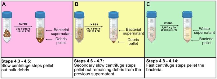
Figure 1: Bacterial superwash protocol. (A) Hydrate the stool and perform the slow centrifugation steps outlined in steps 4.3–4.5. (B) Pellet any remaining debris, leaving a bacteria-containing supernatant ready for further washing. (C) Pellet the bacteria using faster centrifugation, leaving behind waste supernatant and a cleaned bacterial pellet ready for downstream steps, including lysis and activity-based protein profiling. Please click here to view a larger version of this figure.

Figure 2: Overall workflow of chemoproteomic applications with human stool bacterial lysates. Starting with a human fecal sample, perform bacterial superwash steps followed by bacterial lysis to generate bacterial lysate for probe labeling. Perform copper-catalyzed azide-alkyne cycloaddition “click” chemistry, streptavidin enrichment of probe-labeled proteins, and tryptic digestion. Analyze the resulting peptides by liquid chromatography-tandem mass spectrometry and identify proteins using the respective database, resulting in a collection of proteins enriched by the activity-based probe. Please click here to view a larger version of this figure.
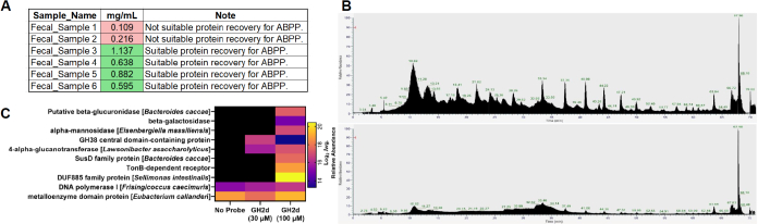
Figure 3: Data generated from fecal bacterial samples prepared using the chemoproteomic workflow. (A) Representative protein concentrations from the fecal lysate bicinchoninic acid assay. (B) Total ion chromatograms of a probe-labeled peptide sample over a 70 min LC-MS/MS gradient (top panel) and a no-probe sample over the same 70 min gradient (lower panel). The y-axis indicates the relative abundance of the detected ion signal at a given time (x-axis) during the LC-MS gradient. (C) Heatmap of LC-MS/MS chemoproteomic data from human stool labeled with the activity-based probe GH2d targeting glycoside hydrolases. Data represent the log2 average relative abundance of detected proteins. Raw ion intensities of peptides matched to a protein were summed. For a protein to be considered detected, it had to be present in three out of three replicates. These values were log2-transformed, and the average of the three values was calculated. Black cells indicate undetected proteins. The no-probe column (far left) shows off-target protein hits, and the other columns show GH2d applied at final concentrations of 30 µM and 100 µM. Please click here to view a larger version of this figure.
讨论
Samples derived from the human gut microbiome, such as stool, represent a complex matrix in which careful sample preparation and protocols need to be implemented. Specifically, for chemoproteomic applications, human stool needs to be handled and processed with minimally invasive steps that account for maintaining bacterial activities, so that chemoproteomic probes can capture live activities within the sample. Traditional sample processing methods for global proteomic or similar applications are generally not completely compatible with the specific needs of samples for chemoproteomic applications. Traditional preparation methods including extensive processing, activity inhibitors such as protease inhibitors, chemical lysis, detergent-assisted lysis, freeze/thaw-assisted lysis, or other similar sample processing methods all pose incompatibilities for ABPP with LC-MS/MS analysis. Additionally, the amount of sample, in terms of bacterial protein, needed for chemoproteomic applications is significantly higher than what is needed for other analyses, such as global proteomics. To ensure robust analyses with adequate protein quantities and adequate replicates, maximum recovery of functional bacterial lysate must be achieved from the processing steps. Because of these limitations, careful fecal sample preparation must be implemented to maintain the integrity of the sample, as well as maximize recovery of usable bacterial protein.
To process the stool in a way to minimize any natural sample degradation, careful planning is used at every step to make the protocol take only the minimum amount of time needed to complete it safely and properly. Setup of the BSC, preparation of materials, buffers, and adequate ice for maintaining the sample at cool temperatures is essential. Critical steps of this protocol include completing the protocol with minimal freeze-thaw cycles to maximize bacterial activities and functions. Keeping the freeze-thaw cycles limited before probe-labeling and CuAAC “click” chemistry is necessary to maintain bacterial activities24,25. Often in this protocol, freeze-thaw cycles are limited to two cycles before probe-labeling takes place; this ensures that bacterial activities are kept intact as best as feasible.
To maximize bacterial protein recovery, multiple washes of the bulk fecal sample (protocol steps 4.3–4.5) are important to repeat until the pellet is small, and the supernatant is mainly clear, indicating that most bacteria have been dislodged and washed out of the bulk of the stool debris. Initially during method development, this step did not involve any repeated resuspensions of the pellet, and the rest of the superwash was conducted after one wash. After troubleshooting, expanding this step to include multiple resuspensions and centrifugations was found to greatly increase bacterial recovery.
Another critical step is to accurately quantify protein after bacterial lysis. Taking lysate directly after lysis steps and using this as a sample for a BCA assay proved to falsely inflate the protein concentration of fecal lysates. To combat this, an acetone precipitation of a small portion of the collected lysate was used to precipitate proteins from the bacterial lysate and quantify those proteins that had been isolated from their color-tinted fecal matrix. This precipitation step proved to be critical in more accurately quantifying bacterial proteins from stool lysate, allowing for the colorimetric assay to perform normally.
The protocol is limited to the biological nature of the samples. While this protocol has been extensively tested on different fecal sample types, including varying Bristol Stool Scale scores, varying volumes of initial donation, and various health states, no two samples are the same. The difficulty of working with a complex, unpredictable matrix is only loosely translatable to handling traditional in vitro bacterial chemoproteomic work, in terms of sample preparation. Understanding which steps are critical throughout the protocol can help account for some of the shortcomings of the protocol, where even with best practices, the sample might have low protein recovery. A limitation of this protocol is that it is conducted aerobically. Given how many human gut bacteria are anaerobes, loss of some enzymatic activities is possible; however, activity assays tested in-house showed insignificant loss of the targeted enzymatic functions.
Significant advances in taxonomic approaches have allowed the gut microbiome field to make strides towards understanding microbes within complex matrices like the gut microbiome, but identifying function of active microbes within complex communities has remained a challenge. Chemoproteomics is a powerful strategy for probing bacterial activities and protein functions that has enabled deeper understanding of bacterial functions in complex systems, allowing for targeted inhibitor development of antibiotics, identifications of post-translational modifications (PTMs) in complex samples, and discovery of active enzymes within biological systems such as the gut microbiome11,12,13,14,26. This protocol, and ABPs in general, have direct applications in profiling enzymatic functions in healthy and diseased patients. For patients that undergo FMT, this protocol has unrealized applications for tracking metabolic functions following an FMT. ABP design is a careful balance applying probe design and chemical synthesis that allows for target capture of active enzymes, and careful design must be used as to not change the intrinsic structure of the targeting group which could lead to off-target labeling or poor reactivity in a complex system. Use of competitive labeling to identify true target proteins can validate probe targeting, as well as concentration-dependent labeling. Choosing the concentration of probe to deploy in an active system is not a one-size-fits-all – it is a balance that needs careful optimization. Previous work supports this, where concentrations are ABP-dependent, as well as biological system-and target-protein dependent. These obstacles when paired with complex biological systems like the gut microbiome, highlight the importance of careful chemoproteomic workflows from starting fecal matter through the analysis of targeted functional proteins.
披露
The authors have no conflicts of interest to declare.
致谢
We thank Agne Sveistyte and Whitney L. Garcia for help with early protocol development and testing, as well as advice on sample preparation. We acknowledge the use of core facilities provided at Baylor University. We acknowledge funding from the NIH NICCH grant #R01AT013241. We acknowledge the use of BioRender to prepare certain figures.
材料
| 姓名 | 公司 | 目录编号 | 评论 |
|---|---|---|---|
| 0.22 µm filter | Fisher Scientific | 14-578-816 | For filtering 1x PBS |
| 1.5 mL low-bind tubes | Fisher Scientific | PI90410 | |
| 1.5 mL tubes | Fisher Scientific | 21-402-902 | |
| 1.5 mL/2 mL rotor for centrifuges | Fisher Scientific | 58-207-60001 | |
| 1.5 mL individually wrapped tubes | Fisher Scientific | 02-681-331 | |
| 10x PBS | Fisher Scientific | BP39920 | |
| 2 mL cryovials | Fisher Scientific | 03-391-523 | |
| 2 mL snap-lock tubes | Fisher Scientific | E0030123620 | For flash freezing lysates |
| 25 mL serological pipettes | Fisher Scientific | 02-923-205 | |
| 5 mL cryovials | Fisher Scientific | 10-500-27 | |
| 50 mL conical tubes | Fisher Scientific | 12-565-270 | |
| 50 mL rotor for centrifuges | Fisher Scientific | 05-401-512 | |
| 70% ethanol in water | Fisher Scientific | 50-005-74341 | |
| Acetone | Fisher Scientific | A18-20 | CAS: 67-64-1 |
| Acetonitrile | Fisher Scientific | A996-4 | CAS: 75-05-8 |
| Amber MS vials with glass insert chamber | Fisher Scientific | 03-452-575 | |
| Ammonium bicarbonate | Sigma-Aldrich | A6141-500G | CAS: 1066-33-7 |
| BCA Protein Assay Kit | Fisher Scientific | PI23225 | |
| Bead mill homogenizer | Fisher Scientific | 17-100-864 | |
| Biological safety cabinet (BSC) | Labconco | 76317-638 | |
| Biotin alkyne | Fisher Scientific | 2615-100mg | |
| Biotin azide | Fisher Scientific | B10184 | |
| Bleach | Fisher Scientific | 50371500 | |
| Centrifuge | Fisher Scientific | 02-262-0603 | |
| Copper (II) sulfate | Fisher Scientific | LC134051 | CAS: 7758-99-8, CuSO4 |
| Dimethyl sulfoxide | Fisher Scientific | AAJ66650AE | CAS: 67-68-5, DMSO |
| Disposable lab coat | Fisher Scientific | 22-770-147 | |
| Disposable plastic scoop | Fisher Scientific | 18-001-017 | |
| Fecal donation buckets | Fisher Scientific | 02-544-208 | |
| Filter pipette tips | Fisher Scientific | 12-111-001, 12-111-126, 12-111-132 | |
| Formic acid | Fisher Scientific | A117-50 | CAS: 64-18-6 |
| GH2d ABP | Synthesized in-house; not commercially available | See comment/description for citation | J. Am. Chem. Soc. 2012, 134 (50), 20521–20532. https://doi.org/10.1021/ja309790w |
| Gloves | Fisher Scientific | 19-167-17 | |
| HPLC-grade water | Fisher Scientific | 7732-18-5 | CAS: 7732-18-5 |
| Ice bucket | Fisher Scientific | 07-210-128 | |
| LC-MS column | Thermo Scientific | ES75500PN | |
| LC-MS trap column | Thermo Scientific | 174500 | |
| Liquid chromotography system | Thermo Scientific | VN-S10-A-01 | |
| Liquid nitrogen | N/A (in-house) | N/A | |
| Mass spectrometer (MS) | Thermo Scientific | BRE725533 | |
| Mixing thermoblock | Fisher Scientific | 05-414-311 | |
| Paraffin film | Fisher Scientific | STLPM996 | |
| Pipet controller | Fisher Scientific | FB14955202 | |
| Pipettes (0.5–10 µL, 20–200 µL, 100–1,000 µL, 8 x 20–200 µL) | IKA | 0020011211, 0020011215, 0020011216, 0020017945 | |
| Polypropylene manifold columns | Fisher Scientific | 7326008EDU | |
| Repeat pipettor | Fisher Scientific | 13-683-550 | Optional, useful during enrichment steps |
| Repeat pipettor tips | Fisher Scientific | 13-683-726 | Optional, useful during enrichment steps (for repeat pipettor) |
| Silica glass beads | Sigma-Aldrich | BMSD113101-200G | |
| Sodium ascorbate | Sigma-Aldrich | A7631-500G | CAS: 134-03-2 |
| Sodium dodecyl sulfate | Sigma-Aldrich | 436143-100G | CAS: 151-21-3, SDS |
| Sonicator | Fisher Scientific | FB120110 | |
| Streptavidin agarose beads | VWR | 78781-823 | |
| Tris(3-hydroxypropyltriazolylmethyl)amine | Sigma-Aldrich | 762342-100MG | CAS: 760952-88-3, THPTA |
| Trypsin | Fisher Scientific | PRV5111 | |
| Tube membranes | Fisher Scientific | 50-189-9351 | |
| Tube rotator | Fisher Scientific | 88-861-122 | |
| Ultrapure water | Fisher Scientific | 14-558-729 | |
| Urea | Sigma-Aldrich | U5128-500G | CAS: 57-13-6 |
| Vacuum manifold | Fisher Scientific | PR-A7231 | |
| Vacuum concentrator | Fisher Scientific | 07-748-13 | |
| Vortex mixer | Fisher Scientific | 02-215-418 | |
| Vortex mixer | Fisher Scientific | 02-215-418 |
参考文献
- Proctor LM et al. The Integrative Human Microbiome Project. Nature. 2019;569(7758):641-648.
- Huttenhower C et al. The Human Microbiome Project Consortium. Structure, Function and Diversity of the Healthy Human Microbiome. Nature. 2012;486(7402):207-214.
- Van Den Bossche T et al. The Metaproteomics Initiative: A Coordinated Approach for Propelling the Functional Characterization of Microbiomes. Microbiome. 2021;9(1):243.
- Verberkmoes NC et al. Shotgun Metaproteomics of the Human Distal Gut Microbiota. ISME J. 2009;3(2):179-189.
- Franzosa EA et al. Relating the Metatranscriptome and Metagenome of the Human Gut. Proc Natl Acad Sci U S A. 2014;111(22):E2329-E2338.
- Zhang X et al. MetaPro-IQ: A Universal Metaproteomic Approach to Studying Human and Mouse Gut Microbiota. Microbiome. 2016;4(1):31.
- Li W et al. Posttranslational Modifications Remodel Proteome-Wide Ligandability. Nat Chem Biol. 2026:1-13.
- Whidbey C. The Right Tool for the Job: Chemical Biology and Microbiome Science. Cell Chem Biol. 2025;32(1):83-97.
- Killinger BJ et al. Activity-Based Protein Profiling Identifies Alternating Activation of Enzymes Involved in the Bifidobacterium Shunt Pathway or Mucin Degradation in the Gut Microbiome Response to Soluble Dietary Fiber. NPJ Biofilms Microbiomes. 2022;8(1):60.
- Cravatt BF, Wright AT, Kozarich JW. Activity-Based Protein Profiling: From Enzyme Chemistry to Proteomic Chemistry. Annu Rev Biochem. 2008;77:383-414.
- Han L, Chang PV. Activity-Based Protein Profiling in Microbes and the Gut Microbiome. Curr Opin Chem Biol. 2023;76:102351.
- Wright AT, Hudson LA, Garcia WL. Activity-Based Protein Profiling - Enabling Phenotyping of Host-Associated and Environmental Microbiomes. Isr J Chem. 2023;63(3-4):e202200099.
- Russo J et al. Synthesis and Application of a Suite of 2,5-Aryl Tetrazole Photoaffinity-Based Probes for Profiling Microbial Carbohydrate and Mucin Metabolism in Gut Microbiota. ACS Chem Biol. 2026;21(6):1496-1506.
- Adhikari A et al. Development of a Covalent Inhibitor of Gut Bacterial Bile Salt Hydrolases. Nat Chem Biol. 2020;16(3):318-326.
- Thuy-Boun PS et al. Quantitative Metaproteomics and Activity-Based Protein Profiling of Patient Fecal Microbiome Identifies Host and Microbial Serine-Type Endopeptidase Activity Associated With Ulcerative Colitis. Mol Cell Proteomics. 2022;21(3):100197.
- Gao S et al. Comparing and Optimizing Protein Extraction Methods with Different Lysis Buffers for the Analyses of Human Fecal Microbiome via Metaproteomics Approach. J Microbiol Methods. 2025;237:107244.
- Chauvigne-Hines LM et al. Suite of Activity-Based Probes for Cellulose-Degrading Enzymes. J Am Chem Soc. 2012;134(50):20521-20532.
- Kim S, Pevzner P. MS-GF+ makes progress towards a universal database search tool for proteomics. Nat Commun. 2014;5:5277.
- Eng JK et al. An approach to correlate tandem mass spectral data of peptides with amino acid sequences in a protein database. J Am Soc Mass Spectrom. 1994;5(11):976-989.
- Perkins DN et al. Probability-based protein identification by searching sequence databases using mass spectrometry data. Electrophoresis. 1999;20:3551-3567.
- Cox J, Mann M. MaxQuant enables high peptide identification rates, individualized p.p.b.-range mass accuracies and proteome-wide protein quantification. Nat Biotechnol. 2008;26(12):1367-1372.
- Wegner CD, Coon JJ. A proteomics search algorithm specifically designed for high-resolution tandem mass spectra. J Proteome Res. 2013;12(3):1377-1386.
- Keller A et al. Empirical statistical model to estimate the accuracy of peptide identifications made by MS/MS and database search. Anal Chem. 2002;74(20):5383-5392.
- Tang Q et al. Chemical Tagging of Protein Lipoylation. Angew Chem Int Ed Engl. 2021;60(8):4028.
- Karatza E et al. The impact of handling and storage of human fecal material on bacterial activity. J Pharm Sci. 2016;105(11):3458-3461.
- Sangermani M et al. Effects of repeated freeze and thaw cycles on the stability of faecal microbiome composition. Sci Rep. 2026:9880.
重印与许可
本文已发表
视频即将推出