This case reports a refractory Descemet membrane detachment (DMD) following phacoemulsification in a patient with prior pterygium surgery. Repeated AS-OCT-guided descemetopexy achieved successful reattachment, highlighting that tailored, image-guided intervention is critical for managing complex DMD, especially in scarred corneas.
病例报告
Treatment of Refractory Descemet Membrane Detachment via Repeat Descemetopexy: A Case Report
本文内容
摘要
摘要
Descemet membrane detachment (DMD) is a relatively uncommon but potentially vision-threatening complication of phacoemulsification that can lead to persistent corneal edema and visual impairment. We report a case of refractory DMD following cataract surgery in an elderly patient with a history of bilateral pterygium surgery and associated corneal scarring. An 82-year-old man with age-related cataract underwent phacoemulsification and intraocular lens implantation. DMD was identified intraoperatively, and initial intracameral air injection failed to achieve stable reattachment. Postoperative slit-lamp examination and anterior segment optical coherence tomography (AS-OCT) demonstrated persistent and subsequently recurrent DMD with corneal stromal edema. Based on the size, location, and configuration of the detached membrane, two additional reattachment procedures were performed using air injection combined with mechanical repositioning. The injection site and direction were adjusted according to AS-OCT findings to facilitate reattachment of the detached membrane. Following the final intervention, the Descemet membrane was completely reattached, corneal transparency was restored, and visual acuity improved to 0.3 at 1-month follow-up. This case suggests that previous pterygium surgery and corneal scarring may increase the risk of persistent DMD after phacoemulsification. For refractory DMD, individualized treatment guided by AS-OCT, with precise air injection directed toward the origin of the detachment and mechanical repositioning when necessary, may facilitate successful anatomical and functional recovery.
引言
In recent years, Descemet membrane detachment (DMD) has been a known common complication secondary to the extensive application of phacoemulsification1,2. The incidence of DMD during phacoemulsification cataract surgery is approximately 0.5% to 2.2%, with significant visual impact observed in about 2.2% of these cases3. DMD can lead to persistent corneal edema and vision loss. If not managed promptly and appropriately, it may result in irreversible corneal changes and visual sequelae4. In severe cases, portions of the detached Descemet's membrane may be observed floating in the anterior chamber4. Early diagnosis and treatment are pivotal for the visual prognosis of patients with moderate/severe DMD, particularly when spontaneous reattachment is unlikely. Most DMD may be identified intraoperatively by observing a transparent membrane attached to the cornea in the anterior chamber. Meticulous slit-lamp examination remains essential for identifying DMD and assessing its extent and configuration when planning treatment. Postoperative imaging techniques such as anterior segment optical coherence tomography (AS-OCT)5,6, ultrasound biomicroscopy7, confocal microscopy8, and Scheimpflug imaging6 are used for timely and accurate diagnosis and evaluation of DMD, thus contributing to the selection of an appropriate treatment strategy. In particular, AS-OCT can be performed preoperatively to characterize the location, extent, and configuration of DMD, while intraoperative OCT (iOCT) may provide additional real-time information to assist surgeons in planning the site and direction of intracameral air injection. Current treatment strategies for DMD include conservative observation in selected cases and active interventions, such as descemetopexy, suture fixation, and keratoplasty, with the choice of treatment depending on the size, location, and severity of the detachment9,10,11,12. Descemetopexy with intracameral air injection is a relatively simple and minimally invasive approach that promotes direct reattachment of the detached Descemet's membrane to the underlying stroma, although repeated intervention may be required in refractory cases13.
In the present case, a refractory DMD following phacoemulsification in a patient with a history of pterygium surgery and corneal scarring is reported. The overall aim of this case report is to describe the clinical course and management of this refractory DMD and to highlight the potential value of AS-OCT in guiding individualized repeated reattachment procedures. This case is particularly relevant because the initial air injection failed to achieve stable reattachment, requiring subsequent interventions based on the size and location of the detachment. Compared with more invasive surgical approaches, repeated intracameral air injection provides a relatively simple and minimally invasive option for selected refractory DMD cases. This case therefore provides additional clinical experience regarding the individualized management of complex DMD and the role of image-guided treatment.
CASE PRESENTATION:
An 82-year-old man presented with bilateral decreased visual acuity for more than 10 years and was diagnosed with age-related cataracts. He was otherwise healthy but had undergone bilateral pterygium surgery approximately 10 years earlier. On admission, visual acuity was 0.1 in the right eye (corrected, 0.16) and 0.02 in the left eye (corrected, 0.2). Intraocular pressure was 14 mmHg in the right eye and 15 mmHg in the left eye. A corneal macula was present in the bilateral nasal cornea and largely covered the left pupil. The remaining cornea was relatively transparent, and the lens showed a nuclear hardness grade of IV. The endothelial cell counts (Corneal endothelial cell analyzer, Table of Materials) were 2171.8 cells/mm2 in the right eye and 2271.4 cells/mm2 in the left eye.
The patient underwent phacoemulsification (Phacoemulsification system, Table of Materials) combined with foldable intraocular lens implantation in the left eye under superficial anesthesia. During cortical removal, a transparent membranous structure was observed floating in the anterior chamber and partially connected to the cornea, suggesting DMD. Intracameral air was injected through the 2 o'clock incision to facilitate reattachment of the Descemet membrane. After completion of cortical removal and intraocular lens implantation, the pupil was constricted, and the eye was covered with a bandage.
On the first postoperative day, slit-lamp examination (Table of Materials) revealed mild stromal edema near the superior-nasal corneal incision and a linear abnormality at the posterior surface of the inferior cornea, indicating persistent non-reattachment of the Descemet membrane. The membrane was detached and folded downward in a semicircular configuration, extending from approximately the 8 o'clock peripheral region toward the 12 o'clock central corneal region. AS-OCT (Table of Materials) demonstrated a double-layer hyperintense signal corresponding to the detached membrane and confirmed persistent DMD.
Because the corneal stromal edema was increasing, repeat reattachment was performed on the second postoperative day. A puncture was made at 5 o'clock from the opposite direction toward the detached membrane. Mechanical manipulation was used to reposition the folded Descemet membrane, followed by intracameral air injection to maintain its reattached position. After the procedure, approximately two-thirds of the anterior chamber remained filled with air, and the patient was closely monitored for postoperative complications. Intraocular pressure remained within the normal reference range, although mild stromal edema persisted.
On the fourth postoperative day, corneal stromal edema remained evident and the patient reported decreased visual acuity. Slit-lamp examination and AS-OCT demonstrated recurrent separation of the Descemet membrane from the stroma, although the membrane was no longer folded. A third reattachment procedure was therefore performed, with repeat intracameral air injection. Following this intervention, stromal edema gradually improved and the Descemet membrane became completely reattached to the posterior corneal surface.
At the 1-month outpatient follow-up, the corneal stroma had become transparent, and visual acuity in the affected eye had improved to 0.3. No further intervention was required.
DIAGNOSIS, ASSESSMENT, AND PLAN:
The patient was diagnosed with refractory Descemet membrane detachment (DMD) following phacoemulsification in the left eye. The diagnosis was based on intraoperative visualization of a transparent membranous structure partially detached from the corneal surface and confirmed postoperatively by slit-lamp examination and anterior segment optical coherence tomography (AS-OCT). AS-OCT demonstrated persistent separation of the Descemet membrane from the posterior corneal stroma, with a folded configuration of the detached membrane.
The refractory nature of the DMD was considered to be associated with several factors, including previous bilateral pterygium surgery, corneal scarring, limited visualization of the surgical field, and the extensive size and configuration of the detached membrane. AS-OCT was used to assess the location, extent, and configuration of the detachment and to guide subsequent treatment decisions.
Because persistent corneal edema and non-reattachment of the Descemet membrane were observed after initial intracameral air injection, repeat descemetopexy was planned. The subsequent procedures were performed according to AS-OCT findings, with adjustment of the puncture site and air injection direction toward the origin of the detachment. Mechanical repositioning was performed when membrane folding prevented adequate reattachment. Postoperative monitoring included slit-lamp examination, intraocular pressure measurement, and AS-OCT evaluation until stable anatomical reattachment was achieved.
方案
This single retrospective case report describes the clinical course of one patient and was exempt from formal ethical review under institutional policy and applicable national regulations governing medical research involving humans. The Institutional Review Board/Research Ethics Committee confirmed that ethical approval was not required; therefore, no approval number or date was issued. Written informed consent was obtained from the patient for participation and publication of this case report and any accompanying clinical images.
1. Preoperative assessment
- Evaluate the patient for potential risk factors for DMD before phacoemulsification, including previous corneal surgery, corneal scarring, and other conditions that may interfere with visualization of the anterior chamber.
- Perform routine ophthalmic examination (Ophthalmic surgical microscope, Table of Materials), including visual acuity, intraocular pressure, slit-lamp examination, and corneal endothelial cell density.
- Assess the corneal condition and identify any pre-existing corneal opacity or macular changes that may limit visualization of the surgical field.
2. Intraoperative identification and initial management of DMD
- Standard phacoemulsification: under topical anesthesia, create a 2.8-3.2 mm clear corneal main incision and side ports; inject ophthalmic viscosurgical device (OVD), perform a 5-6 mm capsulorhexis, hydrodissection, and hydrodelineation; emulsify and aspirate the nucleus using divide-and-conquer or chop techniques; remove residual cortex and polish the posterior capsule; implant a foldable intraocular lens (IOL) in the capsular bag; aspirate OVD, hydrate the incisions, and confirm a sutureless watertight closure with a stable anterior chamber.
- Carefully inspect the anterior chamber during cortical removal for a transparent membranous structure floating within the anterior chamber or remaining partially attached to the cornea, which may indicate DMD.
- When DMD is identified, assess the apparent location and extent of the detached Descemet membrane and determine an appropriate site for intracameral air injection.
- Inject air into the anterior chamber through the selected corneal incision to promote reattachment of the detached Descemet membrane to the underlying stroma. The puncture site is selected from the direction opposite to the origin or extension of the detachment. Air is injected into the anterior chamber through the selected corneal incision using a blunt anterior chamber cannula attached to a syringe, and is slowly directed toward the detached Descemet membrane under direct visualization to promote reattachment to the underlying stroma. The filling endpoint is formation of an air bubble large enough to tamponade the detached membrane; approximately two-thirds of the anterior chamber is filled with air, and care is taken to avoid overfilling. Postoperatively, the patient is kept supine, and intraocular pressure is monitored.
- Complete the remaining cataract procedure and maintain appropriate postoperative positioning and observation.
3. Postoperative evaluation using AS-OCT
- Perform slit-lamp examination on the first postoperative day to evaluate corneal edema, visual axis involvement, and persistent non-attachment of the Descemet membrane.
- Perform AS-OCT when persistent or recurrent DMD is suspected.
- Use AS-OCT to determine the location, extent, configuration, and relationship of the detached Descemet membrane to the underlying corneal stroma.
- Use the AS-OCT findings to determine the direction and site of subsequent reattachment procedures rather than relying solely on the original incision site.
4. Repeat descemetopexy for persistent or recurrent DMD
- When the detached membrane remains non-adherent and corneal edema is increasing, perform repeat reattachment based on the AS-OCT findings.
- Select the puncture site from the direction opposite to the origin or extension of the detachment when appropriate, with the aim of directing the air toward the detached membrane.
- When the Descemet membrane is folded, use gentle mechanical manipulation to reposition and flatten the detached membrane before or during air injection. When the Descemet membrane is folded or inverted, gently manipulate the membrane using a blunt ophthalmic instrument or cannula to unfold and reposition it against the posterior corneal stroma. In this case, the puncture site was selected at the 5 o'clock position opposite to the origin of the detachment, allowing the instrument and air flow to approach the detached membrane from the opposite direction. Excessive mechanical force should be avoided to prevent further endothelial or Descemet membrane injury.
- Inject intracameral air to maintain contact between the repositioned Descemet membrane and the posterior corneal stroma. Inject sterile air into the anterior chamber through the selected incision to provide tamponade and maintain contact between the repositioned Descemet membrane and the posterior corneal stroma. In this case, air was injected until approximately two-thirds of the anterior chamber was filled, while avoiding excessive pressure elevation. After air injection, intraocular pressure was monitored and the patient was positioned supine to facilitate sustained contact between the Descemet membrane and the underlying stroma.
- Monitor intraocular pressure and corneal clarity after the procedure and evaluate the persistence or recurrence of DMD using slit-lamp examination and AS-OCT when clinically indicated.
5. Management of recurrent DMD
- If AS-OCT demonstrates recurrent separation of the Descemet membrane after initial reattachment, reassess the membrane configuration and determine whether repeat intervention is required.
- Repeat mechanical repositioning and intracameral air injection when persistent separation is present, and the membrane remains amenable to reattachment.
- Continue postoperative monitoring until stable anatomical reattachment of the Descemet membrane is confirmed.
6. Follow-up assessment
- Assess corneal transparency, visual acuity, and the anatomical status of the Descemet membrane during follow-up.
- Confirm complete reattachment and resolution of corneal stromal edema before discontinuing further intervention.
- Record the final visual outcome and any treatment-related complications during follow-up.
结果
Following the initial intracameral air injection, the Descemet membrane remained detached, with persistent corneal stromal edema observed on the first postoperative day (Figure 1). AS-OCT confirmed persistent DMD and demonstrated a folded configuration of the detached membrane (Figure 2). A second reattachment procedure was therefore performed using mechanical repositioning followed by intracameral air injection. Although the membrane was initially reattached, recurrent DMD was identified on the fourth postoperative day by slit-lamp examination and AS-OCT. A third reattachment procedure with intracameral air injection was subsequently performed. Following the final intervention, the Descemet membrane remained completely reattached, corneal stromal edema gradually resolved, and corneal transparency was restored. At the 1-month follow-up, visual acuity in the affected eye had improved to 0.3, and no further intervention was required.
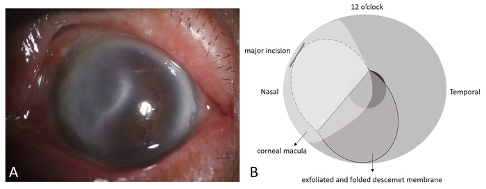
Figure 1: Slit-lamp findings of Descemet membrane detachment after phacoemulsification. (A) Large-scale corneal macula adjacent to the nasal region covering the pupil area, with slight stromal edema near the major incision at the superior-nasal corneal margin, observed on the first postoperative day. (B) Schematic illustration of Descemet membrane detachment. Please click here to view a larger version of this figure.
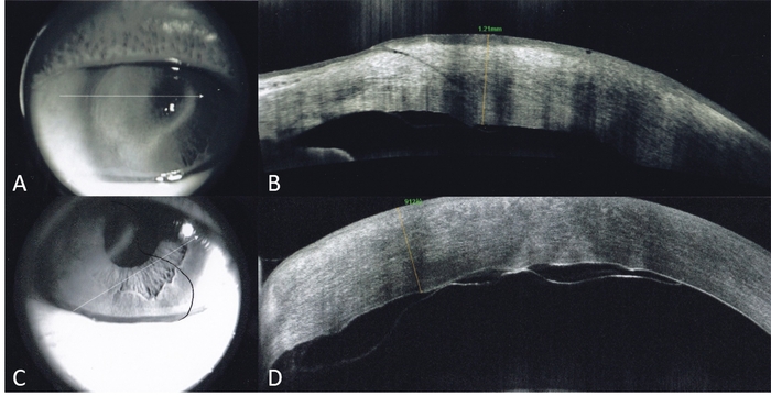
Figure 2: AS-OCT findings of Descemet membrane detachment. (A) Scanning direction corresponding to panel B. (B) Increased corneal thickness and Descemet membrane detachment at the major incision. (C) Scanning direction corresponding to panel D. (D) Detached Descemet membrane folded downward, appearing as a double-layer hyperintense signal in the inferior cornea on AS-OCT. Please click here to view a larger version of this figure.
讨论
DMD is a potential vision-threatening complication related to intraocular surgeries, including extracapsular cataract extraction, small incision cataract surgery, phacoemulsification, iridectomy, trabeculectomy, pars plana vitrectomy, viscocanalostomy, and keratoplasty1. The classification and treatment of DMD depend mainly on the size, location, and configuration of the detachment2. Small and peripheral detachments may undergo spontaneous reattachment through aqueous humor circulation, whereas active intervention is generally considered for larger detachments, particularly when there is a substantial separation between the Descemet membrane and stroma, membrane folding, or involvement of the optical axis2. Descemetopexy9,10, suture fixation11, and keratoplasty12 are indicated for moderate/severe DMD when over 1/4 of the corneal region with a tremendous gap (>1 mm) between the Descemet membrane and the stroma are detected, as well as the folded margin and jeopardized optical axis region is present. Descemetopexy is commonly acknowledged as the option of priority for treating DMD as long as the puncture position is accurately located, even though repetitive injection is necessary for DMD over 1/2 of the corneal region13. Successful air descemetopexy also depends on adequate maintenance of intracameral tamponade and wound integrity, because postoperative air leakage may reduce the tamponade effect and potentially contribute to persistent or recurrent DMD.
In the present case, the detached Descemet membrane was initially overlooked because the surgical field was obscured by a corneal macula, making the direction of the enlarged detachment difficult to identify. Consequently, a viscoelastic substance was applied from the distal site, accompanied by intracameral air injection through the former incision rather than close-range reattachment. Local corneal edema and a Descemet membrane detachment with laceration from the stroma at the former incision at 2 o'clock, occupying approximately one-quarter of the corneal region, including the pupil area, were detected by AS-OCT on the first postoperative day. The detached membrane was folded downward toward the inferior cornea. During the first-stage treatment, air was injected from the lateral incision rather than from the opposite direction toward the origin of the DMD, resulting in reverse folding and unsatisfactory reattachment of the Descemet membrane. In contrast, during the subsequent procedure, a puncture was prepared at 5 o'clock toward the origin of the detachment. A small amount of air was injected along the direction of the detachment to unfold the folded flap, followed by repeated rinsing with the needle to facilitate anatomical reattachment. The anterior chamber was then filled with air to maintain the reattached membrane. Other interventions were not selected because viscoelastic substances may remain between the Descemet membrane and stroma and impede reattachment, whereas inert gas may cause complications including pupillary block and ocular hypertension14. However, recurrent detachment occurred because the intracameral air was absorbed within a short time and could not maintain sufficient pressure for complete healing of the extensive DMD. The membrane therefore became partially detached again, and a third intracameral air injection was performed until visual acuity was satisfactorily rehabilitated. In retrospect, factors affecting the duration and effectiveness of intracameral tamponade, including wound integrity and potential air leakage, should be considered in recurrent DMD after air descemetopexy. The available information does not allow determination of whether wound leakage contributed to the recurrence; therefore, this possibility cannot be excluded. When intracameral air or gas is used, prevention of pupillary block and appropriate postoperative positioning should also be considered. An inferior peripheral iridotomy may be considered when clinically appropriate to reduce the risk of pupillary block, while supine positioning may help maintain contact between the detached Descemet membrane and posterior stroma15. These measures should be individualized according to the extent of DMD, the amount of intracameral air or gas, and the patient's clinical condition.
In summary, several factors may have contributed to the development of DMD following phacoemulsification in this case. Previous pterygium surgery and corneal scarring may have altered the local corneal structure and reduced the tolerance of the Descemet membrane to surgical manipulation. Incision placement in an area with corneal abnormalities may have increased the risk of membrane injury during intraocular instrument passage. In addition, prolonged surgical manipulation and higher ultrasound energy required for hard nuclear cataract removal may have increased the likelihood of Descemet membrane trauma. Furthermore, corneal opacity and edema may have impaired visualization of the anterior chamber and complicated intraoperative identification of DMD. Based on these observations, preventive strategies were adopted during subsequent cataract surgery on the contralateral eye, including selecting an incision site with relatively normal corneal tissue, minimizing unnecessary intraocular manipulation, and optimizing ultrasound parameters. The subsequent procedure was associated with satisfactory visual recovery and no occurrence of DMD. These findings suggest that careful preoperative assessment, individualized surgical planning, and meticulous intraoperative management may help reduce the risk of DMD, particularly in eyes with previous corneal surgery or scarring.
There is no unified guideline for the treatment of refractory DMD16. Meticulous slit-lamp examination may reveal this type of DMD, particularly when the detached Descemet membrane has become folded or rolled, and the surgeon may attempt to unroll and reattach it. A "double-bubble pneumo-descemetopexy" technique has been described, in which a small air bubble is used to unroll the Descemet membrane, followed by a larger air bubble for descemetopexy, with successful reattachment reported15. Another technique, termed the "pull technique," uses a bent-tip 27-gauge cannula to pull and disimpact the inferior border of an impacted DMD, after which an intracameral air bubble can unfold and reattach the Descemet membrane17. These techniques provide additional minimally invasive approaches for selected cases of folded or impacted DMD. Although neither technique was used in the present case, the treatment similarly involved mechanical repositioning of the folded membrane followed by intracameral air injection to facilitate reattachment. For persistent or recurrent DMD, other reported options include full-thickness corneal sutures to anchor the detached Descemet membrane to the underlying stroma and longer-acting gas tamponade with sulfur hexafluoride (SF6) or perfluoropropane (C3F8). These approaches may be considered in selected cases when conventional air descemetopexy is insufficient, although they were not used in the present case.
This case, through a cyclical "assessment-intervention-reassessment" model, validated the necessity of individualized management. For instance, during the second repositioning procedure, it was observed that the DMD was unfolded but not fully adhered, suggesting that pneumatic tamponade alone might be insufficient and should be combined with mechanical flattening3,18. Furthermore, postoperative monitoring of intraocular pressure, visual function, and corneal clarity is important after intracameral air descemetopexy to assess anatomical recovery and detect potential complications14. However, the present case does not establish the superiority of this approach over other treatment strategies.
This case report indicates that for refractory DMD associated with corneal scarring, repeated Descemet membrane repositioning guided by AS-OCT may be a useful, individualized treatment approach. The purpose of this case report is not to propose a novel or unique surgical technique, as both intracameral air descemetopexy and mechanical repositioning are established approaches. Rather, this case illustrates the stepwise management of persistent and recurrent DMD with a folded membrane and the role of serial Anterior Segment Optical Coherence Tomography (AS-OCT) assessment in guiding subsequent treatment decisions. This study further summarizes the following key points for clinical practice: First, the identification and assessment of preoperative high-risk factors (such as a history of pterygium surgery) should be emphasized. Second, AS-OCT should be utilized as an important imaging tool for preoperative assessment, treatment planning, and postoperative evaluation in such cases. Finally, for refractory cases, a flexible combination of mechanical repositioning and intracameral air or, when clinically appropriate, longer-acting gas tamponade may be considered to facilitate reattachment. The aforementioned experience provides practical insights into the individualized clinical management of complex Descemet Membrane Detachment (DMD), rather than evidence of a novel or uniquely superior treatment.
披露
The authors have nothing to disclose. No artificial intelligence (AI) tools were used in the preparation of this manuscript or figures.
致谢
The authors received no financial support for the research, authorship, and/or publication of this article.
材料
| 姓名 | 公司 | 目录编号 | 评论 |
|---|---|---|---|
| Anterior segment optical coherence tomography (AS-OCT) system | Heidelberg Engineering, Germany | Spectralis OCT | Used for postoperative anterior segment examination and detection of corneal stromal edema, Descemet membrane detachment, and air bubble presence. |
| Corneal endothelial cell analyzer | Konan Medical, Japan | CellChek D | Used to assess the corneal endothelial cells. |
| Disinfectant air (sterile filtered air) | Hospital pharmacy / operating room supply | N/A | Injected into the anterior chamber for pneumatic tamponade to reattach the detached Descemet membrane. |
| Foldable intraocular lens (IOL) | Not specified | N/A | Implanted into the capsular bag after phacoemulsification for visual rehabilitation. |
| Ophthalmic surgical microscope | Carl Zeiss Meditec, Germany | OPMI Lumera | Provided intraoperative visualization during ophthalmic surgery. |
| Phacoemulsification system | Alcon, USA | Centurion Vision System | Used for cataract removal via ultrasound energy during the primary surgery. |
| Pilocarpine | Not specified (miotic agent) | N/A | Used to constrict the pupil before repeat descemetopexy to facilitate surgical access. |
| Slit-lamp microscope | Haag-Streit, Switzerland | BQ 900 | Used for postoperative anterior segment examination and detection of corneal stromal edema, Descemet membrane detachment, and air bubble presence. |
参考文献
- Singhal D, et al. Descemet membrane detachment. Surv Ophthalmol. 2020;65(3):279-293.
- Benatti CA, Tsao JZ, Afshari NA. Descemet membrane detachment during cataract surgery: etiology and management. Curr Opin Ophthalmol. 2017;28(1):35-41.
- Gab-Alla AA. A comparative study between air bubble tamponade alone versus air bubble tamponade with internal fluid aspiration for nonplanar Descemet's membrane detachment after phacoemulsification. J Ophthalmol. 2021;2021:9953418.
- Keye P, Reinhard T, Maier P. Therapie der Descemetolyse nach Kataraktoperation: Erfolgreiche Visusrehabilitation durch wiederholte intrakamerale Lufteingabe [Treatment of Descemet's membrane detachment after cataract surgery: successful visual recovery by repeated injection of air into the anterior chamber]. Ophthalmologe. 2020;117(7):700-703. German.
- Guo P, et al. Study on the classification of Descemet membrane detachment after cataract surgery with AS-OCT. Int J Med Sci. 2018;15(11):1092-1097.
- Li F, et al. A simple repair algorithm for Descemet's membrane detachment performed at the slit lamp. J Clin Med. 2022;11(23):7001.
- Osman EA. The benefit of ultrasound biomicroscopy (UBM) in management of total Descemet's membrane detachment after deep sclerectomy surgery. Int Ophthalmol. 2011;31(4):345-348.
- Patel DV, Phang KL, Grupcheva CN, Best SJ, McGhee CNJ. Surgical detachment of Descemet's membrane and endothelium imaged over time by in vivo confocal microscopy. Clin Exp Ophthalmol. 2004;32(5):539-542.
- Jain R, Mohan N. Outcomes of repeat descemetopexy in post-cataract surgery Descemet membrane detachment. Am J Ophthalmol. 2014;157(3):571-575.e2.
- Weng Y, Ren YP, Zhang L, Huang XD, Shen-Tu XC. An alternative technique for Descemet's membrane detachment following phacoemulsification: case report and review of literature. BMC Ophthalmol. 2017;17(1):109.
- Das M, Begum Shaik M, Radhakrishnan N, Prajna VN. Descemet membrane suturing for large Descemet membrane detachment after cataract surgery. Cornea. 2020;39(1):52-55.
- Kim JJ, Kim HK. Descemet membrane stripping endothelial keratoplasty for Descemet membrane detachment following phacoemulsification. Can J Ophthalmol. 2015;50(1):73-76.
- Ti SE, Chee SP, Tan DTH, Yang YN, Shuang SL. Descemet membrane detachment after phacoemulsification surgery: risk factors and success of air bubble tamponade. Cornea. 2013;32(4):454-459.
- Jain R, Murthy SI, Basu S, Ali MH, Sangwan VS. Anatomic and visual outcomes of descemetopexy in post-cataract surgery Descemet's membrane detachment. Ophthalmology. 2013;120(7):1366-1372.
- Sharma A, Sharma R, Kulshreshta A, Nirankari VS. Double bubble pneumo-descemetopexy for the management of Descemet membrane detachment: an innovative technique. Indian J Ophthalmol. 2023;71(5):2234-2236.
- Chudzinski R, et al. Decollement descemetique post-phakoemulsification: a propos de 9 cas [Descemet's membrane detachment after phacoemulsification: series of 9 cases]. J Fr Ophtalmol. 2020;43(10):1002-1008.
- Sharma A, Sharma R, Kulshreshta A, Nirankari V. Manual schism and intracameral air injection for impacted Descemet's membrane detachment. BMJ Case Rep. 2023;16(8):e253252.
- Fernandez Lopez E, et al. Descemet stripping only and ripasudil for the treatment of traumatic Descemet's membrane ruptures. Eur J Ophthalmol. 2023;33(4):NP13-NP18.