High-temperature exposure severely degrades deoxyribonucleic acid (DNA) in burned bones, limiting conventional short tandem repeat profiling. This review examines thermal damage mechanisms, current forensic DNA workflows, and emerging approaches for highly degraded remains, with particular emphasis on the potential and current limitations of hybridization capture for forensic identification.
综述文章
Forensic DNA Analysis of Burned Bones: Current Research Status and Future Perspectives
* These authors contributed equally
本文内容
摘要
摘要
Burned skeletal remains are often critical evidence for human identification in fires, deliberate burning, aircraft accidents, explosions, and similar events because bone persists under extreme thermal conditions. However, extensive DNA degradation caused by high-temperature burning poses a major challenge to conventional short tandem repeat (STR) profiling. In recent years, DNA hybridization capture has emerged as a promising molecular approach for enriching target fragments from highly degraded samples, offering new opportunities to overcome key limitations in DNA analysis of burned bone. This review summarizes the mechanisms by which high-temperature burning alters the physicochemical properties of bone and affects DNA preservation. Particular emphasis is placed on multilevel thermal changes across temperature ranges, including collagen denaturation, hydroxyapatite crystal reorganization, DNA strand fragmentation, and chemical modification. On this basis, we review the current workflow for forensic DNA analysis of burned bone, including skeletal element selection, decontamination and bone powder preparation, decalcification strategies, silica-based extraction and purification, STR profiling, and high-throughput sequencing (HTS). We further discuss the differential applicability of complete and incomplete decalcification protocols for burned bone samples, the advantages of ancient DNA (aDNA) extraction methods for highly charred remains, and the forensic utility of automated extraction systems. Given the extremely low DNA yield, severe fragmentation, inhibitor interference, and lack of standardized protocols associated with extensively burned samples, this review also evaluates the potential of DNA hybridization capture and targeted enrichment technologies. Because hybridization capture does not require intact double-stranded DNA (dsDNA) templates and can efficiently recover target sequences from ultrashort DNA fragments, it may be particularly well suited to burned skeletal material, although the current forensic evidence base remains limited. This review provides an integrated overview of burned bone DNA analysis, from thermal damage mechanisms to targeted enrichment strategies, and highlights how hybridization capture may extend forensic practice beyond identification toward DNA phenotyping.
引言
In forensic casework, burned skeletal remains are often among the most important forms of biological evidence for reconstructing events. In fires, deliberate burning of human remains, aircraft accidents, explosions, and similar incidents, soft tissues may be completely destroyed, leaving only fragmented skeletal material for identification1. Recovering informative genetic data from highly charred and fragmented bone has therefore remained a persistent technical challenge.
The value of bone as a DNA source after thermal exposure is closely related to its structure. Bone contains osteocytes, osteoblasts, and osteoclasts, which carry the individual’s complete genetic information; hydroxyapatite provides high density and resistance to degradation, allowing bone to retain extractable DNA even when its external morphology is severely damaged2. Traditionally, identification of burned bone has relied largely on histomorphological features such as colour changes and fracture patterns. However, burning causes irreversible structural damage. Burning temperature, exposure duration, and oxygen availability jointly determine the final state of DNA preservation; DNA analysis may remain feasible at 250°C–400°C under certain conditions, depending strongly on exposure duration, oxygen availability, tissue type, and the experimental model used, whereas success rates decrease sharply at higher temperatures. Collagen denatures at approximately 155°C and is largely lost by 400°C3, and hydroxyapatite undergoes marked crystal growth and phase transition above 500°C, changes that both damage DNA directly and indirectly interfere with its extraction and detection4.
Forensic approaches have shifted from histomorphological assessment toward molecular analysis. Earlier studies used microscopic changes in bone tissue, such as lamellar bone disruption and carbonization of osteocytes, to determine whether bone had been burned5. Morphological methods nevertheless have inherent limitations: diagenetic alteration may produce similar changes and complicate interpretation, and morphology cannot provide individual genetic information6. Although short tandem repeat (STR) profiling remains the gold standard, the severe DNA fragmentation caused by burning frequently leads to profiling failure7. DNA hybridization capture, which enriches target fragments from highly degraded samples, offers a promising strategy for overcoming this bottleneck.
Although burned bone identification has been studied for decades, much of the literature has focused on histomorphological observations or practical experience with conventional STR analysis, and an integrated understanding of thermal damage mechanisms and optimization strategies across the complete DNA workflow remains insufficient8. This review therefore summarizes how high-temperature burning affects the physicochemical properties of bone and the preservation of DNA, critically evaluates current methods and optimization strategies from sampling to genotyping, and discusses emerging technologies for extremely degraded samples.
Several reviews have recently addressed DNA analysis of thermally altered or skeletal remains, most notably the systematic review by Sessa et al., which followed Preferred Reporting Items for Systematic Reviews and Meta-Analyses (PRISMA) 2020, searched Scopus and Web of Science to June 5, 2026, and included 37 studies on genetic identification of burned human remains9, and the review by Cainé et al. on human skeletal remains more broadly2. The present review differs in both focus and purpose. Sessa et al. address “what works,” cataloguing which approaches have been validated in degraded or burned remains; the present review addresses “why and how the workflow fails,” tracing the causal chain from heat-induced physicochemical damage to specific methodological bottlenecks and arguing that sampling, decalcification, library preparation, and targeted enrichment must be optimized as a continuous chain rather than as independent steps. Our distinctive contribution is a mechanistic, workflow-level appraisal of hybridization capture for burned bone—including the evidence that currently supports, and currently tempers, expectations for this technology—together with an explicit account of the practical barriers to its adoption in routine forensic laboratories.
This review was conducted as a narrative review of the current research status. Relevant literature was identified through searches of PubMed/MEDLINE, Web of Science Core Collection, and Scopus, with the China National Knowledge Infrastructure (CNKI) used to retrieve Chinese-language publications. The search covered records from database inception to June 30, 2026. Keyword combinations combined burned-bone terms (“burned bone,” “burnt bone,” “burned human remains,” “thermally altered bone,” “cremated remains,” and “fire damage”) with DNA- and methodology-related terms (“DNA,” “DNA degradation,” “forensic DNA,” “STR,” “mitochondrial DNA,” “high-throughput sequencing,” “next-generation sequencing,” “hybridization capture,” “targeted enrichment,” “ancient DNA,” “decalcification,” and “petrous bone”). The reference lists of relevant reviews and included articles were also screened manually. Studies were considered eligible if they addressed DNA preservation, extraction, typing, sequencing, or targeted enrichment in thermally damaged or burned skeletal remains. Studies were excluded if they focused exclusively on bone histology or anthropology without DNA analysis, involved non-skeletal tissues or non-thermal degradation, or were conference abstracts lacking sufficient data. No language restrictions were applied. The final references were selected according to their relevance to forensic DNA analysis of burned bones.
综述与观点
Effects of high-temperature incineration on bone structure and DNA preservation
Bone structure and its natural protective mechanisms for DNA:
Bone is a hierarchically organized biocomposite comprising compact and cancellous bone, osteons (Haversian systems) and lacunae, and nanoscale collagen fibers and hydroxyapatite crystals10. DNA resides mainly in osteocytes within lacunae and in other bone-associated cells11, where it is electrostatically bound to calcium ions on hydroxyapatite surfaces and embedded in a dense collagen network; the mineral barrier limits microbial invasion, while collagen physically isolates DNA and slows hydrolysis12. This composite structure makes bone one of the most decomposition-resistant tissues and explains why burned bone can remain a source of DNA evidence in extreme environments13. Burning disrupts this protection in a multilevel, nonlinear manner—through protein denaturation, mineral phase transition and recrystallization, and, ultimately, DNA strand breaks and chemical modification—with different mechanisms dominating at different temperature ranges.
Destruction of the organic components of bone by high temperature:
Type I collagen accounts for approximately 90% of the organic matrix and is an important physical barrier for DNA. At approximately 155°C, its triple helix begins to denature into a random coil14, marking the onset of barrier loss. Between 300°C and 400°C, extensive pyrolysis and carbonization sharply reduce collagen content, and by approximately 400°C, the organic components are almost completely lost15; DNA then loses its main physical protection, and thermal damage shifts from gradual degradation to rapid destruction. Non-collagenous proteins (osteocalcin, osteopontin, and osteonectin) also denature and may indirectly alter DNA–mineral binding. Loss of organic matter causes volumetric shrinkage of up to approximately 20%16, deforming lacunae and osteonal structure and further compromising the DNA microenvironment.
Reorganization of the inorganic components of bone by high temperature:
Hydroxyapatite, Ca10(PO4)6(OH)2, exists at room temperature as nanoscale crystallites with a large specific surface area, forming a close organic–inorganic interface with collagen and providing binding sites for DNA17. Burning markedly alters crystal size and structure. Below 500°C, changes are limited: using X-ray diffraction, McKinnon et al. found no significant change in the crystallinity index of fresh, burned (100°C–1000°C), and archaeological bone below 500°C18. Above 500°C, crystals grow markedly and nonlinearly19, with increased crystallinity and reduced specific surface area; larger crystals may protect residual DNA, whereas grain-boundary migration and recrystallization may desorb or physically damage surface-bound DNA20. Above 700°C, hydroxyapatite dehydrates and decomposes into β-tricalcium phosphate21, and the final mineral composition depends strongly on oxidizing versus reducing conditions22. Crystal thickness increases linearly with temperature, while width and length change more complexly23, suggesting that DNA-binding stability may differ across crystal faces.
Macroscopic and microscopic morphological changes in burned bone:
Colour is the most intuitive macroscopic indicator. Shipman et al. demonstrated a regular progression with temperature—ivory white to pale yellow (20°C–285°C), reddish-brown to dark gray-brown (285°C–525°C), black (525°C–645°C), gray-white (645°C–940°C), and pure white above 940°C—with oxidizing conditions accelerating and reducing conditions prolonging blackening24. The a* (red–green) coordinate correlates significantly with DNA recovery and STR success, with black, gray, and white calcined bone yielding significantly less DNA25; therefore, colour can help predict analytical success. Microscopically, tissue structure is largely preserved below 400°C, lamellar structure is enhanced and microcracks propagate at 400°C–500°C, osteonal architecture deforms with carbon accumulation in lacunae at 600°C–900°C, and crystals fuse at higher temperatures26. Changes are more pronounced in cancellous than in compact bone27, and burning-induced alterations must be distinguished from diagenetic changes during burial6.
DNA preservation status across different temperature ranges:
Integrating these changes, the effect of temperature on DNA preservation can be described as three idealized zones (Table 1). In the low-temperature zone (<300°C), collagen begins to denature, but the organic framework persists and crystals change little, so DNA remains protected; Imaizumi et al. detected mtDNA amplification products from bone incinerated at 100°C but none at 250°C28. In the moderate-temperature zone (300°C–600°C), pyrolysis and carbonization occur, organic matter essentially disappears by 400°C, crystals grow markedly above 500°C, and DNA shifts from protected degradation to direct thermal fragmentation, with STR success declining as temperature rises and longer loci failing first29. In the high-temperature zone (>600°C), organic matter is fully oxidized, bone becomes gray-white and fragile, DNA is highly fragmented, and conventional STR typing is essentially ineffective. Nevertheless, short analyzable fragments may persist: Thibodeau-Cadieux et al. recovered DNA from bone exposed to temperatures up to 800°C, with recovery decreasing as thermal exposure increased30; this result should be interpreted cautiously, as it refers to total extracted DNA in a porcine rib model without confirmatory STR profiling.
| Study | Substrate / model | Temperature and duration | Atmosphere | Quantification / assay | Key outcome |
| Trębacz et al.14; Kronick et al.3 | Bone collagen | ~155°C | — | Thermal analysis | Onset of collagen triple-helix denaturation |
| Harbeck et al.15 | Cremated remains | ~400°C | — | Trace analysis | Near-complete loss of organic matter |
| McKinnon et al.18 | Fresh, burned, and archaeological bone | 100°C–1000°C | — | XRD crystallinity index | No significant change below 500°C |
| Piga et al.19 | Burned bone | >500°C | — | XRD | Nonlinear crystal growth above 500°C |
| Marques et al.22 | Human burned bone | Oxidizing vs reducing | Controlled | FTIR / XRD | Atmosphere determines final mineral phase |
| Imaizumi et al.28 | Burned bone | 100 vs 250°C | — | mtDNA amplification | Amplification at 100°C; none at 250°C |
| Adserias-Garriga et al.29 | Burned teeth | 300°C–400°C | — | STR typing | Success declines with temperature; longer loci fail first |
| Macias et al.25 | Burned bone | — | — | CIE L*a*b* vs DNA yield | a* correlates with DNA yield and STR success |
| Thibodeau-Cadieux et al.30 | Porcine rib | Up to 800°C, 30 min | Muffle furnace | UV spectrophotometry (total DNA) | DNA detectable at 800°C; no STR/qPCR confirmation |
Table 1: Thermal thresholds and associated DNA outcomes reported in controlled experimental and forensic studies. This table summarizes reported temperature-associated changes in collagen, bone mineral structure, and DNA recovery or profiling across different experimental models and analytical methods. Temperature-associated outcomes reflect the specific conditions of individual studies and should not be interpreted as universal predictive thresholds. DNA detected after exposure to 800°C represents total extracted DNA measured by ultraviolet–visible spectrophotometry in a porcine rib model without confirmatory STR or qPCR profiling.
This framework idealizes controlled laboratory conditions. At real fire scenes, temperature distribution is highly uneven; different skeletal elements—or even regions of the same bone—may have very different thermal histories, and incineration duration, oxygen supply, accelerant use, and initial bone state all influence DNA preservation. Assessment therefore requires multidimensional information rather than a single temperature indicator.
Forensic DNA analysis of burned bones: optimization of the workflow from sampling to genotyping
Skeletal element selection and sampling strategy:
Because skeletal elements experience different degrees of thermal damage and vary in bone density and structure, DNA preservation differs markedly among elements, making sampling-site selection a primary determinant of identification success. Dense bone provides a stronger physical barrier, slowing heat conduction and oxidative damage31, and weight-bearing long bones (femur and tibia) and enamel-protected teeth have long been preferred sites32. Recent studies have revised this view: the optimal substrate is now understood to depend on the degree and pattern of burning and the condition of the remains (Table 2).
| Substrate | DNA yield / preservation | Degradation | Practical constraints |
| Long bones | Good; dense cortical bone³² | Moderate | May be extensively fragmented in severe burns |
| Petrous portion of temporal bone | Endogenous DNA 14.61%–66.89%³³ | May exceed that in long bones | Restricted accessibility; specialized sampling; fragmentation in severe burns; medico-legal considerations |
| Small bones of hands and feet | Variable; sometimes higher than long bones in non-burned cases³⁴ | High under burning | Small size; easily destroyed; pugilistic fractures |
| Teeth | Consistently good; cementum is DNA-rich³⁵ | Low | Enamel bursting; heat conduction via canals |
| Intervertebral disc fibrocartilage | 0.57–17.51 ng/µL; complete profiles in 9/9 cases³⁶ | Lowest reported (DI 0.80–1.99) | Requires preserved tissue |
| Blood | Most complete STR profiles of all tissues³⁷ | — | Availability limited by burning and post-mortem interval |
Table 2: Comparison of skeletal and non-skeletal substrates for DNA analysis of burned remains. This table compares reported DNA yield or preservation, degradation, and practical constraints across hard, soft, and cartilaginous tissues. The optimal substrate depends on the degree and pattern of burning and the condition of the remains. Petrous bone and teeth can perform well among hard tissues, while blood and intervertebral disc fibrocartilage may provide valuable alternatives when preserved. Parallel sampling of multiple tissue types may therefore improve substrate selection for downstream DNA analysis.
The petrous portion of the temporal bone, one of the densest skeletal regions, has attracted attention because of its compact structure and low porosity. In charred forensic remains, its endogenous DNA proportion ranged from 14.61% to 66.89%—significantly higher than that of other elements—and some samples yielded whole-genome single nucleotide polymorphism (SNP) data enabling inference of sex and biogeographic ancestry33. It is therefore a preferred substrate when anatomically accessible and not extensively fragmented, although limitations must be acknowledged, including restricted accessibility, specialized sampling procedures, extensive fragmentation in some burned remains, and medico-legal considerations in certain jurisdictions. Moreover, petrous-portion DNA may show greater degradation than DNA from other hard tissues, including long bones.
The value of the small bones of the hands and feet remains debated. In non-burned routine cases, they sometimes yield more DNA than long bones34, but under burning conditions, they are easily destroyed or lost because of their small size and limited protection, and the “pugilistic posture” may fracture the distal metacarpals and proximal phalanges. They should therefore be regarded not as generally less suitable, but as substrates whose value depends on the burning pattern and condition of the remains. Teeth likewise deserve a balanced appraisal: although root canals can conduct heat and enamel may burst35, dental tissues—especially DNA-rich cementum, whose thickness increases with age—consistently perform well in burned remains and are often more readily available.
Because the optimal substrate is case-dependent and evidence under burning conditions remains limited, we recommend analyzing two or three different hard-tissue types in parallel and selecting the extract with the best quantitative and qualitative parameters, as assessed by real-time polymerase chain reaction (PCR), for subsequent multiplex PCR.
Soft and cartilaginous tissues recovered from fire scenes:
Burned remains may also retain soft and cartilaginous tissues, including rib-associated tissues and intervertebral discs, which are frequently overlooked yet can be decisive when skeletal material is extensively destroyed. Tomsia et al. analyzed intervertebral disc fibrocartilage from nine severely charred cadavers and obtained complete profiles in all nine, with DNA concentrations of 0.57–17.51 ng/µL, degradation indices of only 0.80–1.99, and resolution by likelihood-ratio calculation and kinship comparison36. In a multicenter study of 19 soft and hard tissues from 102 altered bodies (949 samples), Senst et al. found that blood yielded the most complete STR profiles even in profoundly degraded corpses, vertebral discs showed the lowest degradation, and massively parallel sequencing (MPS)-based profiles were more complete than capillary electrophoresis (CE)-based profiles37. Such tissues—especially blood and intervertebral discs—should therefore be considered alongside hard tissue when planning sampling, subject to availability. Gianfreda et al.’s experience with highly carbonized remains reinforces this point: identification of 11 victims rested on a systematic strategy that prioritized the best-preserved biological material available, with soft tissues (muscle and organs) preferred over bone, rather than on typing of high-temperature burned bone as such38.
Pretreatment techniques for decontamination and bone powder preparation:
Burned bone requires rigorous pretreatment before extraction. During burning, the surface may be coated with soot, charred organic material, environmental microorganisms, and exogenous human DNA, and further contamination may be introduced during recovery and storage. Decontamination typically combines mechanical abrasion—removing the outer 1–2 mm with a dental drill or grinding wheel—with chemical cleaning using sodium hypochlorite or ethanol. Sodium hypochlorite must be used cautiously: Korlević et al. showed that it can cause the loss of approximately 63% of target endogenous DNA39, so brief wiping followed by immediate drying, or ethanol alone, is preferable for trace samples. Bone is then ground into a fine powder to increase the surface area for decalcification and lysis; frictional heat must be avoided, and low-temperature grinding under liquid nitrogen or in a cryogenic mill is recommended40. Grinding to a particle size below 1 mm improves DNA yield, and powdered bone reportedly provides higher recovery and more complete STR profiles than bone fragments41.
Decalcification methods and strategies for releasing mineral-bound DNA:
Bone powder must be decalcified to release mineral-bound DNA; incomplete decalcification is an important cause of extraction failure because unreleased DNA is discarded during purification and residual calcium ions inhibit PCR. Ethylenediaminetetraacetic acid (EDTA), the most widely used reagent, chelates calcium ions to disrupt hydroxyapatite and, being milder than strong acids, causes less chemical damage to DNA42. Two strategies remain under discussion. In the complete decalcification protocol of Loreille et al., EDTA is incorporated directly into the lysis buffer, and bone powder is incubated in an EDTA–proteinase K system until fully dissolved, eliminating washing and supernatant-discarding steps; it is widely regarded as one of the most effective methods for challenging skeletal samples42. The Dabney–Meyer protocol instead adds bone powder directly to EDTA/proteinase K buffer for simultaneous decalcification and digestion, recovering fragments as short as 35 bp43—potentially advantageous for burned bone, in which short fragments predominate, although it has not yet been validated for burned material. Duration must balance recovery against throughput: decalcification over days or weeks suits rare ancient specimens but not mass fatality identification, so modified protocols that shorten decalcification while maintaining high yield are an important goal.
DNA separation and purification from lysates:
DNA must then be separated and purified from lysates containing cellular debris, proteins, and other impurities. Silica-based extraction—the selective binding of DNA to silica surfaces under high-salt conditions in a bind–wash–elute workflow—has largely replaced phenol/chloroform extraction and is the mainstream approach for skeletal DNA because it is simple, free of toxic reagents, effective at removing inhibitors, and suitable for low-copy-number samples44. It uses silica membranes or magnetic beads; Rothe and Nagy found that membranes yielded significantly more DNA but co-extracted more PCR inhibitors, whereas beads are easier to automate45. For burned bone, which typically contains very low amounts of highly fragmented DNA, silica membranes may aid short-fragment recovery. Commercial kits perform poorly on extensively burned bone because they are designed for fresh or mildly degraded samples; Emery et al. found that an aDNA protocol with partial decalcification and silica suspension extraction recovered significantly more DNA from highly charred bone than a routine forensic protocol46, indicating that extraction should draw on aDNA practice rather than rely solely on standard kits. Automation is increasingly used, including an automated method for small amounts of bone powder47 and the validated InnoXtract system48, improving efficiency and reducing contamination risk. Burned bone often contains PCR inhibitors—Maillard reaction products, metal ions, and humic and fulvic acids—that compromise amplification if co-purified49; although silica extraction removes most, additional steps such as ethanol precipitation, inhibitor-removal columns, and inhibitor-tolerant polymerases may further improve success.
Limitations of STR typing and the application of high-throughput sequencing:
STR profiling remains the primary method for forensic identification because of its discriminatory power, standardization, and database compatibility, and autosomal STR profiling is the first-line methodology worldwide. However, DNA from burned bone is highly degraded, and traditional STR kits generate amplicons of 100–400 bp; when burning fragments DNA below this size, amplification becomes inefficient, causing profiling failure or allele dropout. Temperature is the most critical factor: samples burned below 300°C generally yield relatively complete profiles, those burned at 300°C–600°C show a marked decline, with longer loci failing first, and those burned above 600°C usually fail. MiniSTR technology shortens amplicons to typically under 150 bp and improves success with degraded DNA50, and multiplex miniSTR systems outperform conventional STR systems51; nevertheless, miniSTRs still require intact core repeat and flanking sequences and perform poorly below 100 bp. Mitochondrial DNA (mtDNA) supplements nuclear profiling when it fails, principally because of its high copy number—hundreds to thousands of mitochondrial genomes per cell—rather than its circular structure, but it is maternally inherited and far less discriminatory, generally providing exclusionary rather than definitive information. High-throughput sequencing (HTS) offers new routes for degraded DNA: unlike CE, which relies on fragment-size separation, HTS reads sequences directly and tolerates short fragments, and Van Neste et al. first demonstrated forensic STR profiling by MPS52. HTS has also enabled forensic SNP genotyping with amplicons shorter than 50 bp, simultaneously supporting human identification, externally visible trait inference, and biogeographic ancestry analysis; Gaudio et al. obtained genome-wide SNP profiles from charred petrous bone where STR profiling had failed33, and Emery et al. reported a genome-wide SNP enrichment strategy for severely burned bone using double-stranded libraries and biotinylated RNA probes32. These advances should not, however, be interpreted as indicating an imminent replacement of conventional STR analysis: autosomal STR profiling remains first-line worldwide, and MPS-based approaches currently serve as complementary tools. An overview of the forensic DNA analysis workflow for burned remains and the major analytical challenges is shown in Figure 1.
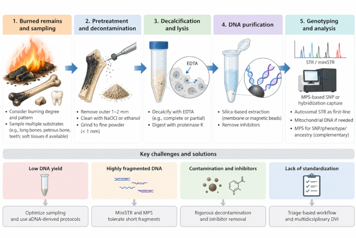
Figure 1: Overview of the forensic DNA analysis workflow for burned skeletal remains and associated analytical challenges. Burned-remains analysis begins with selection of appropriate skeletal or preserved soft-tissue substrates, followed by surface decontamination, bone-powder preparation, decalcification and lysis, DNA purification, and genotyping. Autosomal short tandem repeat (STR) profiling remains the first-line approach, while mitochondrial DNA and massively parallel sequencing (MPS)-based methods, including single nucleotide polymorphism (SNP) analysis and hybridization capture, may provide complementary options for highly degraded samples. Major challenges include low DNA yield, extensive fragmentation, contamination and polymerase chain reaction inhibitors, and the lack of standardized workflows. Current strategies include optimized sampling, ancient DNA-derived extraction approaches, methods tolerant of short DNA fragments, rigorous decontamination, inhibitor removal, and triage-based multidisciplinary workflows. Abbreviations: EDTA, ethylenediaminetetraacetic acid; NaOCl, sodium hypochlorite; STR, short tandem repeat; miniSTR, reduced-size STR; MPS, massively parallel sequencing; SNP, single nucleotide polymorphism; aDNA, ancient DNA; DVI, disaster victim identification. Please click here to view a larger version of this figure.
Technical bottlenecks and cutting-edge advances in burned bone DNA identification
Core challenges in burned bone DNA identification:
Four major challenges remain. First, DNA yield is extremely low: burning removes much of the organic matrix, sharply reduces bone cell numbers, and degrades DNA, so extractable endogenous DNA is often in the picogram-to-nanogram range. Although Thibodeau-Cadieux et al. recovered more than 250 ng of DNA from bone burned at 800°C, this represents total extracted DNA measured by ultraviolet–visible spectrophotometry in a porcine rib model, not endogenous human DNA, and the endogenous proportion relative to microbial DNA or exogenous contamination remains unclear30; in most real cases, it is far lower and may approach the detection limit of conventional PCR. Second, DNA is highly fragmented: average fragment length—depending on thermal damage and the analytical platform—is often below 100 bp and sometimes below 50 bp, making long-amplicon STR profiling difficult and even miniSTR systems ineffective below the primer distance; HTS is more tolerant, but library preparation itself selects against ultrashort fragments. Third, contamination and inhibitor interference persist: fire scenes are complex environments where combustion products such as polycyclic aromatic hydrocarbons and carbon particles, environmental microorganisms, and exogenous DNA from recovery personnel may all be present, while Maillard reaction products and metal ions act as strong PCR inhibitors53. Fourth, standardization is lacking: every burned bone sample has a unique “thermal history” determined by a highly variable combination of factors, producing substantial heterogeneity in DNA preservation32, and no protocol based on graded burning severity yet guides stepwise decision-making in casework.
DNA hybridization capture and targeted enrichment technologies:
Among emerging technologies, DNA hybridization capture has shown particular potential (Table 3). Biotin-labeled RNA or DNA probes complementary to target regions are hybridized with denatured DNA libraries and captured on streptavidin-coated magnetic beads, after which the enriched targets are sequenced. Its core advantage is independence from intact double-stranded templates: even fragments only a few dozen bases long can be captured efficiently if sufficient probe complementarity remains54. Widely applied in ancient DNA research, the technique enabled Fu et al. to capture the nuclear genome of the approximately 40,000-year-old Oase 1 individual from Romania54. Forensically, Emery et al. developed a genome-wide SNP targeted enrichment strategy for severely burned bone using double-stranded DNA libraries with biotinylated RNA probes32.
| Strategy | Principle | Fragment tolerance | Validation status |
| Complete decalcification42 | EDTA in lysis buffer; complete dissolution | Moderate | Widely used for challenging skeletal samples |
| Partial decalcification / direct lysis43 | Simultaneous decalcification and digestion | Recovers fragments ≥35 bp | Not yet systematically validated for burned bone |
| Silica membrane extraction45 | Selective DNA binding under high-salt conditions | Good for short fragments | Mainstream approach; higher DNA yield but greater co-extraction of inhibitors |
| Magnetic bead extraction45 | Selective DNA binding under high-salt conditions | — | Automatable and suited to high-throughput processing |
| Ancient DNA extraction protocol46 | Partial decalcification and silica suspension extraction | High recovery from charred bone | Demonstrated on severely burned remains |
| Hybridization capture32,55 | Probe-based target enrichment | Tolerates ultrashort fragments with sufficient complementarity | Limited forensic evidence; performance comparable to STR profiling in the reported application to highly degraded forensic bone |
Table 3: Comparison of extraction, library, and enrichment strategies for degraded skeletal DNA. This table compares the principles, fragment tolerance, and validation status of approaches used or proposed for highly degraded skeletal DNA. Ancient DNA-derived extraction methods can improve DNA recovery from severely burned remains, while hybridization capture can enrich highly fragmented target DNA. However, evidence supporting routine forensic application of hybridization capture to burned bone remains limited, and findings from ancient or otherwise highly degraded DNA should not be considered equivalent to direct validation in burned forensic samples.
Evidence in forensic casework nevertheless remains limited and must be clearly distinguished from extrapolation based on ancient DNA. The first forensic application of genome-wide capture to highly degraded human bones reported that capture and conventional STR profiling performed comparably on six highly degraded bones from the Spanish Civil War, with the principal added value lying in authentication of endogenous DNA rather than increased profiling success55—a finding that should temper expectations that capture will routinely rescue burned samples that fail conventional typing. Targeted panels for core forensic genetic markers are also being developed: Xiong et al. constructed a hybridization capture-based SNP panel optimized for the Han Chinese population and validated it on trace and degraded DNA56; although burned bone was not directly tested, the low-quantity, highly fragmented validation DNA resembles the preservation state of burned skeletal remains, supporting the design's potential applicability to burned bone. In addition, the European VISAGE project developed a tool integrating 41 phenotype-associated and 115 ancestry-informative SNPs, enabling simultaneous inference of externally visible characteristics and biogeographic origin from degraded DNA and extending burned bone identification toward investigative intelligence57.
Combining single-stranded DNA library preparation with hybridization capture is also noteworthy. Because a large proportion of DNA molecules in burned bone may be single-stranded as a consequence of severe thermal damage, conventional double-stranded library preparation can lose this information, whereas single-stranded preparation directly converts such molecules into sequenceable libraries and substantially improves template utilization58,59,60; success nevertheless depends on the preparation method, since standard double-stranded protocols systematically lose molecules bearing damaged termini. This combination may extend the sensitivity limits of burned bone DNA analysis, although it remains a reasoned expectation based on highly fragmented ancient DNA rather than an established forensic finding. Finally, the maturity of hybridization capture should not be overstated: most supporting evidence derives from ancient DNA research or specialized experimental studies, and evidence for routine implementation in real forensic burned-remains investigations is very limited, with barriers including high costs, extensive validation requirements, accreditation challenges, bioinformatic complexity, a lack of standardized forensic protocols, and limited availability in routine laboratories.
Integration into operational forensic practice: DVI and mass fatality incidents:
Burned bone analysis is ultimately embedded in operational identification workflows. In disaster victim identification (DVI) and mass fatality incidents—including aviation disasters, explosions, and large fires—large numbers of severely altered remains must be identified within a compressed timeframe through multidisciplinary frameworks combining anthropology, odontology, fingerprint examination, DNA analysis, and contextual information. Sessa et al. reported identification rates exceeding 90%–95% under structured multidisciplinary DVI workflows and emphasized triage-based allocation of samples and methods according to expected information yield9. Within such workflows, success depends not only on laboratory performance but also on reference sample management, the design of kinship-based inference chains (direct versus collateral relatives), and the use of likelihood ratios to integrate DNA results with other lines of evidence; platforms such as Familias, Bonaparte, and DNA-View are widely used for these calculations. Gianfreda et al.’s identification of 11 highly carbonized helicopter-crash victims—through systematic sampling and kinship analysis using the DVI module of Familias—exemplifies this complete chain from sampling through molecular analysis to kinship-based identification38. For burned remains, the practical implication is that sampling, extraction, and genotyping should be planned from the outset in relation to the reference samples available and the identification framework in use.
Research prospects:
Research on burned bone DNA analysis should advance along three lines. First, controlled burning experiments generating standardized datasets of DNA degradation levels, fragment-length distributions, and damage patterns under defined temperatures, durations, and atmospheres would provide a scientific basis for predicting DNA preservation in real cases. Second, the workflow itself should be further optimized: the DNA preservation advantages of unconventional sampling sites under burning conditions require validation; decalcification protocols balancing recovery against throughput should be developed; and the suitability of single-stranded versus double-stranded library preparation for burned bone should be systematically evaluated, together with hybridization capture panels integrating human identification, phenotype inference, and ancestry analysis. Third, interdisciplinary collaboration spanning mineralogical analysis, ancient DNA technology, and forensic genetics, with shared standards and technology-transfer mechanisms, will be essential for translating these advances into routine forensic practice.
结论
This review has traced the chain of events by which high-temperature burning alters bone and its DNA and has examined how each stage of the forensic workflow must be adapted in response. Three points emerge. First, thermal damage is a graded, cumulative process rather than a simple threshold phenomenon, and the preservation of DNA in burned bone is a function of temperature together with exposure duration, oxygen availability, tissue type, and the initial state of the remains; the widely used temperature zones should therefore be treated as a descriptive idealization rather than a predictive rule. Second, no single substrate, extraction method, or genotyping platform is universally optimal. Sampling should be planned across multiple tissues—including soft and cartilaginous tissues such as blood and intervertebral discs where these survive—while the choice of extract and assay should be guided by quantitative and qualitative assessment; extraction strategies developed for ancient DNA have repeatedly proved advantageous for severely charred material. Third, hybridization capture and targeted enrichment are genuinely promising for the ultrashort, damaged fragments characteristic of burned bone, but the current evidence base is narrower than the technology's theoretical appeal might suggest: the only forensic application to highly degraded bones reported performance comparable to conventional STR profiling, and cost, validation requirements, accreditation, bioinformatic complexity, and the absence of standardized protocols remain substantial barriers to routine use. Autosomal STR profiling therefore remains the first-line method, with MPS-based approaches serving as complementary tools. Progress will depend less on any single breakthrough than on the systematic integration of sampling, extraction, library preparation, and enrichment into a validated, triage-based workflow.
披露
The authors have no conflicts of interest to disclose.
致谢
This study was financially supported by the Fundamental Research Funds for the Central Public Welfare Research Institutes (Grant No. 2023JB004) and the Liaoning Provincial Science and Technology Plan Project (Grant No. 2025JH2/101800069).
参考文献
- McKinnon M, Henneberg M, Higgins D. A review of the current understanding of burned bone as a source of DNA for human identification. Sci Justice. 2021;61(4):332-8.
- Cainé L, et al. Genetic identification of human skeletal remains in forensic context: a review. Genes. 2026;17(4):492.
- Kronick PL, Cooke P. Thermal stabilization of collagen fibers by calcification. Connect Tissue Res. 1996;33(4):275-82.
- Piga G, et al. Understanding the crystallinity indices behavior of burned bones and teeth by ATR-IR and XRD in the presence of bioapatite mixed with other phosphate and carbonate phases. Int J Spectrosc. 2016;2016:4810149.
- Xu GC, Ren F, Hou XW, Yuan LB. Application of the burned bone morphology and DNA technology in human identification. J Forensic Med. 2007;23(5):370-2, 379.
- Sluis I, Duijst W, Krap T. Bone histology for forensic anthropology: a technical review on the advances in microstructural analysis of taphonomically altered buried or subaerially exposed bone. Int J Legal Med. 2025;139(6):2921-38.
- Grela M, et al. Effectiveness of various methods of DNA isolation from bones and teeth of animals exposed to high temperature. J Forensic Leg Med. 2021;78:102131.
- Imaizumi K. Forensic investigation of burnt human remains. Res Rep Forensic Med Sci. 2015;5:67-74.
- Sessa F, et al. Genetic identification of burned human remains: a systematic review. Genes. 2026;17(8):881.
- Weiner S, Wagner HD. The material bone: structure-mechanical function relations. Annu Rev Mater Sci. 1998;28(1):271-98.
- Inkret J, Zupanič Pajnič I. Bone type selection for human molecular genetic identification of skeletal remains. Genes (Basel). 2025;16(8):872.
- Revilla-López G, et al. Modeling biominerals formed by apatites and DNA. Biointerphases. 2013;8(1):10.
- Schwark T, Heinrich A, Preusse-Prange A, von Wurmb-Schwark N. Reliable genetic identification of burnt human remains. Forensic Sci Int Genet. 2011;5(5):393-9.
- Trębacz H, Wójtowicz K. Thermal stabilization of collagen molecules in bone tissue. Int J Biol Macromol. 2005;37(5):257-62.
- Harbeck M, et al. Research potential and limitations of trace analyses of cremated remains. Forensic Sci Int. 2010;204(1-3):191-200.
- Thompson T. Heat-induced dimensional changes in bone and their consequences for forensic anthropology. J Forensic Sci. 2005;50(5):1008-15.
- Campos PF, et al. DNA in ancient bone—where is it located and how should we extract it? Ann Anat. 2011;194(1):7-16.
- McKinnon M, Henneberg M, Simpson E, Higgins D. A comparison of crystal structure in fresh, burned and archaic bone—implications for forensic sampling. Forensic Sci Int. 2020;313:110328.
- Piga G, Thompson TJU, Malgosa A, Enzo S. The potential of X-ray diffraction in the analysis of burned remains from forensic contexts. J Forensic Sci. 2009;54(3):534-9.
- Grunenwald A, et al. Novel contribution on the diagenetic physicochemical features of bone and teeth minerals, as substrates for ancient DNA typing. Anal Bioanal Chem. 2014;406(19):4691-704.
- Végh EI, Márquez-Grant N, Schulting R. Physicochemical changes in bone bioapatite during the late postmortem interval pre- and post-burning. Appl Spectrosc. 2022;76(9):1080-99.
- Marques MPM, et al. Profiling of human burned bones: oxidising versus reducing conditions. Sci Rep. 2021;11(1):1361.
- Hiller JC, et al. Bone mineral change during experimental heating: an X-ray scattering investigation. Biomaterials. 2003;24(28):5091-7.
- Shipman P, Foster G, Schoeninger M. Burnt bones and teeth: an experimental study of color, morphology, crystal structure and shrinkage. J Archaeol Sci. 1984;11(4):307-25.
- Macias E, Hartline K, Buzzini P, Hughes S. Quantitative color analysis of burned bone to predict DNA quantity, quality, and genotyping success. J Forensic Sci. 2024;69(3):836-46.
- Hanson M, Cain CR. Examining histology to identify burned bone. J Archaeol Sci. 2007;34(11):1902-13.
- Fernández Castillo R, et al. Effects of temperature on bone tissue: histological study of the changes in the bone matrix. Forensic Sci Int. 2013;226(1-3):33-7.
- Imaizumi K, Taniguchi K, Ogawa Y. DNA survival and physical and histological properties of heat-induced alterations in burnt bones. Int J Legal Med. 2014;128:439-46.
- Adserias-Garriga J, Ubelaker DH, Zapico SC. Evaluation of macroscopic changes and the efficiency of DNA profiling from burnt teeth. Sci Justice. 2016;56(6):437-42.
- Thibodeau-Cadieux E, Beauchamp AM, Currie KA. Effects of thermal exposure on bone surface characteristics and DNA recovery. Forensic Sci Int Synerg. 2025;11:100644.
- Kendall C, et al. Diagenesis of archaeological bone and tooth. Palaeogeogr Palaeoclimatol Palaeoecol. 2017;491:21-37.
- Emery MV, et al. Targeted enrichment of whole-genome SNPs from highly burned skeletal remains. J Forensic Sci. 2024;69(5):1558-77.
- Gaudio D, et al. Genome-wide DNA from degraded petrous bones and the assessment of sex and probable geographic origins of forensic cases. Sci Rep. 2019;9:8226.
- Otagiri T, et al. An optimal skeletal element for DNA testing: evaluation of DNA quantity and quality from various bone types in routine forensic practice. Leg Med (Tokyo). 2024;68:102415.
- Roshin CN, et al. Forensic DNA analysis of heat-exposed unrestored teeth: temperature and duration effects. J Oral Maxillofac Pathol. 2025;29(3):478-85.
- Tomsia M, et al. The intervertebral discs’ fibrocartilage as a DNA source for genetic identification in severely charred cadavers. Forensic Sci Med Pathol. 2022;18(4):442-9.
- Senst A, et al. Recommendations for the successful identification of altered human remains using standard and emerging technologies: results of a systematic approach. Forensic Sci Int Genet. 2022;62:102790.
- Gianfreda D, et al. Experience based efficient approach for DNA-led identification of highly carbonized human remains. Forensic Sci Int. 2025;367:112359.
- Korlević P, et al. Reducing microbial and human contamination in DNA extractions from ancient bones and teeth. Biotechniques. 2015;59(2):87-93.
- Sweet D, Hildebrand D. Recovery of DNA from human teeth by cryogenic grinding. J Forensic Sci. 1998;43(6):1199-202.
- Zupan E, et al. Effect of bone grinding on the quantity and quality of DNA obtained from aged trabecular bones. Forensic Sci Int. 2026;383:112884.
- Loreille OM, et al. High efficiency DNA extraction from bone by total demineralization. Forensic Sci Int Genet. 2007;1(2):191-5.
- Dabney J, Meyer M. Extraction of highly degraded DNA from ancient bones and teeth. Methods Mol Biol. 2019;1963:25-9.
- Vinueza-Espinosa DC, et al. Human DNA extraction from highly degraded skeletal remains: how to find a suitable method? Electrophoresis. 2020;41(24):2149-58.
- Rothe J, Nagy M. Comparison of two silica-based extraction methods for DNA isolation from bones. Leg Med (Tokyo). 2016;22:36-41.
- Emery MV, et al. Reconstructing full and partial STR profiles from severely burned human remains using comparative ancient and forensic DNA extraction techniques. Forensic Sci Int Genet. 2020;46:102272.
- Zupanič Pajnič I, et al. A fast and highly efficient automated DNA extraction method from small quantities of bone powder from aged bone samples. Forensic Sci Int Genet. 2023;65:102882.
- Snedeker J, Hughes S, Houston R. Optimization of InnoXtract extraction and purification system for DNA extraction from skeletal samples. Int J Legal Med. 2023;137(4):949-59.
- Vajpayee K, et al. PCR inhibitors and facilitators—their role in forensic DNA analysis. Forensic Sci Int. 2023;349:111773.
- Butler JM, Shen Y, McCord B. The development of reduced size STR amplicons as tools for analysis of degraded DNA. J Forensic Sci. 2003;48(5):1054-64.
- Grubwieser P, et al. A new "miniSTR-multiplex" displaying reduced amplicon lengths for the analysis of degraded DNA. Int J Legal Med. 2006;120(2):115-20.
- Van Neste C, et al. Forensic STR analysis using massive parallel sequencing. Forensic Sci Int Genet. 2012;6(6):810-6.
- Alaeddini R. Forensic implications of PCR inhibition—a review. Forensic Sci Int Genet. 2012;6(3):297-305.
- Fu Q, et al. An early modern human from Romania with a recent Neanderthal ancestor. Nature. 2015;524(7564):216-9.
- Haarkötter C, et al. Exploring the potential of genome-wide hybridization capture enrichment for forensic DNA profiling of degraded bones. Genes (Basel). 2025;16(1):23.
- Xiong X, et al. Evaluation of a hybridization capture-based hereditary panel for forensic DNA analysis using next-generation sequencing. Forensic Sci Int. 2025;376:112594.
- Freire-Aradas A, et al. Development and inter-laboratory evaluation of the VISAGE Enhanced Tool for Appearance and Ancestry inference from DNA. Forensic Sci Int Genet. 2022;61:102779.
- Gansauge MT, Meyer M. Single-stranded DNA library preparation for the sequencing of ancient or damaged DNA. Nat Protoc. 2013;8(4):737-48.
- Gansauge MT, Aximu-Petri A, Nagel S, Meyer M. Manual and automated preparation of single-stranded DNA libraries for the sequencing of DNA from ancient biological remains and other sources of highly degraded DNA. Nat Protoc. 2020;15(8):2279-300.
- Kapp JD, Green RE, Shapiro B. A fast and efficient single-stranded genomic library preparation method optimized for ancient DNA. J Hered. 2021;112(3):241-9.