Der Einsatz von Schweinen in der Forschung hat in den letzten Jahren zugenommen. Dennoch zeichnen sich Schweine durch eine schwierige Atemwegsanatomie aus. Durch die Demonstration der Durchführung einer endoskopisch gesteuerten endotrachealen Intubation zielt das vorliegende Protokoll darauf ab, die Sicherheit von Labortieren weiter zu erhöhen, um Tierleid und unnötigen Tod zu vermeiden.
Ein Abonnement von JoVE ist erforderlich, um diesen Inhalt anzuzeigen. Melden Sie sich an oder beginnen Sie noch heute mit einer kostenlosen Testphase.
Methodenartikel
Endotracheale Intubation mit einem flexiblen Intubationsendoskop als standardisiertes Modell für ein sicheres Atemwegsmanagement bei Schweinen
In diesem Artikel
Erratum-Hinweis
Important: There has been an erratum issued for this article. View Erratum Notice
Zusammenfassung
Zusammenfassung
Die endotracheale Intubation ist oft eine Grundvoraussetzung für die translationale Forschung in schweinen Modellen für verschiedene Eingriffe, die einen gesicherten Atemweg oder hohe Beatmungsdrücke erfordern. Die endotracheale Intubation ist eine anspruchsvolle Fähigkeit, die eine minimale Anzahl erfolgreicher endotrachealer Intubationen erfordert, um unter optimalen Bedingungen eine hohe Erfolgsrate zu erzielen, die für Nicht-Anästhesiologieforscher oft unerreichbar ist. Aufgrund der spezifischen Anatomie der porcinen Atemwege kann in der Regel von einem schwierigen Atemweg ausgegangen werden. Die Unmöglichkeit, einen sicheren Atemweg herzustellen, kann zu Verletzungen, unerwünschten Ereignissen oder zum Tod des Labortiers führen. Unter Verwendung eines prospektiven, randomisierten, kontrollierten Bewertungsansatzes konnte gezeigt werden, dass die faseroptische endotracheale Intubation länger dauert, aber eine höhere First-Pass-Erfolgsrate aufweist als die konventionelle Intubation, ohne klinisch relevante Abfalle der Sauerstoffsättigung zu verursachen. Dieses Modell stellt ein standardisiertes Schema für die endoskopisch gesteuerte endotracheale Intubation dar, das einen gesicherten Atemweg bietet, insbesondere für Forscher, die mit der Technik der endotrachealen Intubation durch direkte Laryngoskopie unerfahren sind. Dieses Verfahren soll Tierleid und unnötige Tierverluste minimieren.
Einleitung
Die endotracheale Intubation ist oft eine Grundvoraussetzung für die translationale Forschung in Schweinemodellen für verschiedene Interventionen, die einen gesicherten Atemweg oder einen hohen Beatmungsdruck erfordern (wie z.B. Beatmung während der Herz-Lungen-Wiederbelebung1 oder akutes Atemnotsyndrom2) oder erfordern, dass der zerebrale Blutfluss nicht durch interne Kompression durch supraglottische Atemwegsgeräte beeinträchtigt wird3 , die gelegentlich als Alternativen im Zusammenhang mit einer zu erwartenden schwierigen Atemwegserkrankung bei Schweinen vermehrt werden ....
Zugriff eingeschränkt. Bitte melden Sie sich an oder starten Sie eine Testversion, um diesen Inhalt anzuzeigen.
Protokoll
Die Versuche in diesem Protokoll wurden vom Landesuntersuchungsamt Rheinland-Pfalz (Koblenz, Deutschland; Zulassungs-Nr. G20-1-135) genehmigt. Die Experimente wurden nach den ARRIVE-Richtlinien durchgeführt. Insgesamt wurden 10 anästhesierte männliche Schweine (Sus scrofa domestica) mit einem Durchschnittsgewicht von 30 kg ± 2 kg und einem Alter von 12-16 Wochen für die vorliegende Studie verwendet.
1. Tierische Vorbereitung
- Sorgen Sie für eine normale Umgebung für die Tiere, um Stress zu minimieren. Halten Sie die Nahrung 6 Stunden vor dem geplanten Experiment zurück, um das Aspirationsrisiko zu verringe....
Zugriff eingeschränkt. Bitte melden Sie sich an oder starten Sie eine Testversion, um diesen Inhalt anzuzeigen.
Ergebnisse
Die endotracheale Intubation wurde an 10 männlichen Schweinen (Alter 12-16 Wochen, Gewicht 30 kg ± 3 kg) in einer prospektiven, randomisierten, kontrollierten Studie durchgeführt. Die Schweine wurden randomisiert in zwei Gruppen eingeteilt: eine wurde konventionell larynoskopisch intubiert (CI-Gruppe) und die andere Gruppe wurde mit Hilfe eines flexiblen Intubationsendoskops intubiert, wie im Protokoll beschrieben (FIE-Gruppe). Die Gruppenzuordnung erfolgte durch Ziehen versiegelter Umschläge. Der Ermittler wurd.......
Zugriff eingeschränkt. Bitte melden Sie sich an oder starten Sie eine Testversion, um diesen Inhalt anzuzeigen.
Diskussion
In früheren Studien hat unsere Forschungsgruppe bereits spezifische Details zu den translationalen Vorteilen des porcinen Modells 2,27,32,33 beschrieben. Im Allgemeinen sollte die Reduzierung des Stressniveaus des Tieres und unnötiger Schmerzen ein integraler Bestandteil jedes Studienprotokolls sein und ist von größter Bedeutung, um zuverlässig reproduzierbare Daten zu generieren. Daher ist ein.......
Zugriff eingeschränkt. Bitte melden Sie sich an oder starten Sie eine Testversion, um diesen Inhalt anzuzeigen.
Offenlegungen
Das flexible Intubationsendoskop und sein Zubehör wurden vom Hersteller vorbehaltlos nur zu Forschungszwecken zur Verfügung gestellt. Die Autoren erklären keine weiteren finanziellen oder sonstigen Interessenkonflikte.
Danksagungen
Die Autoren bedanken sich bei Dagmar Dirvonskis für die hervorragende technische Unterstützung.
....Zugriff eingeschränkt. Bitte melden Sie sich an oder starten Sie eine Testversion, um diesen Inhalt anzuzeigen.
Materialien
| Name | Unternehmen | Katalognummer | Kommentare |
|---|---|---|---|
| Ambu aScope Regular | Ambu GmbH, Medizinprodukte, Bad Nauheim, Deutschland | Einweg-LWL-Außendurchmesser 5 mm | |
| Ambu aView Monitor | Ambu GmbH, Medizinprodukte, Bad Nauheim, Deutschland | Monitor | |
| Atracurium Hikma 50 mg/5mL | Hikma Pharma GmbH, Martinsried | Atracurium | |
| Azaperon (Stresnil) 40mg/mL | Lilly Deutschland GmbH, Bad Homburg, Deutschland | Azaperon | |
| BD Discardit II Spritze 2, 5, 10, 20 mL | Becton Dickinson S.A. Carretera, Mequinenza Fraga, Spanien | Spritze | |
| BD Luer Connecta | Becton Dickinson Infusion Therapy, AB Helsingborg, Schweden | 3-Wege-Absperrhahn | |
| BD Microlance 3 20 G | Becton Dickinson S.A. Carretera, Mequinenza Fraga, Spanien | Kanüle | |
| Curafix i.v. classics | Lohmann & Rauscher International GmbH & Co. KG, Rengsdorf, Deutschland | Kanülen-Retentionsverband | |
| Engströ m Carestation | GE Heathcare, Madison USA | Beatmungsgerät | |
| Fentanyl-Janssen 0,05 mg/mL | Janssen-Cilag GmbH, Neuss | Fentanyl | |
| Fü hrungsstab, Durchmesser 4.3 | Rü sch | Endotrachealtubus Einführhilfe | |
| IBM SPSS Statistics für Windows, Version 20 | IBM SPSS Statistics für Windows, Version 20.0. Armonk, NY: IBM Corp.) | Statistiksoftware | |
| Incetomat-line 150 cm | Fresenius, Kabi Deutschland, GmbH | Perfusor line | |
| Intrafix Primeline | B. Braun Melsungen AG, Melsungen, Germany | Infusionslinie | |
| JOZA Einmal Nitril Untersuchungshandschuhe | JOZA, Mü nchen, Deutschland | Einmalhandschuhe | |
| Laryngoskop, 45.48.50, KL 2000 | Medicon | Laryngoskop Griff | |
| Littmann Classic III Stethoskop | 3M Deutschland GmbH, Neuss, Deutschland | Stethoskop | |
| Luer Lock | B.Braun Melsungen AG, Deutschland | ||
| Verstümmelt Vlieskompresse | Maiming GmbH, Neuenkirchen, Deutschland | Fleece-Kompresse zur Fixierung der Zunge | |
| Masimo LNCS Adtx SpO2 Sensor | Masimo Corporation Irvine, Ca 92618 USA | Sättigungsclip für den Schwanz | |
| Masimo LNCS TC-I SpO2 Ohrclip Sensor | Masimo Corporation Irvine, Ca 92618 USA | Sättigungsclip für das | |
| Ohr Masimo Radical 7 | Masimo Corporation Irvine, Ca 92618 USA | periphere Sauerstoffsättigung | |
| Midazolam 15 mg/3 mL | Hameln Pharma GmbH, Hameln, Deutschland | Midazolam | |
| Midmark Hundemaske Kleinplastik mit Diaphragma FRSCM-0005 | Midmark Corp., Dayton, Ohio, USA | Beatmungsmaske für Hunde | |
| Octeniderm farblos | Schü LKE & Mayr GmbH, Nordenstedt, Deutschland | Alkoholisches Desinfektionsmittel | |
| Original Perfusor Spritze 50 mL | B.Braun Melsungen AG, Deutschland | Perfusor | |
| Spritze Perfusor FM Braun | B.Braun Melsungen AG, Deutschland | Spritzenpumpe | |
| Propofol 2% 20 mg/mL (50 mL Kolben) | Fresenius, Kabi Deutschland, GmbH | Propofol | |
| RÜ SCH Fü hrungsstab fü r Endotrachealtubus (ID 5,6 mm) | Teleflex Medical Sdn. Bhd, Malaysia | PVC-beschichteter Schlauchführungsdraht | |
| Rü Schelit Super Safety Clear >ID 6/6,5 /7,0 mm | Teleflex Medical Sdn. Bhd, Malaysia | Endotrachealtubus | |
| Edelstahl Macintosh Grö ß e 4 | Welch Allyn69604 | Spatel für Laryngoskop | |
| Sterofundin | B.Braun Melsungen AG, Melsungen, Deutschland | Ausgewogene Elektrolytlösung | |
| Ultrastop Antibeschlagmittel Flasche mit Pipette 25 mL | Sigmapharm Arzneimittel GmbH, Wien, Österreich | Antifogmittel | |
| Vasofix Safety 22 G-16 G | B.Braun Melsungen AG, Deutschland | Venenkatheter | |
| VBM Manschettenmanometer | VBM Medizintechnik GmbH, Sulz a.N., Deutschland | Manschettenmanometer | |
| Zelette | Lohmann & Rauscher International GmbH & Co. KG, Rengsdorf, Deutschland | Gewebeabstrich |
Referenzen
- Kleinman, M. E., Oh, W., Stonestreet, B. S. Comparison of intravenous and endotracheal epinephrine during cardiopulmonary resuscitation in newborn piglets. Critical Care Medicine. 27 (12), 2748-2754 (1999).
- Rissel, R., et al.
Zugriff eingeschränkt. Bitte melden Sie sich an oder starten Sie eine Testversion, um diesen Inhalt anzuzeigen.
Nachdrucke und Genehmigungen
Erratum
Formal Correction: Erratum: Endotracheal Intubation Using a Flexible Intubation Endoscope As a Standardized Model for Safe Airway Management in Swine
Posted by JoVE Editors on 4/03/2023. Citeable Link.
An erratum was issued for: Endotracheal Intubation Using a Flexible Intubation Endoscope As a Standardized Model for Safe Airway Management in Swine. The Protocol, Representative Results, and Discussion sections were updated.
In the Protocol, step 1.5 was updated from:
Disinfect the skin with a disinfectant (alcoholic) before inserting a peripheral vein cannula (22 G) into an ear vein. Spray the area, wipe once, then spray again, and allow the disinfectant to dry.
to:
Disinfect the skin with a disinfectant (alcoholic) before inserting a peripheral vein cannula (22 G) into an ear vein. Spray the area, wipe once, then spray again, and allow the disinfectant to dry. Secure the ear cannula with a band-aid (See Table of Materials).
In the Protocol, step 3.7 was updated from:
While maintaining the position of the endoscope, advance the endotracheal tube until it becomes visible in the camera image.
NOTE: If the endotracheal tube cannot be advanced through the glottic plane, there is a possibility that it has become caught on the arytenoid cartilage. In this case, the endotracheal tube must be withdrawn 1 cm and rotated by 90° before gently advancing again. If necessary, this maneuver can be repeated. Similar calibers of flexible intubation endoscope and endotracheal tube can minimize the risk of this issue occurring. If the endotracheal tube cannot be advanced despite this maneuver, it is likely that the subglottic narrowness-the narrowest part of the porcine larynx-cannot be passed. In this case, a smaller endotracheal tube size needs to be selected. Regular commercially available endotracheal tubes in sizes 6.5 cm or 7.0 cm ID should be able to pass the glottis as long as no anatomic abnormalities are present.
to:
While maintaining the position of the endoscope, advance the endotracheal tube until it becomes visible in the camera image.
NOTE: If the endotracheal tube cannot be advanced through the glottic plane, there is a possibility that it has become caught on the arytenoid cartilage. In this case, the endotracheal tube must be withdrawn 1 cm and rotated by 90° before gently advancing again. If necessary, this maneuver can be repeated. Similar calibers of flexible intubation endoscope and endotracheal tube can minimize the risk of this issue occurring. If the endotracheal tube cannot be advanced despite this maneuver, it is likely that the subglottic narrowness-the narrowest part of the porcine larynx-cannot be passed. In this case, a smaller endotracheal tube size needs to be selected. Regular commercially available endotracheal tubes in sizes 6.5 cm or 7.0 cm ID should be able to pass the glottis as long as no anatomic abnormalities are present. Endotracheal tube size requirements vary depending on the piglet size and breed.
In the Representative Results, the sixth paragraph was updated from:
Statistical analyses were performed using commercially available software (see Table of Materials). Normal distribution was examined using the Kolmogorov-Smirnoff test28. If a normal distribution was determined, group differences were analyzed using t-tests of independent samples29 or the Mann-Whitney U test30 for the non-parametric version. Data are presented as mean (± standard deviation). Correlations of ordinal-scale data were examined using Spearman's correlation coefficient31. A significance level of p < 0.05 was assumed.
to:
Statistical analyses were performed using commercially available software (see Table of Materials). Normal distribution was examined using the Kolmogorov-Smirnoff test28. If a normal distribution was determined, group differences were analyzed using t-tests of independent samples29 or the Mann-Whitney U test30 for the non-parametric version. Data are presented as mean (± standard deviation). Correlations of ordinal-scale data were examined using Spearman's correlation coefficient31. A significance level of p < 0.05 was assumed. All tests were performed with exploratory intention; therefore p-values are descriptive. Nevertheless, p < 0.05 was accepted as indicative of statistical significance.
In the Representative Results, the legend for figure 1 was updated from:
Figure 1: Number of intubation attempts in group comparison. For the group that was intubated using a flexible intubation endoscope, every intubation attempt was successful; in the group that was conventionally intubated, it took an average of 1.4 attempts before the endotracheal tube could be placed correctly. Error bars show the standard deviation. Please click here to view a larger version of this figure.
to:
Figure 1: Number of intubation attempts in group comparison. For the group that was intubated using a flexible intubation endoscope, every intubation attempt was successful; in the group that was conventionally intubated, it took an average of 1.4 attempts before the endotracheal tube could be placed correctly. Error bars show the standard deviation. n = 5 (for each group). Please click here to view a larger version of this figure.
In the Representative Results, figure 2 was updated from:
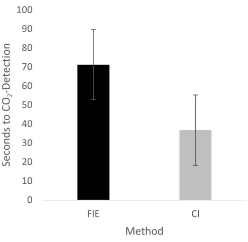
Figure 2: Time until CO2 detection in group comparison. For the group that was intubated using a flexible intubation endoscope, it took significantly longer until end-tidal CO2 could be detected, depicted as mean and standard deviation. Please click here to view a larger version of this figure.
to:
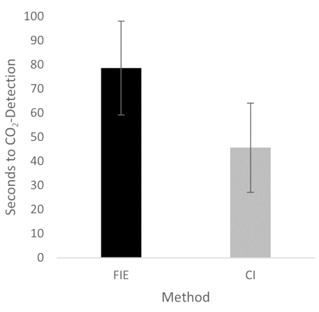
Figure 2: Time until CO2 detection in group comparison. For the group that was intubated using a flexible intubation endoscope, it took significantly longer until end-tidal CO2 could be detected, depicted as mean and standard deviation. n = 5 (for each group). Please click here to view a larger version of this figure.
In the Discussion, the fifth paragraph was updated from:
The increased duration had no clinical significance in this cohort. At no time was the termination criterion-a saturation of less than 93%-reached. This is shown in the results because a procedure change was unnecessary at any time. Prior adequate mask ventilation is a critical step to allow sufficient time for fiberoptic endotracheal tube placement to avoid rapid desaturation34. These results are consistent with previous studies comparing conventional intubation and endoscopically assisted intubations with inexperienced providers35.
to:
The increased duration had no clinical significance in this cohort. At no time was the termination criterion-a saturation of less than 93%-reached. This is shown in the results because a procedure change was unnecessary at any time. Prior adequate mask ventilation is a critical step to allow sufficient time for fiberoptic endotracheal tube placement to avoid rapid desaturation34. These results are consistent with previous studies comparing conventional intubation and endoscopically assisted intubations with inexperienced providers35. We attribute the prolonged duration of fiberoptic intubation to the fact that one must first reorient again after insertion, whereas with conventional intubation, one retains a view of the glottis. It is also important to avoid contact with the mucosa with the flexible intubation endoscope during advancement. This requires occasional corrective maneuvers. Last but not least, after successful placement, retraction of the relatively long endoscope is required, which increases the time to CO2 detection slightly.
