Serum miR-508-3p is significantly downregulated in acute ischemic stroke, correlating with disease severity, inflammation, and poor 90-day prognosis. It demonstrates strong diagnostic performance (AUC 0.864). Functional studies show that miR-508-3p protects brain microvascular endothelial cells by targeting SOCS3, highlighting its potential as a biomarker and therapeutic target.
Forschungsartikel
Diagnostic and Prognostic Significance of Aberrant miR-508-3p Expression in Acute Ischemic Stroke
In diesem Artikel
Zusammenfassung
Zusammenfassung
The objectives were to investigate serum miR-508-3p expression in patients with acute ischemic stroke (AIS), determine its value for early diagnosis and prognosis of AIS, and preliminarily explore its underlying mechanism. A total of 131 AIS patients (case group) admitted between January 2023 and December 2024, and 125 healthy individuals (control group) were enrolled. Quantitative real-time polymerase chain reaction (qRT-PCR) was performed to detect serum miR-508-3p expression, and an enzyme-linked immunosorbent assay (ELISA) was conducted to assess the concentrations of IL-6, IL-1β, TNF-α, and CRP in serum samples. The clinical value of miR-508-3p was analyzed using correlation, receiver operating characteristic (ROC), and Kaplan-Meier methods. Multivariate logistic regression was used to identify independent predictors of 90-day outcome. In vitro, human brain microvascular endothelial cells (HBMECs) were subjected to OGD/R; cell viability, injury, and apoptosis were determined by CCK-8, LDH release, and flow cytometry, and the miR-508-3p/SOCS3 targeted binding was confirmed using a dual-luciferase reporter system. Serum miR-508-3p was significantly decreased in AIS patients compared with controls (p < 0.001) and negatively correlated with National Institutes of Health Stroke Scale (NIHSS) score and levels of IL-6, IL-1β, TNF-α, and CRP (all p < 0.001). ROC analysis yielded an area under the curve (AUC) of 0.8641 (95%CI: 0.8184-0.9098), with 87.79% sensitivity and 72.80% specificity. Multivariate analysis confirmed low miR-508-3p as an independent predictor of poor 90-day prognosis (OR 0.002, 95% CI 0.000–0.581, p = 0.032). In vitro, miR-508-3p overexpression enhanced HBMEC viability and reduced LDH release and apoptosis after oxygen-glucose deprivation/reoxygenation (OGD/R). Dual‑luciferase reporter assay confirmed that miR-508-3p targets the 3'UTR of SOCS3. Downregulated serum miR-508-3p is associated with AIS severity, inflammation, and poor prognosis, suggesting its diagnostic and prognostic potential. Its protective effect may involve targeting suppressor of cytokine signaling 3 (SOCS3) and alleviating brain microvascular endothelial cell injury.
Einleitung
Accounting for 80–85% of all stroke cases worldwide, acute ischemic stroke (AIS) is the predominant subtype of stroke and the second leading cause of death globally, as well as the primary cause of mortality and disability among adults in China, featuring the "four highs": high incidence, high disability rate, high mortality, and high recurrence rate1. At present, the definitive diagnosis of AIS relies on cranial DWI-MRI as the gold standard, yet its reach in primary hospitals is limited, and false negatives may occur in the hyperacute period2. Intravenous thrombolysis and mechanical thrombectomy are the core reperfusion therapies for acute AIS, and efficacy is strictly time‑dependent3. Unfortunately, there is a lack of noninvasive biomarkers for the rapid identification of AIS in the prehospital or emergency ultra-early phase. Meanwhile, the prognosis of AIS patients varies greatly among individuals, and the predictive performance of existing clinical scoring systems for poor prognosis remains to be improved. Thus, novel biomarkers that allow for accurate early diagnosis and prognostic stratification of AIS are in urgent demand.
Endogenous noncoding RNAs of 18–25 nucleotides, known as microRNAs (miRNAs), control gene expression post-transcriptionally by targeting the 3'UTR of target gene mRNAs4. These molecules are widely implicated in diverse cellular processes, ranging from proliferation and apoptosis to inflammation, oxidative stress, and vascular homeostasis5. Notably, miRNAs can be stably preserved in peripheral biofluids such as serum and plasma, with inherent resistance to RNase-mediated degradation, repeated freeze-thaw cycles, and pH variations; this robust stability is a core superiority over conventional mRNA and labile protein biomarkers, making them ideal candidates for non-invasive diagnostic biomarkers6,7,8. Moreover, these molecules exhibit characteristic abnormal expression in the early stage of disease, earlier than most traditional serological markers, and can be reliably quantified via minimally invasive venous blood sampling, making them promising noninvasive diagnostic biomarkers with high clinical translation potential9,10. Accumulating studies have confirmed that various miRNAs show differential expression profiles in AIS and participate in core pathological processes such as post‑ischemic inflammatory brain injury, blood‑brain barrier disruption, and neuronal apoptosis, thus possessing value in diagnosis, prognosis, and therapeutic targeting11,12.
miR‑508‑3p is a functional miRNA identified in recent years. Existing evidence has revealed that this miRNA exhibits dysregulated expression patterns in multiple pathological conditions, ranging from malignant tumors (gastric cancer, ovarian cancer) to inflammatory and ischemic diseases (rheumatoid arthritis)13,14. It participates in disease progression by targeting inflammatory pathways, apoptosis, and other processes, and can serve as an early diagnostic biomarker for multiple diseases. miR-508-3p was selected as the target molecule based on integrated bioinformatics analyses (TargetScan, miRDB, miRTarBase), which identified suppressor of cytokine signaling 3 (SOCS3), a key regulator of JAK/STAT inflammatory signaling, as a high-confidence conserved target. Furthermore, a pilot screening of 10 AIS patients versus 10 controls revealed a marked reduction in serum miR-508-3p, providing initial clinical evidence to pursue this candidate. However, the expression pattern, diagnostic and prognostic value of miR‑508‑3p in AIS have not been systematically reported, and its correlation with inflammation - the core pathological process of AIS - remains unclear.
SOCS3 is a key member of the SOCS family and a critical negative regulator of the JAK/STAT signaling pathway. It is widely expressed in central nervous system cells including brain microvascular endothelial cells, microglia, and neurons15,16. Evidence has shown that SOCS3 can attenuate the post‑ischemic inflammatory cascade, inhibit brain microvascular endothelial cell apoptosis, and maintain blood‑brain barrier integrity by suppressing excessive activation of the JAK2/STAT3 pathway, acting as an important endogenous neurovascular protective factor in AIS17. According to predictions from bioinformatics tools like TargetScan and miRDB, SOCS3 may be a direct target gene of miR-508-3p; however, whether this regulatory interaction exists in AIS remains unverified.
Accordingly, this study measured the serum expression level of miR‑508‑3p in AIS patients and healthy controls, investigated its early diagnostic efficacy for AIS, analyzed its correlation with inflammatory factors and neurological deficit severity, and clarified its predictive value for 90‑day poor functional prognosis in AIS patients. Mechanistically, we verified the targeted regulatory relationship between miR-508-3p and SOCS3 in an OGD/R cell model. This study adopted a standardized experimental system from clinical sample detection to in vitro mechanism validation, with good reproducibility, aiming to provide a new serological biomarker and theoretical basis for the early and precise diagnosis and treatment of AIS.
Protokoll
This study was approved by the Ethics Committee of QianWei Hospital of Jilin Province (approval number: JPQWIRB-LW260623). All research procedures complied with the ethical requirements of the Declaration of Helsinki, and all enrolled patients and their families signed written informed consent. All the materials used in this study are listed in the Table of Materials.
Study subjects
A total of 131 inpatients with AIS admitted to our hospital from January 2023 to December 2024 were enrolled as the case group. Inclusion criteria: Inclusion criteria were as follows: participants met the diagnostic criteria of Chinese Guidelines for the Diagnosis and Treatment of Acute Ischemic Stroke 2023 and diagnosis was confirmed by cranial DWI/MRI; participants were admitted to hospital within 72 h of onset, with blood samples collected prior to pharmacotherapy; participants were aged ≥ 18 years with complete clinical data and able to complete 90-day follow-up.
Exclusion criteria: Participants were excluded if they had other cerebrovascular diseases, such as cerebral hemorrhage, subarachnoid hemorrhage, and transient ischemic attack (TIA), or if they presented with concurrent severe infection, autoimmune disease, malignant tumor, severe hepatic and renal insufficiency, or hematological disease. Individuals with a history of major surgery, trauma, or myocardial infarction within the previous 3 months, or those who had used immunosuppressants or glucocorticoids prior to admission, were also excluded. In addition, participants with a prior stroke history accompanied by severe neurological deficit (modified Rankin Scale score ≥ 3) were not eligible for enrollment, nor were pregnant or lactating individuals, patients who were lost to follow-up, or those with incomplete clinical data.
Meanwhile, 125 healthy individuals with age- and sex-matched characteristics were enrolled from the Physical Examination Center of our hospital as the control group. Inclusion criteria for controls: Eligible healthy controls had no history of cerebrovascular disease or neurological symptoms, and no abnormal findings were identified on cranial computed tomography or magnetic resonance imaging. All control participants had no severe underlying systemic diseases, and all routine physical examination indicators were within the normal reference ranges. None of the controls had a history of infection, trauma, or surgery within the previous 3 months, and the age and sex distributions of the control group were matched with those of the case group.
Sample collection and processing
For the case group, 5 mL of fasting peripheral venous blood was collected within 24 h after admission (before thrombolysis, antiplatelet, statin, and other drug treatments). For the control group, 5 mL of fasting peripheral venous blood was collected on the day of physical examination. All blood samples were collected in EDTA anticoagulant tubes. Within 2 h post-collection, samples were subjected to centrifugation at 1000 × g for 15 min under 4 ˚C conditions. The supernatant serum was separated, subdivided into RNase-free EP tubes, and stored in a -80 ˚C ultra-low temperature freezer to avert repeated freeze-thaw events.
Detection of serum miR-508-3p expression by qRT-PCR
Total RNA was extracted from a 100 µL serum sample using TRIzol reagent following the manufacturer’s instructions. RNA concentration and purity were measured using a UV spectrophotometer. RNA with an OD260/OD280 ratio between 1.8 and 2.0 was considered qualified. RNA was reverse-transcribed into cDNA using a commercial miRNA first-strand synthesis kit according to the protocol, and stored at 4 ˚C. qRT-PCR amplification was performed using cDNA as the template. Each sample was run in triplicate, with U6 as the internal reference.
Primer sequences: miR-508-3p forward: 5'-GCCGAGTTCCTGATTTTGT-3', reverse: 5'-GTGCAGGGTCCGAGGT-3', U6 forward: 5'-CTCGCTTCGGCAGCA CA-3', reverse: 5'-AACGCTTCACGAATTTGCGT-3'. To quantify the relative expression of serum miR-508-3p, we adopted the 2⁻ΔΔCt method, with U6 serving as the internal reference and the average expression of the healthy control group as the calibrator for normalization.
Detection of serum inflammatory factor levels by ELISA
The levels of IL-6, IL-1β, TNF-α, and CRP in serum samples from both groups were determined using ELISA, with all operations performed strictly following the instructions supplied by the kit manufacturer. Each sample was tested in duplicate. The absorbance at the corresponding wavelength was measured using a microplate reader. A standard curve was plotted based on the standard products, and the actual concentrations of each inflammatory factor were calculated.
Clinical data collection and follow-up
A standardized case report form (CRF) was designed to collect baseline information for case group patients, covering age, sex, medical history (hypertension, diabetes, hyperlipidemia, atrial fibrillation, smoking, alcohol use), as well as clinical measures, including NIHSS score at admission and routine lab findings.
All AIS patients were followed up for 90 days after onset via outpatient revisit or telephone interview. Using the modified Rankin Scale (mRS), functional prognosis was evaluated at 90 days after symptom onset, which was set as the study’s follow-up endpoint. Favorable functional prognosis was defined as an mRS score of 0–2, while an mRS score ≥3 was classified as poor functional prognosis, representing moderate to severe disability or death. The loss-to-follow-up rate was kept below 10%, and no patients were lost to follow-up in this study.
Cell culture
Human brain microvascular endothelial cells (HBMEC) were cultured in endothelial cell-specific ECM medium (containing 5% FBS, 1% endothelial cell growth supplement, and 1% penicillin-streptomycin double antibody) in a cell incubator at 37 ˚C with 5% CO₂ and saturated humidity. Cells were subcultured at 2–3 day intervals, and cells at passages 3–8 in the exponential growth phase and with optimal viability were selected for subsequent experiments.
Establishment of the OGD/R model
Ischemia‑reperfusion injury in AIS was modeled by subjecting cells to oxygen‑glucose deprivation and subsequent reoxygenation (OGD/R). Cells were subjected to removal of the original culture medium, washed twice gently with glucose‑free DMEM, and subsequently incubated in glucose‑free and serum‑free DMEM. Cells were cultured in a tri-gas incubator (37 ˚C, 94% N₂, 5% CO₂, 1% O₂) for 6 h of hypoxia. After hypoxia, the glucose-free medium was removed, replaced with normal complete medium, and cells were reoxygenated in a normal 5% CO₂ incubator at 37 ˚C for 24 h to establish the OGD/R model. Normal control group: cells were cultured in normal serum-containing medium in a conventional CO₂ incubator without hypoxia treatment.
Cell transfection
HBMECs seeded in 6-well plates (3 × 10⁵ cells/well) were transfected with miR-508-3p mimics/inhibitors and matched NCs at 50 nmol/L using a commercial liposome transfection reagent. After 6 h of transfection, the medium was replaced with complete medium, and the cells were cultured for an additional 48 h. Transfection efficiency was verified by qRT-PCR, and only cells with transfection efficiency ≥ 80% were used for subsequent OGD/R experiments.
Verification of miR-508-3p expression after OGD/R
To verify whether the expression changes of miR-508-3p in the OGD/R model were in accordance with those observed in clinical specimens, cells were collected at 24 h after OGD/R intervention, and the relative expression of miR-508-3p was determined by qRT-PCR.
CCK-8 assay for cell viability
HBMEC cells were plated in 96‑well plates and subjected to group‑specific interventions. Then, 10 µL of CCK‑8 reagent was introduced to each well, followed by a 2‑hour incubation at 37 ˚C. The absorbance at 450 nm was measured using a microplate spectrophotometer, and relative cell viability was calculated from the resulting readings.
LDH release assay for cell injury
Cell culture supernatants from each group were collected. LDH release was detected according to the LDH kit instructions. The absorbance at 490 nm was measured using a microplate reader, and the LDH release rate was calculated.
Flow cytometry for cell apoptosis
For subsequent flow cytometric detection, cells in each group were first collected, washed twice with pre-cooled PBS, and then resuspended in 500 µL of binding buffer. After resuspension in binding buffer, 5 µL Annexin V-FITC and 5 µL PI were added to the samples in sequence, the mixture was gently mixed, and then incubated at room temperature in the dark for 15 min. The apoptotic rate was detected by flow cytometry.
Dual-luciferase reporter assay
The binding sites between miR-508-3p and SOCS3 3'UTR were predicted using the TargetScan database. Wild-type (WT) and mutant (MUT) SOCS3 3'UTR dual-luciferase reporter vectors were constructed, commercially synthesized, and verified by Sanger sequencing. 293T cells were seeded in 24-well plates and divided into four co-transfection groups when cell confluence reached 70%: WT vector with mimics NC, WT vector with miR-508-3p mimics, MUT vector with mimics NC, and MUT vector with miR-508-3p mimics. 48 h after transfection, dual-luciferase activity was assayed according to the manufacturer’s protocol, and relative activity was calculated using Renilla luciferase as an internal control.
Statistical analysis
Data were analyzed using SPSS 26.0 and GraphPad Prism 8.0. Measurement data were expressed as mean ± standard deviation, and compared using t-test / ANOVA (continuous variables) or χ2 test (categorical variables). Prior to formal analysis, outlier detection was performed for all continuous variables using descriptive statistics, boxplots, and Grubbs’ test at a significance threshold of p < 0.05. Cook’s distance was calculated to assess individual sample leverage, and a sensitivity analysis was conducted by re-running the core analyses after excluding extreme values to verify robustness of the results. The association of miR-508-3p expression with clinical indicators and inflammatory factors was systematically analyzed using the Spearman rank correlation test. The diagnostic efficacy of miR-508-3p was evaluated through ROC curve analysis with corresponding AUC calculation. Univariate logistic regression was first conducted for the following candidate variables: age, gender, overweight/obese (BMI‑derived categorical variable), hypertension, diabetes, hyperlipidemia, atrial fibrillation, drinking history, smoking history, admission NIHSS score, and miR‑508‑3p expression level. Variables with p<0.20 in univariate analysis were entered into the subsequent multivariate logistic regression model, including age, sex, hypertension, diabetes, hyperlipidemia, atrial fibrillation, admission NIHSS score, and miR‑508‑3p expression level. Multivariate logistic regression was performed to identify independent predictors of 90‑day poor functional outcome. p < 0.05 was considered statistically significant.
Ergebnisse
Comparison of baseline data between the two groups
131 AIS patients in the case group (70 males, 61 females; age range 41–84 years, mean 62.54 ± 8.27 years) were enrolled in this study. Among the 125 healthy subjects in the control group, 65 were male, and 60 were female, aged 40-85 years, with a mean age of (62.31 ± 8.52) years. There was no significant between-group difference in age, sex, smoking history, or drinking history, with all p values exceeding 0.05. Increases in the proportions of hypertension, hyperlipidemia, and atrial fibrillation were observed in the case group relative to the control group (all p < 0.05). The proportion of diabetes mellitus was higher in AIS patients but did not reach statistical significance (p = 0.051), as indicated in Table 1.
Serum miR-508-3p expression levels in AIS patients and the control group
Serum miR-508-3p expression was markedly reduced in AIS patients, with a statistically significant difference (Figure 1A). It is worth noting that all serum samples were collected within 24 hours of disease onset, before drug treatment. miR-508-3p showed strong diagnostic potential for AIS, with an AUC of 0.8641, a sensitivity of 87.79%, a specificity of 72.80%, 95%CI = 0.8184–0.9098, and a cutoff value of 0.8150 (relative expression, Figure 1B). In addition, serum miR-508-3p expression was significantly inversely correlated with the admission NIHSS score of AIS patients (r = -0.9006, p < 0.001, Figure 1C), indicating a strong link between miR-508-3p levels and disease severity in this study cohort.
miR-508-3p as an independent predictor of 90-day outcome
According to Kaplan-Meier curves, the 90-day poor-prognosis cumulative incidence was significantly higher in the low miR-508-3p expression group than in the high expression group (Figure 1D, 27.84% vs. 13.46%, χ2 = 4.159, p < 0.05). To control for potential confounders, multivariate logistic regression was conducted with poor 90-day functional outcome (mRS ≥ 3) as the dependent variable. After adjusting for age, sex, hypertension, diabetes, hyperlipidemia, atrial fibrillation, and admission NIHSS score, low miR-508-3p expression remained an independent predictor of poor prognosis (OR 0.002, 95% CI 0.000–0.581, p = 0.032; Table 2).
Correlation analysis between serum miR-508-3p and inflammatory factors in AIS patients
As determined by Spearman rank correlation analysis, serum miR-508-3p expression was inversely correlated with serum concentrations of IL-6, IL-1β, CRP, and TNF-α in AIS patients, with all correlations being statistically significant (r = -0.9092, -0.9216, -0.8208, -0.9016, respectively; all p < 0.001; Figure 2A-D).
Effects of miR-508-3p on OGD/R‑induced HBMEC injury
Compared with the control group, miR-508-3p expression was significantly decreased in HBMECs from the OGD/R group (p < 0.001; Figure 3A), consistent with changes observed in clinical samples. Cell viability was significantly decreased (Figure 3B), and LDH release rate and apoptosis rate were significantly increased in the OGD/R group (p < 0.001, Figure 3C-D). Compared with the mimics NC group, cell viability was significantly increased, and LDH release rate and apoptosis rate were significantly decreased in the miR-508-3p mimics group (Table 3, p < 0.001). Compared with the inhibitor NC group, cell viability was further decreased, and LDH release rate and apoptosis rate were further increased in the miR-508-3p inhibitor group (p < 0.001).
Dual‑luciferase reporter assay
Four miRNA target prediction databases, StarBase, miRDB, TargetScan, and miRTarBase, were used to screen 10 common potential target genes of miR-508-3p. Combined with literature reports and correlation with AIS pathological processes (inflammatory response), SOCS3 was finally selected as the research target (Figure 4A). TargetScan database predictions showed conserved binding sites between miR-508-3p and the 3'UTR of SOCS3 (Figure 4B). Dual-luciferase reporter assay results demonstrated that, relative to the mimics NC group, miR-508-3p mimics markedly decreased the relative luciferase activity of the SOCS3 wild-type (WT) vector (p < 0.01, Figure 4C), whereas no significant change was observed in the SOCS3 mutant (MUT) vector (p > 0.05, Figure 4C), verifying that SOCS3 is a direct target gene of miR-508-3p via binding to its 3'UTR.
DATA AVAILABILITY:
All raw data supporting the findings of this study are provided in Supplementary Table 1, including the full anonymized clinical dataset and raw experimental quantification data (qRT-PCR, ELISA, CCK-8, LDH, flow cytometry).
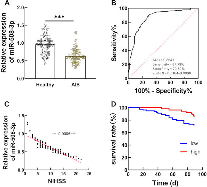
Figure 1: Expression characteristics and clinical value of serum miR-508-3p in patients with AIS. (A) Scatter plot of relative serum miR-508-3p expression in healthy controls and AIS patients. (B) ROC curve evaluating the diagnostic efficacy of serum miR-508-3p for AIS. (C) Spearman correlation between miR-508-3p expression and NIHSS scores in AIS patients. (D) Event‑free survival of AIS patients, shown by Kaplan‑Meier curves and grouped by miR-508-3p expression level (low vs. high). Data for panel A is presented as individual values with mean ± SD. Statistical significance was determined by Student’s t-test. ***p < 0.001 vs. healthy group. For panel C, Spearman rank correlation analysis was used, r = -0.9006, ***p < 0.001. Please click here to view a larger version of this figure.
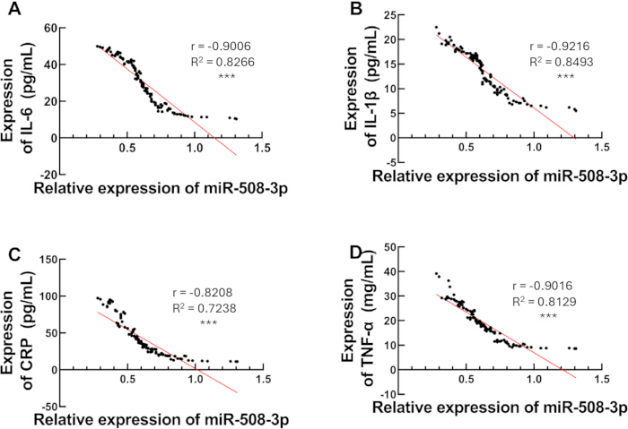
Figure 2: Correlation between serum miR-508-3p and inflammatory factors in AIS patients. (A) Correlation between miR-508-3p and IL-6; (B) Correlation between miR-508-3p and IL-1β; (C) Correlation between miR-508-3p and CRP; (D) Correlation between miR-508-3p and TNF-α. All panels were analyzed using Spearman's rank correlation. (A) r = -0.9006, ***p < 0.001; (B) r = -0.9216,***p < 0.001; (C) r = -0.8208, ***p < 0.001; (D) r = -0.9016, ***p < 0.001. Please click here to view a larger version of this figure.
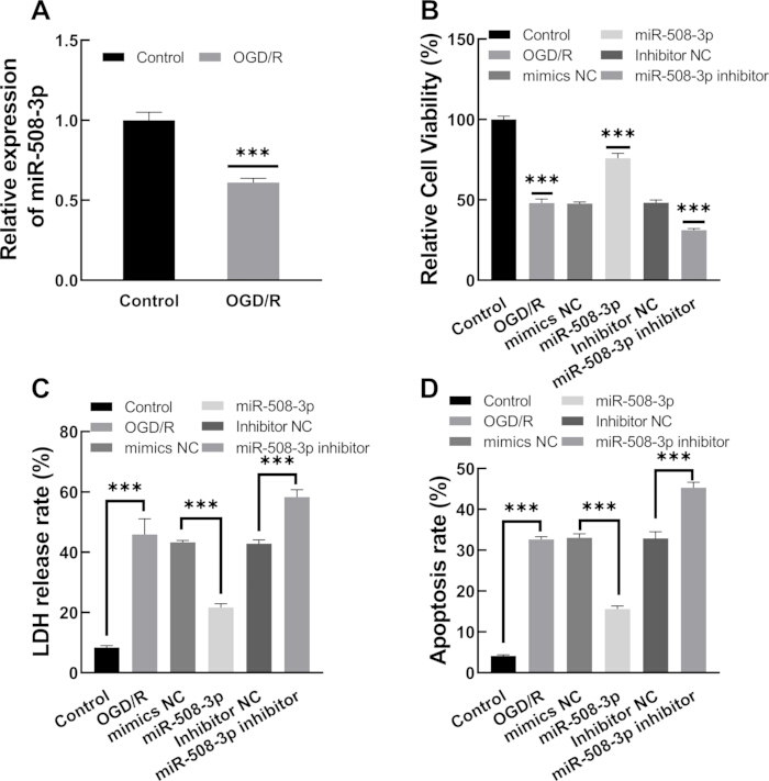
Figure 3: Effects of miR-508-3p on OGD/R-induced injury in HBMECs. (A) Relative expression of miR-508-3p in control and OGD/R-treated HBMECs; (B) Relative cell viability of HBMECs in each group detected by CCK-8 assay; (C) LDH release rate of HBMECs in each group; (D) Apoptosis rate of HBMECs in each group detected by flow cytometry. All quantitative data are presented as mean ± SD from three independent experiments. Statistical significance was determined using one-way ANOVA followed by post hoc comparisons. ***p < 0.001. Please click here to view a larger version of this figure.
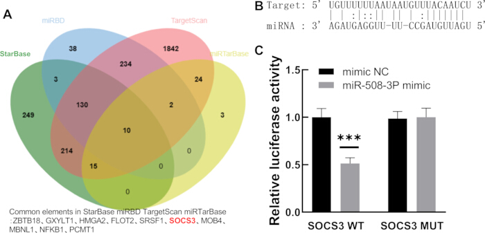
Figure 4: Validation of the targeted binding between miR-508-3p and SOCS3. (A) Venn diagram of common potential target genes of miR-508-3p predicted by StarBase, miRDB, TargetScan, and miRTarBase databases; (B) Schematic of the predicted miR-508-3p binding region in the SOCS3 3'UTR; (C) Verification of the direct miR-508-3p/SOCS3 interaction using a dual-luciferase reporter system. All quantitative data are presented as mean ± SD from three independent experiments. Statistical significance was determined by Student’s t-test. **p < 0.01 vs. mimic NC group. Please click here to view a larger version of this figure.
| Variable | Healthy (n=125) | AIS (n=131) | p value |
| Age (years)a | 62.31 ± 8.52 | 62.54 ± 8.27 | 0.877 |
| Male, n (%) | 65 (52.0) | 70 (53.4) | 0.818 |
| overweight/obese, n (%) | 49 (39.2) | 51 (38.9) | 0.965 |
| Hypertension n (%) | 34 (27.2) | 54 (41.2) | 0.018 |
| Diabetes n (%) | 36 (28.8) | 53 (40.5) | 0.051 |
| Hyperlipemia n (%) | 20 (16.0) | 38 (29.0) | 0.013 |
| atrial fibrillation n (%) | 12 (9.6) | 26 (19.8) | 0.021 |
| drinking history n (%) | 22 (17.6) | 23 (17.6) | 0.993 |
| smoking history n (%) | 15 (12.0) | 17 (13.0) | 0.813 |
Table 1: The baseline characteristics of controls and patients with acute ischemic stroke. Data are presented as mean ± SD or number (percentage). Statistical significance was determined by Student’s t-test for continuous variables and chi-square test for categorical variables. *p < 0.05 indicates a statistically significant difference between groups. Abbreviations: BMI = body mass index.
| Logistic univariate regression analysis | Logistic multivariate regression analysis | |||||
| p | OR | 95% CI | p | OR | 95% CI | |
| Age | 0.156 | 0.537 | 0.227-1.268 | |||
| Gender | 0.314 | 1.532 | 0.668-3.514 | |||
| overweight/obese | 0.220 | 1.685 | 0.732-3.882 | |||
| Hypertension | 0.655 | 0.824 | 0.353-1.924 | |||
| Diabetes | 0.940 | 1.033 | 0.446-2.390 | |||
| Hyperlipemia | 0.495 | 0.718 | 0.278-1.857 | |||
| atrial fibrillation | 0.200 | 1.882 | 0.716-4.950 | |||
| drinking history | 0.611 | 0.738 | 0.229-2.382 | |||
| smoking history | 0.168 | 2.158 | 0.722-6.450 | |||
| NIHSS | 0.038 | 1.098 | 1.005-1.199 | 0.390 | 0.925 | 0.776-1.104 |
| miR-508-3p | 0.007 | 0.015 | 0.001-0.319 | 0.032 | 0.002 | 0.0001-0.581 |
Table 2: Multivariate logistic regression analysis of independent predictors for 90-day poor functional prognosis in AIS patients. OR values and 95% CIs are presented for each variable. Statistical significance was determined using multivariate logistic regression. p < 0.05 indicates independent statistical significance.
| Grouping | Relative Cell Viability (%) | LDH release rate (%) | Apoptosis rate (%) |
| Control | 100 ± 0.00 | 8.26 ± 1.15 | 4.18 ± 0.62 |
| OGD/R | 48.35 ± 4.27 | 42.58 ± 3.64 | 32.75 ± 2.83 |
| mimics NC | 47.92 ± 3.95 | 43.12 ± 3.27 | 33.04 ± 2.56 |
| miR-508-3p | 76.24 ± 5.18 | 21.37 ± 2.45 | 15.62 ± 1.74 |
| Inhibitor NC | 48.06 ± 4.03 | 42.85 ± 3.41 | 32.87 ± 2.69 |
| miR-508-3p inhibitor | 31.25 ± 3.06 | 58.63 ± 4.12 | 47.53 ± 3.25 |
Table 3: Effects of miR-508-3p regulation on viability, LDH release, and apoptosis of HBMECs under OGD/R conditions. Data are presented as mean ± SD from three independent experiments. Statistical significance was determined by one-way ANOVA. *p < 0.05 indicates a statistically significant difference between groups.
Supplementary Table 1: Raw data. All the quantitative raw data values of various experiments, along with basic clinical data.Please click here to download this file.
Diskussion
The present study represents the first systematic investigation into the altered expression and clinical significance of circulating miR-508-3p in AIS. This study demonstrated that miR-508-3p expression showed an extremely strong negative correlation with the admission NIHSS score (r = -0.9006) and a highly strong negative correlation with serum levels of the inflammatory factors IL-6, IL-1β, CRP, and TNF-α (absolute r values > 0.82). These results not only suggest that miR-508-3p can quantitatively reflect the severity of neurological deficits but also reveal its functional association with the core pathological process of AIS, the inflammatory response.
Existing studies have confirmed the inflammation-regulating function of miR-508-3p from multiple aspects. Huang et al.14 demonstrated that miR-508-3p can inactivate the canonical NF-κB signaling pathway in gastric cancer by co‑silencing NFKB1 and RELA. In research on periodontitis with diabetes, miR-508-3p was found to be involved in regulating inflammation-related signaling axes18. Collectively, this cross‑disease evidence suggests that low expression of miR-508-3p may represent a ubiquitous “inflammatory license” signal. In AIS, its downregulation further relieves the inhibition of downstream inflammation‑related target genes, leading to uncontrolled post‑ischemic inflammatory cascades and exacerbated neuronal damage.
In addition, serum miR-508-3p showed favorable diagnostic efficacy for AIS, and low expression levels were closely associated with poor 90‑day functional prognosis in patients. Multivariate regression analysis further confirmed that miR-508-3p was an independent prognostic factor beyond traditional clinical indicators, supporting its clinical application value for prognostic stratification. Regarding diagnostic specificity, a previous study reported altered miR-508-3p levels in cerebrospinal fluid (CSF) from subarachnoid hemorrhage (SAH) patients. However, this study focused on serum samples, which are more suitable for non-invasive rapid screening in pre-hospital and emergency settings. CSF biomarkers reflect direct central nervous system changes but require invasive lumbar puncture, which is not applicable for emergency triage. Furthermore, the pathological mechanisms of AIS and SAH are essentially distinct: SAH is dominated by hemorrhagic injury, cerebral vasospasm, and blood component-induced inflammation, while AIS is characterized by ischemic-reperfusion endothelial injury and an inflammatory cascade. The expression pattern and functional role of miR-508-3p may differ across disease subtypes and sample types. It is acknowledged that the current study only compared AIS patients with healthy controls, and the differential diagnostic value between AIS and other cerebrovascular diseases (hemorrhagic stroke, TIA) or other brain injuries remains to be verified in disease control cohorts.
In this study, dual-luciferase reporter assays demonstrated for the first time that miR-508-3p can directly bind to the 3'UTR region of SOCS3 mRNA. Furthermore, OGD/R cell functional assays showed that overexpression of miR-508-3p alleviated HBMEC injury, whereas inhibition of miR-508-3p exacerbated the damage15. The regulatory effect of the miR-508-3p/SOCS3 axis shows prominent cell specificity. It is well documented that SOCS3 exerts anti-inflammatory and neuroprotective effects in microglia and neurons by inhibiting the overactivated JAK2/STAT3 inflammatory pathway. However, in brain microvascular endothelial cells, moderate STAT3 activation is essential for maintaining cell survival, tight junction integrity, and blood-brain barrier function. After OGD/R injury, excessive upregulation of SOCS3 strongly suppresses endothelial STAT3 activity, leading to increased apoptosis and barrier dysfunction. miR-508-3p directly targets and inhibits the aberrant overexpression of SOCS3, restores physiological STAT3 activity in endothelial cells, and thus alleviates OGD/R-induced HBMEC injury.
In addition, the overall protective effect of miR-508-3p is mediated by multi-target regulation. Previous studies have shown that miR-508-3p directly silences key components of the NF-κB pathway (NFKB1 and RELA), thereby inhibiting the systemic inflammatory cascade. The endothelial protection via SOCS3 inhibition and the systemic anti-inflammatory effect via the NF-κB pathway jointly contribute to the beneficial role of miR-508-3p in AIS. It is worth noting that although the targeting relationship between miR-508-3p and SOCS3 has been previously recorded in databases such as miRTarBase, its functional validation in AIS is reported here for the first time. From a broader perspective, miR-508-3p acts as a tumor suppressor in various cancers. For example, Guo et al.19 showed that miR-508-3p expression is significantly downregulated in triple‑negative breast cancer tissues and cells, and its low expression is significantly associated with lymph node metastasis and distant metastasis. The present study found that miR-508-3p is also downregulated in AIS and targets SOCS3, further expanding the functional landscape of miR-508-3p in non-neoplastic diseases.
These results suggest that miR-508-3p represents a novel potential serum biomarker for early diagnosis and prognostic evaluation of AIS, and may be implicated in the modulation of post-ischemic vascular endothelial injury and inflammatory response. From the perspective of clinical translation, compared with previously reported AIS biomarkers such as miR-140-3p, miR-130a-3p, and miR-320b, miR-508-3p has a diagnostic AUC of 0.8641, combined with prognostic evaluation value, and a stronger correlation with disease severity and inflammatory burden, highlighting its greater potential for clinical translation20,21. Methodologically, this study established a standardized workflow from serum sample processing to in vitro model construction and target validation, with high operational reproducibility. The optimized pre-analytical protocol for serum miRNA detection and the stable OGD/R model of HBMECs are suitable for visual demonstration and can provide a replicable reference for related research on stroke biomarkers, which aligns well with the scope of this journal.
This study also has certain limitations that need to be further improved in future research. First, given the single‑center nature and modest sample size of this work, the broader generalizability of the conclusions awaits verification by prospective, multicenter studies with larger cohorts. Second, only the baseline miR-508-3p expression level at admission was measured, and its dynamic changes across different stages of AIS were not analyzed. Dynamic monitoring of serum miR-508-3p at multiple time points (6 h, 24 h, 7 d, and 14 d after onset) will be performed in our prospective cohort to explore its temporal expression pattern. Further analysis will be performed to examine the correlation between dynamic changes in miR-508-3p and reperfusion effect, neurological recovery, and disease progression, to evaluate its value for disease monitoring and efficacy assessment. Third, the current study only included healthy controls, and the differential diagnostic efficacy of serum miR-508-3p between AIS and other brain injuries (hemorrhagic stroke, TIA) needs further evaluation in disease control cohorts. Meanwhile, no DWI-derived infarct volume data were collected, and therefore could not assess the correlation between miR-508-3p levels and quantitative infarct size. The NIHSS score is known to be influenced by stroke laterality and is not a perfect surrogate for lesion volume; future work should incorporate volumetric imaging analysis to more precisely link miR-508-3p to tissue injury. Fourth, only the direct targeting relationship between miR-508-3p and SOCS3 has been verified; the specific functional role of SOCS3 in mediating the protective effect needs to be confirmed by rescue experiments, and the complete regulatory network of miR-508-3p should also be explored by transcriptomic sequencing. Finally, no in vivo animal experiments were performed to verify the protective effect of miR-508-3p in AIS. Subsequent in vivo validation will be conducted using the MCAO mouse model to provide more sufficient experimental evidence for its clinical translation.
In conclusion, this study demonstrates, for the first time, that miR-508-3p is consistently downregulated in the serum of AIS patients, with its expression significantly negatively correlated with neurological deficit severity and systemic inflammatory burden. miR-508-3p shows favorable early diagnostic efficacy for AIS and may predict a poor 90-day functional prognosis. Mechanistically, this study is the first to demonstrate that miR-508-3p directly targets the 3'UTR of SOCS3. By negatively regulating aberrant SOCS3 activation, it alleviates ischemia-reperfusion-induced brain microvascular endothelial injury, apoptosis, and inflammatory responses, thereby exerting endogenous neurovascular protective effects. Collectively, miR-508-3p is a novel molecular biomarker for AIS early diagnosis, prognostic stratification, and targeted therapy, providing new experimental evidence for precise AIS management.
Offenlegungen
The authors have no relevant financial or non-financial interests to disclose.
AUTHORS’ CONTRIBUTIONS:
Conceptualization was performed by Shitong Zhang (S.Z.) and Qiaomei Yan (Q.Y.). Data curation was carried out by S.Z. and Q.Y. Formal analysis, funding acquisition, and investigation were performed by S.Z. Methodology and software were developed by Wendong Zuo (W.Z.), Dayan Wang (D.W.), and Wenqian Huang (W.H.). Project administration was managed by Q.Y., while resources and supervision were provided by W.Z. Validation and visualization were conducted by S.Z., Zheng Wang (Z.W.), and Yingju Jin (Y.J.). S.Z. prepared the original draft of the manuscript, and Q.Y. reviewed and edited the manuscript.
Danksagungen
Not applicable.
Materialien
| Name | Unternehmen | Katalognummer | Kommentare |
|---|---|---|---|
| Annexin V-FITC/PI apoptosis detection kit | BD Biosciences, USA | 556547 | |
| CCK-8 assay kit | Dojindo, Japan | CK04 | |
| Flow cytometer | BD Biosciences, USA | FACSCalibur | |
| Human brain microvascular endothelial cells (HBMECs) | Procell Life Science & Technology, Wuhan, China | CL-0217 | |
| Human IL-6 / IL-1β / TNF-α / CRP ELISA Kit | Elabscience, Wuhan, China | E-EL-H0102c, etc. | |
| LDH cytotoxicity assay kit | Beyotime, Shanghai, China | C0017 | |
| Lipofectamine 3000 Transfection Reagent | Invitrogen, USA | L3000001 | |
| Microplate reader | Thermo Fisher Scientific, USA | Multiskan FC | |
| miR-508-3p mimics / inhibitor / negative control | GenePharma, Shanghai, China | Custom | |
| miRNA First-Strand Synthesis Kit | Takara Bio, Japan | 638315 | |
| SOCS3 3'UTR WT/MUT dual-luciferase reporter vector | GeneChem, Shanghai, China | Custom | |
| TRIzol reagent | CWBIO, Jiangsu, China | CW0580S |
Referenzen
- Lapchak PA. Emerging therapies: pleiotropic multi-target drugs to treat stroke victims. Transl Stroke Res. 2011;2(2):129-135.
- Krongsut S, Srikaew S, Anusasnee N. Prognostic value of combining 24-hour ASPECTS and hemoglobin to red cell distribution width ratio to the THRIVE score in predicting in-hospital mortality among ischemic stroke patients treated with intravenous thrombolysis. PLoS One. 2024;19(6):e0304765.
- Powers WJ, et al. 2018 Guidelines for the early management of patients with acute ischemic stroke: a guideline for healthcare professionals from the American Heart Association/American Stroke Association. Stroke. 2018;49(3):e46-e110.
- Shukla GC, Singh J, Barik S. MicroRNAs: processing, maturation, target recognition and regulatory functions. Mol Cell Pharmacol. 2011;3(3):83-92.
- O'Brien J, Hayder H, Zayed Y, Peng C. Overview of microRNA biogenesis, mechanisms of action, and circulation. Front Endocrinol (Lausanne). 2018;9:402.
- Balázs K, Antal L, Sáfrány G, Lumniczky K. Blood-derived biomarkers of diagnosis, prognosis, and therapy response in prostate cancer patients. J Pers Med. 2021;11(4):301.
- Mitchell PS, et al. Circulating microRNAs as stable blood-based markers for cancer detection. Proc Natl Acad Sci U S A. 2008;105(30):10513-10518.
- Katayama ES, et al. The stability of microRNAs in serum and plasma suggests they could serve as circulating biomarkers. Noncoding RNA Res. 2025;15:132-141.
- Bejleri J, Jirström E, Donovan P, Williams DJ, Pfeiffer S. Diagnostic and prognostic circulating microRNA in acute stroke: a systematic and bioinformatic analysis of current evidence. J Stroke. 2021;23(2):162-182.
- Felekkis K, Papaneophytou C. The circulating biomarkers league: combining miRNAs with cell-free DNAs and proteins. Int J Mol Sci. 2024;25(6):3189.
- Li S, et al. Non-coding RNAs in acute ischemic stroke: from brain to periphery. Neural Regen Res. 2025;20(1):116-129.
- Burlacu CC, et al. Circulating microRNAs and extracellular vesicle-derived microRNAs as predictors of functional recovery in ischemic stroke patients: a systematic review and meta-analysis. Int J Mol Sci. 2022;24(1):744.
- Guo F, et al. miR-508-3p suppresses the development of ovarian carcinoma by targeting CCNA2 and MMP7. Int J Oncol. 2020;57(1):264-276.
- Huang T, et al. miR-508-3p concordantly silences NFKB1 and RELA to inactivate canonical NF-κB signaling in gastric carcinogenesis. Mol Cancer. 2016;15:9.
- Lu E, Zhou P, Li Y, Chen J, Zhang K, Zhou K. Long noncoding RNA ISA1 protects against ischemic brain damage by promoting the transformation of microglia toward an anti-inflammatory phenotype via the SOCS3/JAK2/STAT3 pathway. Neurochem Res. 2025;50(2):92.
- Carmichael ST. Gene expression changes after focal stroke, traumatic brain and spinal cord injuries. Curr Opin Neurol. 2003;16(6):699-704.
- Liang E, Li X, Fu W, Zhao C, Yang B, Yang Z. COP9 signalosome subunit 3 restricts neuroinflammatory responses during cerebral ischemia/reperfusion injury through stabilizing suppressor of cytokine signaling 3 proteins. Neuropsychiatr Dis Treat. 2021;17:1217-1227.
- He L, et al. Circular RNA hsa_circ_0084054 promotes the progression of periodontitis with diabetes via the miR-508-3p/PTEN axis. J Periodontal Res. 2023;58(4):827-840.
- Guo SJ, Zeng HX, Huang P, Wang S, Xie CH, Li SJ. MiR-508-3p inhibits cell invasion and epithelial-mesenchymal transition by targeting ZEB1 in triple-negative breast cancer. Eur Rev Med Pharmacol Sci. 2018;22(19):6379-6385.
- Ju HY, Tang SS, Li BJ, Luo X, Li Q. The expression levels of circulating miR-140-3p, miR-130a-3p, and miR-320b as diagnostic biomarkers in acute ischemic stroke. Kaohsiung J Med Sci. 2023;39(9):927-935.
- Xie Z, et al. miR-494 in lymphocytes: a promising biomarker for acute ischemic stroke. Rev Neurol. 2025;80(7):37809.