This study presents a standardized protocol for robot-assisted submandibular gland tumor excision via a gasless trans-hairline approach. Using a Chinese-developed robotic platform, the technique emphasizes reproducible docking, sequential anatomical dissection, neurovascular preservation, and intact en bloc tumor removal with favorable early outcomes.
Fallbericht
Robot-Assisted Gasless Trans-Hairline Excision of Submandibular Gland Tumors
In diesem Artikel
Zusammenfassung
Zusammenfassung
Benign submandibular gland tumors are usually resected through a transcervical incision, which provides direct exposure but leaves visible cervical scarring. Remote-access endoscopic techniques improve scar concealment but can be limited by two-dimensional visualization, assistant-dependent camera control, and rigid instrument collision in the narrow submandibular gland space. This article presents a standardized video protocol for robot-assisted submandibular gland excision via a gasless trans-hairline approach for benign tumors. The procedure begins with postauricular trans-hairline incision design, subplatysmal flap elevation, and mechanical retractor-based creation of a gasless working cavity. After robotic docking, a 3D endoscope and articulated instruments are used for sequential dissection of the superior, posterior, inferior, deep, and anterior glandular borders. Critical steps include protection of the marginal mandibular branch of the facial nerve, management of the facial vein and facial artery branches, exposure of the digastric and mylohyoid muscles, identification of the lingual nerve, division of submandibular ganglion branches, clipping of Wharton’s duct, en bloc gland-and-tumor retrieval, hemostasis, drainage, and layered closure. Ten female patients with clinically benign submandibular gland tumors underwent this procedure. All operations were completed robotically without conversion. The mean operative time was 134.5 min ± 36.9 min, the mean estimated blood loss was 16.2 mL ± 7.6 mL, and the mean incision length was 5.6 ± 0.5 cm. Final pathology confirmed pleomorphic adenoma in all patients. No major vascular injury, postoperative marginal mandibular, lingual, or hypoglossal nerve dysfunction, hematoma, seroma, infection, or salivary fistula was observed during the available follow-up. This protocol provides a reproducible workflow for selected patients, although larger studies with longer follow-up are needed.
Einleitung
Salivary gland tumors represent a distinct subset of head and neck neoplasms, accounting for approximately 3% to 6% of all cases1. The submandibular gland (SMG) is the second largest salivary gland, where approximately 75% of the tumors are benign2,3. Among these, pleomorphic adenoma (PA) is the overwhelmingly predominant histological subtype2,3. Currently, the transcervical approach (TCA) remains the standard surgical procedure for SMG tumor excision4. While oncologically safe, TCA inevitably leaves a visible cervical scar and carries the risk of keloid formation or hyperpigmentation5. For young patients and those predisposed to keloids, this can result in a significant psychological burden and aesthetic distress5.
Driven by increasing aesthetic demands, alternative remote-access surgical approaches have been explored. The transoral approach successfully achieves a "scarless" outcome on the skin. However, it is hindered by a narrow, inverted surgical field, rendering hemostasis challenging and carrying a potentially increased risk of lingual nerve injury6,7. In addition, endoscope-assisted trans-hairline approaches emerged to conceal the incision. Yet, traditional endoscopic techniques present several intrinsic limitations. Surgeons struggle with a lack of three-dimensional (3D) depth perception and rely heavily on a skilled assistant for camera stability5,8. Furthermore, rigid endoscopic instruments are highly prone to collisions within the confined SMG workspace9.
Robotic surgical systems may help address several of these technical constraints by providing magnified three-dimensional high-definition visualization, tremor filtration, and articulated instruments with multiple degrees of freedom10,11. These features may improve operative stability and dexterity, reduce dependence on assistant-held visualization, and mitigate instrument interference during fine dissection around the facial vessels, marginal mandibular branch of the facial nerve, lingual nerve, hypoglossal nerve, and Wharton’s duct12. When combined with a gasless trans-hairline approach, robotic surgery allows the creation of a stable working cavity without CO₂ insufflation, thereby avoiding insufflation-related concerns such as gas leakage, subcutaneous emphysema, and hypercarbia13.
Previous studies by Yang and colleagues established the feasibility of robotic trans-hairline SMG resection using standard multiarm and flexible single-port robotic systems12. More recently, Chinese-developed robotic platforms have been evaluated in oral and maxillofacial head and neck procedures, including preclinical parotidectomy, submandibular gland resection, and neck dissection, as well as early clinical applications in salivary gland surgery14. Building on these reports, the present study focuses on the integration of a CO₂-free gasless workflow, a single-port surgical system, and a standardized step-by-step video protocol. The protocol emphasizes reproducible workspace creation, robotic docking, sequential anatomical dissection, and neurovascular preservation. In addition, because PA requires strict capsular safety, the procedure is designed to achieve complete en bloc gland-and-tumor excision while avoiding capsular rupture, tumor spillage, and piecemeal resection. Representative outcomes from ten patients are presented to demonstrate the feasibility of this workflow. For this protocol, eligible patients had a primary, clinically benign, mobile tumor confined to the submandibular gland, with a greatest diameter of ≤4 cm on preoperative imaging. Suspected malignancy, extraglandular extension, involvement of major neurovascular structures, or cervical nodal disease requiring neck dissection were considered contraindications; recurrent tumors, dense fibrosis associated with chronic sialadenitis, and previous neck surgery or radiotherapy were regarded as relative contraindications. Conversion to a transcervical approach was predefined when adequate exposure, anatomic orientation, or hemostasis could not be maintained, when dense adhesions precluded safe dissection, when capsular integrity was threatened, or when intact en bloc resection could not be ensured.
Case presentation:
We present a case series of 10 female patients aged 25–63 years who presented with slowly enlarging, painless submandibular masses (Table 1). The duration of their symptoms varied widely, ranging from 2 months to over 10 years, with no relevant family or social histories, and no previous treatments or radiation to the head and neck region. Physical examination uniformly revealed unilateral, palpable submandibular masses ranging from 1.0 cm to approximately 3.0 cm in diameter. Upon palpation, the tumors were generally characterized as tough or firm in consistency, nodular or smooth, mobile, and non-tender, without any fixation to the overlying skin or surrounding deep tissues. Furthermore, none of the patients exhibited symptoms of facial nerve weakness, and regional cervical lymph nodes were largely impalpable, except for one patient who presented with an incidental 0.6 cm reactive cervical lymph node.
Diagnosis, assessment, and plan:
Initial evaluations included cervical ultrasonography to assess tumor size, echogenicity, borders, and vascularity. Lesion characteristically revealed hypoechoic nodules with clear boundaries and some internal blood flow signals. To accurately delineate the tumors’ relationship with surrounding deep neck structures and evaluate for potential malignancy, additional imaging via contrast-enhanced CT or MRI was utilized. The lesions were characterized by well-demarcated, encapsulated soft-tissue lesions with varying degrees of enhancement. Based on the clinical signs of an indolent, painless, and mobile mass coupled with the imaging findings, the presumptive diagnosis was a benign submandibular gland neoplasm, most likely a PA. The differential diagnosis included Warthin tumor, basal cell adenoma, chronic sialadenitis, and low-grade malignancies. After counseling regarding conventional transcervical surgery and remote-access alternatives, all patients elected the hairline approach because of concern regarding visible cervical scarring. The procedure was selected only when preoperative assessment suggested that the gland and tumor could be removed intact through the remote-access corridor. Scar concealment was discussed as a potential aesthetic benefit, but did not supersede the requirements for safe exposure and appropriate oncological management. Potential complications and side effects associated with this treatment include transient or permanent paresis of the marginal mandibular branch of the facial nerve, lingual or hypoglossal nerve neuropraxia, hematoma, seroma, salivary fistula (sialocele), and the long-term risk of tumor recurrence.
Protokoll
This case series used the SHURUI® Endoscopic Surgical System (SR-ENS-600; Beijing Surgerii Robotics Company Limited, Beijing, China). The procedure described here was approved and conducted following the guidelines of the human research ethics committee of West China Hospital of Stomatology, Sichuan University, Chengdu, China (WCHSIRB-D-2024-346-R2). The reagents and the equipment used are listed in the Table of Materials.
1. Patient preparation and positioning
- Patient positioning
- Under general anesthesia, the patient was placed in the supine position with the neck extended and the head rotated toward the contralateral side (Figure 1A). The position was adjusted to provide an unobstructed operative corridor from the retroauricular hairline to the submandibular triangle.
- Intraoperative neuromonitoring
- Intraoperative neuromonitoring was used in all cases. Recording electrodes were positioned in the ipsilateral orbicularis oris region to monitor the marginal mandibular branch of the facial nerve. The stimulation current was set within a range of 0.2–0.5 mA. Neuromonitoring was used as an adjunct to anatomical identification rather than as a substitute for direct visualization.
- Facial nerve protection was based primarily on maintaining the correct subplatysmal dissection plane and elevating the tissue superficial to the facial vessels with the skin flap. Fixed traction and energy activation near the inferior mandibular border were minimized.
2. Incision design and creation of working space
- Incision infiltration and design
- Approximately 5 mL of 1:100,000 epinephrine-saline solution was infiltrated subcutaneously along the planned incision and flap-elevation area. A curved trans-hairline incision was then made along the natural contour of the retroauricular hairline (Figure 1A).
- Flap elevation under direct vision
- The skin flap was initially elevated under direct vision in a plane superficial to the sternocleidomastoid muscle. Dissection continued until the anterior border of the sternocleidomastoid muscle was reached.
- The greater auricular nerve, external jugular vein, and platysma were identified as landmarks during flap elevation. These structures were preserved when they did not interfere with safe working-space creation.
NOTE: Adequate direct-vision flap elevation was confirmed when the anterior border of the sternocleidomastoid muscle was exposed, and the external jugular vein and platysma could be distinguished clearly.
- Endoscopic flap elevation and gasless working-space creation
- A surgical retractor was inserted after the anterior border of the sternocleidomastoid muscle had been reached. The retractor was used to elevate the flap and maintain the gasless working cavity. Flap elevation continued anteriorly along the deep surface of the platysma.
- The working cavity extended superiorly to the inferior border of the mandible, anteriorly to approximately 1 cm beyond the anterior border of the gland, inferiorly to approximately 1 cm below the inferior gland border, and posteriorly to the anterior border of the sternocleidomastoid muscle.
- The retractor was adjusted until at least 2 cm of vertical clearance was obtained at the entrance to the working cavity. Retractor tension was reduced if excessive blanching or compression of the elevated flap was observed.
NOTE: Successful working-space creation was confirmed when the elevated flap remained stable without obstructing the endoscopic field, and the inferior mandibular border, anterior gland region, and anterior border of the sternocleidomastoid muscle were visible (Figure 1C).
3. Robot docking and instrument setup
- Console configuration
- The robotic system was brought to the operative field and initialized using the manufacturer-recommended standard console configuration. Tremor filtration and motion scaling remained at the standard settings, and no user-defined operating mode or motion-scaling ratio was applied.
- Endoscope and instrument arrangement
- A 0°, 10-mm three-dimensional endoscope was introduced centrally through the hairline incision. The grasping forceps were positioned to the lower left of the endoscope, and the Maryland bipolar forceps were positioned to the lower right. The endoscope and the two working instruments were aligned at angles of approximately 30° to one another to reduce external and intracavitary instrument interference (Figure 1B).
- Before console dissection began, the working ends of both instruments were confirmed within the endoscopic field. The instruments were opened, closed, and moved through the anticipated working range to confirm that no collision or restriction was present.
NOTE: The principal components of the endoscopic surgical system are shown in Supplementary Figure 1.
4. Tumor and gland resection
- Superior border dissection
- The facial vein was identified first. The tissue superficial to the vein was dissected and swept superiorly within the subplatysmal plane to elevate the marginal mandibular branch with the flap.
- Intraoperative neuromonitoring was used when dissection approached the expected course of the marginal mandibular branch. Direct visualization and preservation of the correct tissue plane remained the primary means of nerve protection.
- The facial artery was separated anterior to the facial vein. After the facial vessels and the superior gland border had been adequately exposed, the distal facial vein was ligated with a vascular clip (Figure 1D).
- Posterior and inferior border dissection
- The posterior border of the submandibular gland was dissected from superior to inferior along the anterior border of the sternocleidomastoid muscle. The proximal facial vein was then ligated with a vascular clip.
- The gland was retracted anterosuperiorly until the posterior belly of the digastric muscle and its intermediate tendon were exposed. These structures served as the posterior and inferior landmarks for continued dissection.
- The facial artery was identified along the posterior and inferior gland borders. Depending on its anatomical relationship with the gland, either the proximal arterial trunk was ligated with vascular clips or the glandular branches were coagulated using the Maryland bipolar forceps while the main trunk was preserved (Figure 1E).
NOTE: Posterior and inferior dissection was considered adequate when the posterior belly and intermediate tendon of the digastric muscle were visible, and the facial vessels entering the gland had been controlled.
- Deep border dissection
- The dissection proceeded anteriorly along the deep surface of the submandibular gland until the mylohyoid muscle was exposed. The gland was then retracted inferiorly to expose the posterior border of the mylohyoid muscle.
- The V-shaped course of the lingual nerve was identified deep to the posterior border of the mylohyoid muscle. No submandibular ganglion branch or duct was divided until the lingual nerve had been clearly identified.
- The branches of the submandibular ganglion entering the gland were divided sharply. Wharton’s duct was isolated along its linear course from the lingual nerve, secured with vascular clips, and then divided (Figure 1F).
- Anterior border dissection and en bloc mobilization
- The anterior border of the gland was dissected on the superficial surface of the mylohyoid muscle. The remaining fascial attachments were divided until the gland was fully mobilized (Figure 1G).
- The tumor capsule was not grasped directly. Traction was applied to the surrounding glandular tissue, and dissection proceeded along the glandular capsule.
- The submandibular gland and tumor were mobilized as a single en bloc specimen. The exposed mylohyoid and digastric muscles were inspected to confirm completion of gland removal.
5. Specimen retrieval and wound closure
- Specimen inspection and retrieval
- Before retrieval, the gland-and-tumor specimen was inspected to confirm capsular integrity. Traction was applied to the gland rather than to the tumor surface during extraction. When extraction through the hairline incision produced excessive specimen compression or capsular distortion, the incision was enlarged rather than forcing the specimen through the original opening.
- Drain placement and layered closure
- The operative cavity was irrigated with sterile saline. A negative-pressure suction drain was placed within the surgical bed and secured. The incision was then closed in layers (Figure 1H). Final wound inspection confirmed that the skin edges were approximated without excessive tension and that the drain maintained negative pressure.
6. Postoperative management and outcome assessment
- Postoperative analgesia and pain assessment
- All patients received diclofenac sodium sustained-release capsules orally at 50 mg twice daily as the routine postoperative analgesic regimen. The administration of any additional rescue analgesic was recorded. Postoperative pain was assessed using an 11-point Visual Analogue Scale ranging from 0, indicating no pain, to 10, indicating the worst imaginable pain. Pain scores were recorded on postoperative days 1, 2, and 3, and at 1 week and 1 month.
- Facial and lingual nerve assessment
- Facial nerve function was graded using the House–Brackmann system. Lingual nerve function was evaluated by asking patients about tongue numbness, paresthesia, dysesthesia, perceived taste alteration, and sensory asymmetry, followed by bilateral light-touch comparison over the anterior two-thirds of the tongue.
- Lingual nerve function was considered clinically normal when no sensory disturbance was reported, and light-touch sensation was symmetric between the operated and contralateral sides. Perceived taste changes were recorded by history; no formal gustatory testing was performed.
- Hypoglossal nerve assessment
- Hypoglossal nerve function was assessed by inspecting tongue position at rest and evaluating tongue protrusion, deviation, lateral movement, atrophy, and fasciculation. Function was considered clinically normal when the tongue remained midline during protrusion, mobility was preserved, and no atrophy or fasciculation was observed.
- Scar and patient-reported aesthetic assessment
- Scar quality was evaluated at the final available follow-up using the Vancouver Scar Scale, which assesses vascularity, pigmentation, pliability, and scar height. Patient-reported aesthetic satisfaction was recorded using the institutional 0–100 satisfaction scale.
Ergebnisse
Postoperative neurological function, scar quality, patient-reported aesthetic satisfaction, and pain outcomes are summarized in Table 2, with patient-level data provided in Supplementary Table 1, Supplementary Table 2, Supplementary Table 3, and Supplementary Table 4.
All glands and tumors were removed en bloc with intact surgical capsules. No capsular rupture, tumor spillage, or piecemeal resection occurred. Figure 2A presents a representative en bloc gland-and-tumor specimen. Intraoperative frozen-section analysis performed after specimen removal was interpreted as a benign salivary gland neoplasm without evidence of malignancy in all 10 cases. Permanent histopathological examination confirmed pleomorphic adenoma in all patients. Representative hematoxylin and eosin-stained sections showed duct-like epithelial and myoepithelial components within a chondromyxoid stroma (Figure 2B,C).
At the final available follow-up, with a mean duration of 6.5 ± 0.7 months, all patients had House–Brackmann Grade I facial nerve function. No marginal mandibular branch weakness was identified. None of the patients reported lingual nerve-related sensory or taste disturbance, and bilateral light-touch examination showed symmetric sensation over the anterior tongue. Tongue position, protrusion, and mobility were normal in all patients, without deviation, atrophy, or fasciculation.
The median Vancouver Scar Scale score was 1.0 (range, 0–3). The mean institutional patient- reported aesthetic satisfaction score was 94.6 ± 2.2, with a median of 94.5 (range, 91–98). These findings represent descriptive early scar and patient-reported outcomes; the clinical photographs in Figure 3 show one representative patient.
All patients received diclofenac sodium sustained-release capsules orally at 50 mg twice daily. Under this standardized regimen, the median VAS score decreased from 3.0 on postoperative day 1 to 1.0 on postoperative day 3 and 0.0 at 1 month.
Typical case
A 32-year-old female patient presented to our department with a chief complaint of an indolent, painless mass in the left submandibular region that had been slowly enlarging for over 2 years. The patient's facial nerve function was normal (Figure 4A). On physical examination, a well-circumscribed, oval-shaped mass measuring approximately 2.5 cm × 2.0 cm was palpable in the left submandibular area (Figure 4B). The lesion demonstrated a firm consistency, good mobility, and no tenderness upon palpation. There was no skin involvement, and no abnormal cervical lymphadenopathy was detected in the surrounding neck regions.
Preoperative diagnostic imaging and evaluation included an ultrasound-guided biopsy performed at an external institution. The ultrasound revealed a distinct hypoechoic lesion in the left submandibular region. Histological examination of the biopsied tissue suggested a salivary gland-derived neoplasm. Enhanced CT imaging showed a mass-like soft tissue shadow in the lower part of the left submandibular gland, with uneven mild enhancement in the center of the lesion during contrast administration (Figure 4C).
Surgical details and aesthetic results
The total operative time was 76 min, with the core robotic resection taking 52 min. The procedure was completed with minimal intraoperative blood loss, estimated at only 5 mL. The marginal mandibular branch of the facial nerve, lingual nerve, and hypoglossal nerve were successfully identified and anatomically preserved using intraoperative neuromonitoring. There was no need for conversion to a traditional open transcervical approach. Postoperatively, the patient’s recovery was uneventful. The negative-pressure suction drain was removed on postoperative day 2, and the patient was discharged on the third day after surgery. Final histopathological examination confirmed a pleomorphic adenoma. At the 1-week (Figure 3A) and 1-month (Figure 3B) follow-up, the patient exhibited normal facial and tongue movements with no neurological deficits. The incision healed without wound-related complications. At the 1-month follow-up, the hairline location and surrounding hair made the surgical site less conspicuous in this representative patient.
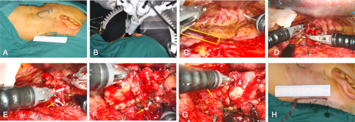
Figure 1: Step-by-step intraoperative visualization of the robot-assisted submandibular gland excision. (A) Preoperative incision design and landmarks. The solid line indicates the trans-hairline incision, the dashed line outlines the extent of the subplatysmal flap elevation, and the circle outlines the palpable submandibular gland and tumor. (B) Robotic system docking. The 3D endoscope and robotic instrument arms are deployed and aligned within the working cavity. (C) Established surgical working space. Endoscopic view of the completed surgical cavity. The yellow box highlights the external jugular vein. (D) Dissection of the superior border. The facial vein (green arrow) is identified and ligated with a vascular clip (yellow arrow). (E) Dissection of the posterior and inferior borders. The glandular branch of the facial artery (green arrow) entering the submandibular gland is isolated and securely ligated with a vascular clip (yellow arrow). (F) Management of the deep border. The submandibular duct (green arrow) is carefully isolated and clamped with a vascular clip (yellow arrow) prior to transection. (G) Dissection of the anterior border. Final detachment of the submandibular gland completes the resection, exposing the underlying mylohyoid muscle (yellow arrow) and the digastric muscle (green arrow). (H) Final wound closure. The image demonstrates the immediate postoperative cosmetic outcome. Please click here to view a larger version of this figure.
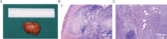
Figure 2: Representative gross and histopathological findings. (A) En bloc resected submandibular gland and tumor specimen. (B) Low-power hematoxylin and eosin-stained section showing a well-circumscribed tumor with a fibrous capsule at its periphery (arrow). (C) Higher-power section showing duct-like epithelial and myoepithelial components within a chondromyxoid stroma (arrow), consistent with pleomorphic adenoma. Scale bars: (B) 200 μm and (C) 100 μm. Please click here to view a larger version of this figure.
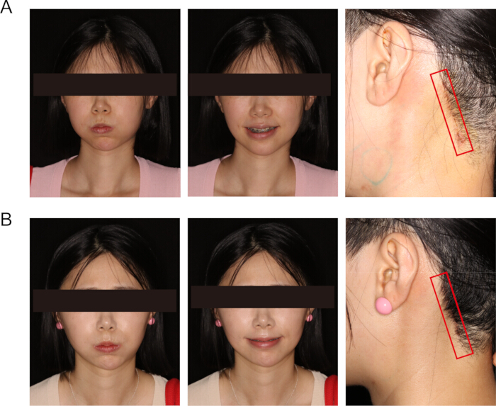
Figure 3: Postoperative clinical and aesthetic outcomes of the representative case. (A) One-week postoperative follow-up showing symmetric facial animation and uncomplicated early wound healing of the trans-hairline incision. (B) One-month postoperative follow-up showing preserved facial nerve function and patient-reported aesthetic satisfaction with the trans-hairline incision by substantial concealment in this representative patient. Please click here to view a larger version of this figure.
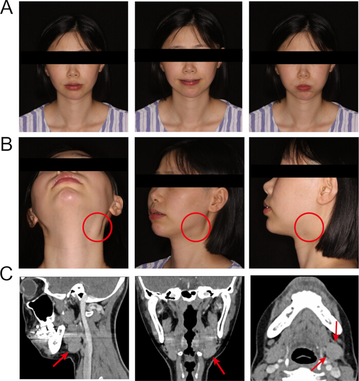
Figure 4: Preoperative clinical presentation and radiological assessment of the representative case. (A) Baseline facial nerve evaluation. The patient demonstrates symmetric and normal facial expressions. (B) Preoperative clinical appearance. The red circle demarcates the externally visible, palpable mass located in the submandibular region. (C) Contrast-enhanced computed tomography (CT) imaging. The scan reveals a well-circumscribed, enhancing soft-tissue lesion within the submandibular gland, supporting the preliminary diagnosis of a benign salivary neoplasm.Please click here to view a larger version of this figure.
| Case Number | Sex | Age (Years) | Side | Tumor dimensions (cm) | Location within the SMG | Surgery Duration (min) | Blood Loss (mL) | Incision Length (cm) | Pathological results |
| 1 | Female | 61 | Right | 2.5×1.6×1.2 | Anterior–inferior | 152 | 20 | 5.6 | PA |
| 2 | Female | 29 | Right | 1.8×1.1×0.8 | Anterior–inferior | 141 | 15 | 6.1 | PA |
| 3 | Female | 31 | Left | 3.7×2.6×1.9 | Central–superior | 142 | 20 | 5.5 | PA |
| 4 | Female | 39 | Left | 1.5×0.9×0.7 | Posterior–superior | 186 | 10 | 4.9 | PA |
| 5 | Female | 25 | Right | 2.7×2.4×1.8 | Central–superior | 123 | 15 | 5.3 | PA |
| 6 | Female | 63 | Left | 2.2×1.4×1.3 | Anterior–inferior | 191 | 30 | 6.3 | PA |
| 7 | Female | 32 | Left | 2.6×2.5×1.9 | Posterior–superior | 76 | 5 | 5.1 | PA |
| 8 | Female | 26 | Right | 3.1×2.7×2.2 | Central–superior | 101 | 10 | 5.6 | PA |
| 9 | Female | 37 | Left | 3.2×2.8×2.1 | Anterior–inferior | 98 | 12 | 6.2 | PA |
| 10 | Female | 31 | Right | 3.1×2.3×1.4 | Posterior–superior | 135 | 25 | 5.1 | PA |
| Mean ± SD | —— | 37.4 ± 13.7 | —— | 2.64 ± 0.67 × 2.03 ± 0.71 × 1.53 ± 0.53 | —— | 134.5 ± 36.9 | 16.2 ± 7.6 | 5.6 ± 0.5 | —— |
Table 1: Clinical characteristics and perioperative outcomes of enrolled patients. Values are presented as individual patient-level data and summarized as mean ± standard deviation. Tumor size was summarized using the tumor dimensions measured on preoperative imaging. The dominant intraglandular location was described according to the anteroposterior position (anterior, central, or posterior third) and craniocaudal position (superior, middle, or inferior third). PA, pleomorphic adenoma.
| Assessment Category | Assessment Method | Time Point | Results |
| Neurological function assessment | Facial nerve | Final follow-up | HB Grade I in 10/10 patients |
| Neurological function assessment | Lingual nerve | Final follow-up | Normal in 10/10 patients |
| Neurological function assessment | Hypoglossal nerve | Final follow-up | Normal in 10/10 patients |
| Vancouver Scar Scale | Vancouver Scar Scale | Final follow-up | Median VSS 1.0 (0–3) |
| Patient-reported aesthetic satisfaction | Patient-reported aesthetic satisfaction score | Final follow-up | Mean score 94.6 ± 2.2 Median 94.5 (91–98) |
| VAS pain score | VAS pain score | POD1 | POD1 median 3.0 (2–4) |
| VAS pain score | VAS pain score | POD3 | POD3 median 1.0 (0–1) |
| VAS pain score | VAS pain score | 1 month | 1 month median 0.0 (0–0) |
| Clinical follow-up | Clinical follow-up | Final follow-up | Mean 6.5 ± 0.7 months Median 6.0 (6–8) months |
Table 2: Quantitative postoperative outcome assessment after surgery.
| Step | Potential Problem | Cause | Recommended Solution |
| Working-space creation | Arm collision | Inadequate flap elevation or poor retractor angle | Expand cavity, adjust retractor, restore 30° instrument separation |
| Superior border dissection | Marginal mandibular nerve risk | Wrong dissection plane | Maintain subplatysmal plane, use IONM, avoid traction on flap |
| Posterior dissection | Bleeding from facial vessels | Incomplete exposure before vessel control | Suction first, identify vessel, apply clips/bipolar under magnified view |
| Deep dissection | Lingual nerve/duct unclear | Insufficient inferior gland traction | Pause, improve traction, expose posterior mylohyoid border |
| Tumor retrieval | Capsule rupture risk | Excessive traction or small incision | Retrieve en bloc, enlarge incision if necessary, consider specimen bag |
| Oncological concern | Unsafe continuation | Suspicious invasion or unclear anatomy | Convert promptly to open transcervical approach |
Table 3: Critical steps, common problems, and troubleshooting strategies.
Supplementary Figure 1: Principal components of the endoscopic surgical system used in this study. (A) Surgical trolley and robotic arms. (B) Surgeon console. (C) Tissue forceps. (D) Maryland bipolar forceps. (E) Distal end of the 3D electronic endoscope. Please click here to download this file.
Supplementary Table 1: Patient-level postoperative neurological assessment. Facial nerve function was graded using the House–Brackmann system. Lingual nerve function was assessed from patient-reported sensory symptoms and bilateral light-touch comparison over the anterior two-thirds of the tongue. Perceived taste alteration was recorded as a patient-reported symptom; no formal gustatory testing was performed. Hypoglossal nerve function was assessed from tongue position at rest, protrusion, deviation, mobility, atrophy, and fasciculation. Please click here to download this file.
Supplementary Table 2: Patient-level scar assessment using the Vancouver Scar Scale. This table presents postoperative scar evaluation for each patient using the Vancouver Scar Scale. The scale includes four domains: vascularity, pigmentation, pliability, and height. The total score ranges from 0 to 13, with lower scores indicating a flatter, softer, less pigmented, and less vascular scar. Scar outcomes were assessed at the available postoperative follow-up visit to quantify incision healing and hairline scar concealment. Please click here to download this file.
Supplementary Table 3: Patient-reported aesthetic outcome assessment. This table summarizes patient-reported aesthetic outcomes after robot-assisted gasless trans-hairline submandibular gland excision. Patient-reported aesthetic satisfaction was recorded using the institutional patient-reported aesthetic satisfaction score (0–100). Higher scores indicate greater patient satisfaction with postoperative appearance, scar concealment, and overall aesthetic outcome. Please click here to download this file.
Supplementary Table 4: Patient-level postoperative pain assessment using the Visual Analogue Scale. This table summarizes postoperative pain intensity for each patient using the Visual Analogue Scale. Pain was scored from 0 to 10, where 0 indicates no pain and 10 indicates the worst imaginable pain. Pain scores were recorded at predefined postoperative time points, including postoperative day 1, postoperative day 2, postoperative day 3, 1 week, and 1 month, to characterize early postoperative recovery after the minimally invasive trans-hairline robotic procedure. Please click here to download this file.
Diskussion
This study establishes a standardized gasless trans-hairline robotic workflow for submandibular gland tumor excision using a single-port robotic endoscopic surgical system. Its main contribution is the integration of patient selection, working-space creation, single-port instrument deployment, anatomy-guided sequential dissection, en bloc specimen retrieval, and predefined conversion criteria into a video-based protocol. In the 10 selected patients treated with this workflow, all procedures were completed without conversion, the gland and tumor were removed en bloc, postoperative neurological function was preserved, and early scar and patient-reported aesthetic outcomes were favorable. These findings support the technical coherence and early clinical feasibility of the protocol in the population for which it was designed.
The procedure depends on maintaining a stable operative corridor and a consistent sequence of anatomical landmarks. Mechanical retraction created an adjustable gasless working cavity, while the approximately 30° separation between the central endoscope and the two working instruments reduced interference at the access site. Dissection then proceeded from the facial vessels and superior gland border to the digastric and mylohyoid landmarks, followed by identification of the lingual nerve before division of the submandibular ganglion branches and Wharton’s duct. This sequence converts a narrow remote-access operation into a series of recognizable anatomical endpoints. Three-dimensional visualization, tremor filtration, and articulated bimanual instruments were most useful during superior-border dissection, facial-vessel control, and deep dissection around the lingual nerve and duct.
The video format gives this protocol a practical role beyond the cases reported here. It can be used for procedure-specific preparation, coordinated training of the console surgeon and bedside assistant, step-based supervision, and structured postoperative video review. Virtual-reality training for endoscope-assisted submandibular gland removal has shown that anatomy-specific simulation can improve novice performance, while validated robotic curricula support the use of video instruction, simulation, and proficiency-based progression in surgical training15,16. Future studies can relate step completion, workflow interruptions, assistance, docking efficiency, conversion, and operative time to surgical experience.
A further application is data-driven analysis of minimally invasive submandibular gland surgery. A recent multicenter study used a standardized annotation framework to divide surgical videos into Creation, Position, Separation, Inspection, and Idle phases and demonstrated the feasibility of automated workflow recognition and exploratory skill assessment17. The more detailed anatomical checkpoints defined in the present protocol could complement such phase-level models by supplying clinically interpretable labels for critical events, including facial-vessel control, lingual-nerve identification, duct division, specimen mobilization, and final inspection. In this context, the standardized video is not only an educational record but also a structured source for future workflow analytics and quality assessment (Table 3).
The use of the surgical system also expands the range of robotic platforms documented in clinical investigations. Chinese robotic systems have undergone preclinical and early clinical evaluation in head and neck surgery, and the system has been evaluated prospectively in other surgical specialties14,18,19. These studies support the technical maturity of Chinese robotic platforms but involve different systems, procedures, and anatomical settings. The present protocol specifies the access route, instrument arrangement, anatomical sequence, checkpoints, and conversion rules required to evaluate this platform in submandibular gland surgery.
The trans-hairline route offers a specific clinical trade-off. It relocates the incision from the exposed cervical region to the postauricular hairline and maintains an external, clean operative pathway. The gasless cavity permits direct adjustment of exposure without insufflation, whereas robotic control provides a stable surgeon-controlled view and articulated bimanual dissection. These advantages are obtained at the cost of a longer operative corridor, docking, and dedicated equipment. The approach is therefore best viewed as a defined remote-access option for appropriately selected patients who place a high value on scar concealment.
For this protocol, eligible patients had a primary, clinically benign, mobile tumor confined to the submandibular gland. Suspected malignancy, extraglandular extension, major neurovascular involvement, or cervical nodal disease requiring neck dissection were contraindications. Selected malignant submandibular gland tumors have been treated through retroauricular or trans-hairline approaches in small observational series, demonstrating that remote-access resection can be technically possible in carefully selected cases12,20. These reports do not, however, establish a general oncological indication. Preoperative assessment and frozen section were therefore used as sequential decision points in the present protocol. If permanent histopathology unexpectedly identifies malignancy, subsequent management should be determined by histological type, grade, margin status, perineural or lymphovascular invasion, and cervical nodal status. Depending on these findings, additional transcervical surgery, neck dissection, or adjuvant treatment may be indicated21,22,23.
The current single-center series of 10 women and the 6–8-month follow-up support early feasibility but do not yet define comparative effectiveness, long-term recurrence, or transferability across surgical teams. The next step is prospective multicenter evaluation using a shared protocol, standardized neurological and patient-reported outcomes, video-derived workflow metrics, and long-term disease surveillance. Such studies would test whether the procedural consistency demonstrated here can be reproduced across surgeons and platforms while preserving the patient-selection and conversion safeguards that underpin the technique.
Offenlegungen
The authors have nothing to disclose.
Danksagungen
This work was supported by West China Hospital of Stomatology, Sichuan University (Grant No. 24CXTD07, No. LCYJ2022-DL-1, No. LCYJ-MS-202303, and 24LCYJZD10) and the State Key Laboratory of Oral Diseases (Grant No. SKLOD-2025KP009).
Materialien
| Name | Unternehmen | Katalognummer | Kommentare |
|---|---|---|---|
| 3-0 antibacterial VICRYL absorbable suture | Johnson (Suzhou) Medical Devices Co., Ltd., Suzhou, China | VCP1772D | Used for layered closure of the trans-hairline incision. |
| DSRobot 4K-Station surgical video-recording system | Digital Vision, China | DSRobot 4K-Station | Used to record the operative procedure for preparation of the standardized video protocol and postoperative video review. |
| GraphPad Prism software | GraphPad Software, Boston, MA, USA | Version 9 | Used for descriptive statistical analysis and data summarization. |
| High-frequency electrosurgical unit | Guangdong Baisheng Medical Equipment Co., Ltd., China | OBS-350A | Used as the electrosurgical energy platform for tissue dissection and bipolar coagulation. |
| SHURUI 3D endoscope | Beijing Surgerii Robotics Company Limited, Beijing, China | Manufacturer safety and performance information: https://www.surgerii.com/images/12.H-15-000104 SafetyandPerformanceInformation.pdf | Flexible 3D electronic endoscope used for robotic visualization. The safety document specifies a working length of 480 mm, a maximum inserted diameter of 10.8 mm, a resolution of 1920 × 1080, and an 85° field of view. |
| SHURUI Endoscopic Surgical System | Beijing Surgerii Robotics Company Limited, Beijing, China | SR-ENS-600; NMPA registration No. 20233010833 | Single-port master–slave robotic surgical system comprising a surgeon console, surgical trolley/patient platform, 3D electronic endoscope, surgical instruments, and accessories. The system was operated using the manufacturer-recommended standard console configuration, without a user-defined operating mode or customized motion-scaling ratio. |
| SHURUI Maryland bipolar forceps | Beijing Surgerii Robotics Company Limited, Beijing, China | NMPA technical review report: https://www.cmde.org.cn/directory/web/cmde/images/1688431479149043688.pdf | Bipolar dissecting and coagulating instrument. The manufacturer document specifies a working length of 529 mm ± 3%, an end-effector length of 22.2 mm ± 2 mm, and an opening angle of 34° ± 20%. |
| SHURUI multichannel trocar/access port | Beijing Surgerii Robotics Company Limited, Beijing, China | Manufacturer product information: https://en.surgerii.com/ | Multichannel access device for introduction of the 3D endoscope and robotic instruments. |
| SHURUI tissue forceps | Beijing Surgerii Robotics Company Limited, Beijing, China | Manufacturer product information: https://en.surgerii.com/ | Tissue-grasping instrument used for gland traction. The manufacturer document specifies a working length of 535 mm ± 3%, an end-effector length of 28.4 mm ± 2 mm, and an opening angle of 58° ± 20%. |
Referenzen
- Dou K, Zhang T, Yang B, Zhao Z. Comparisons between partial and total sialoadenectomy for benign submandibular gland tumors: a systematic review and meta-analysis. J Craniomaxillofac Surg. 2025;53(7):989-998.
- Menon G, Winters R. Pleomorphic adenoma. In: StatPearls. Treasure Island (FL): StatPearls Publishing; 2026.
- Yu G, Peng X. Conservative and functional surgery in the treatment of salivary gland tumours. Int J Oral Sci. 2019;11(3):22.
- Ma S et al. Surgical robot-assisted submandibular gland excision through retroauricular hairline approach: three case reports. Hua Xi Kou Qiang Yi Xue Za Zhi. 2026;44(2):286-292.
- Yang F et al. Gasless endoscopic submandibular gland excision through hairline approach. J Craniofac Surg. 2023;34(5):1563-1569.
- Cammaroto G et al. Submandibular gland excision: from external surgery to robotic intraoral and extraoral approaches. Oral Dis. 2020;26(5):853-857.
- Singh PP, Goyal M. Our experience with intraoral submandibular gland excision. Indian J Otolaryngol Head Neck Surg. 2020;72(3):297-301.
- Seif-Elnasr M, Magdy EA, Fung E, Deot NS, Marzouk MF. Intraoral microscopic versus robot-assisted sialolithotomy and sialendoscopy for submandibular stones. Laryngoscope. 2024;134(5):2170-2176.
- Lee HS et al. Robot-assisted versus endoscopic submandibular gland resection via retroauricular approach: a prospective nonrandomized study. Br J Oral Maxillofac Surg. 2014;52(2):179-184.
- Douglas JE, Wen CZ, Rassekh CH. Robotic management of salivary glands. Otolaryngol Clin North Am. 2020;53(6):1051-1064.
- Park YM, Koh YW. Current issues in treatment of parotid gland cancer and advanced surgical technique of robotic parotidectomy. Curr Oncol Rep. 2022;24(2):203-208.
- Yang TL. Robotic surgery for submandibular gland resection through a trans-hairline approach: the first human series and comparison with applicable approaches. Head Neck. 2018;40(4):793-800.
- Yang TL, Li H, Holsinger FC, Koh YW. Submandibular gland resection via the trans-hairline approach: a preclinical study of a novel flexible single-port surgical system and the surgical experiences of standard multiarm robotic surgical systems. Head Neck. 2019;41(7):2231-2238.
- Ma Z et al. Evaluation of a newly developed robotic system for head and neck surgery: a prospective study. Gland Surg. 2025;14(7):1250-1262.
- Miki T et al. Development of a virtual reality training system for endoscope-assisted submandibular gland removal. J Craniomaxillofac Surg. 2016;44(11):1800-1805.
- Thomaschewski M et al. Conception and prospective multicentric validation of a robotic surgery training curriculum (RoSTraC) for surgical residents: from simulation via laboratory training to integration into the operation room. J Robot Surg. 2024;18(1):53.
- Ma Z et al. Intelligent surgical workflow recognition-based skill assessment for minimally invasive submandibular gland resection. NPJ Digit Med. 2026; doi: 10.1038/s41746-026-02976-w.
- Ma Z et al. Evaluation of a newly developed oral and maxillofacial surgical robotic platform (KD-SR-01) in head and neck surgery: a preclinical trial in porcine models. Int J Oral Sci. 2024;16(1):51.
- Chang R et al. Effectiveness and safety of SR-ENS-600 endoscopic surgical system in benign and malignant gynecological diseases: a prospective, multicenter, clinical trial with 63 cases. J Robot Surg. 2024;18(1):210.
- de Brito Neves CP, Lira RB, Chulam TC, Kowalski LP. Retroauricular endoscope-assisted versus conventional submandibular gland excision for benign and malignant tumors. Surg Endosc. 2020;34(1):39-46.
- Luksic I, Mamic M, Suton P. Management of malignant submandibular gland tumors: a 30-year experience from a single center. Oral Surg Oral Med Oral Pathol Oral Radiol. 2022;134(3):302-309.
- Atula T, Panigrahi J, Tarkkanen J, Mäkitie A, Aro K. Preoperative evaluation and surgical planning of submandibular gland tumors. Head Neck. 2017;39(6):1071-1077.
- Bolooki A, Johnson F, Stenzl A, Zhu Z, Hofauer BG. Frozen section analysis in submandibular gland tumors: optimizing intraoperative decision-making. Cancers (Basel). 2025;17(5):895.
Nachdrucke und Genehmigungen
Dieser Artikel wurde veröffentlicht
Video demnächst verfügbar