Dieses Protokoll beschreibt in-vivo- und in-vitro-Ansätze, um zu untersuchen, wie N-Lactoyl-Phenylalanin die diabetische Kardiomyopathie, den kardialen Lipidstoffwechsel und die Cholesterinsynthese beeinflusst, wobei der Fokus auf dem Adenosinmonophosphat-aktivierten Proteinkinase-alpha-1/3-Hydroxy-3-methylglutaryl-Coenzym-A-Reduktase-Signalweg liegt.
Ein Abonnement von JoVE ist erforderlich, um diesen Inhalt anzuzeigen. Melden Sie sich an oder beginnen Sie noch heute mit einer kostenlosen Testphase.
Forschungsartikel
N-Lactoyl-phenylalanin verbessert die Herzfunktion in einem Mausmodell der diabetischen Kardiomyopathie durch Regulierung des Lipidstoffwechsels
In diesem Artikel
Zusammenfassung
Zusammenfassung
Diabetische Kardiomyopathie (DCM) ist eine der schwerwiegendsten Komplikationen des Diabetes. N-Lactoyl-Phenylalanin (Lac-Phe), ein endogener Metabolit, kann die Gewichtsabnahme fördern und die Glukosehomöostase verbessern. Diese Studie sollte den therapeutischen Effekt von Lac-Phe auf die DCM untersuchen und den zugrundeliegenden Wirkmechanismus weiter aufklären. Mäuse mit Typ-2-Diabetes und DCM wurden mit Lac-Phe behandelt. Lac-Phe verbesserte die kontraktile Herzfunktion, milderte die pathologische Remodeling und verringerte die Lipidakkumulation im Herzen. In vitro wurden die Wirkungen von Lac-Phe in H9c2-Zellen untersucht, die mit hohem Glukosespiegel und Palmitinsäure stimuliert worden waren. Lac-Phe verringerte die Lipidakkumulation, indem es die Cholesterinsynthese in H9c2-Zellen senkte. Mechanistisch förderte Lac-Phe die Phosphorylierung und Aktivierung der adenosinmonophosphat-aktivierte Proteinkinase-alpha-1 (AMPKα1). Daraufhin phosphorylierte die aktivierte AMPKα1 ihr downstream liegendes Zielprotein, die 3-Hydroxy-3-methylglutaryl-Coenzym-A-Reduktase (HMGCR), und hemmte es, was zu einer verminderten Cholesterinsynthese und letztlich zur Verbesserung des gestörten Lipidstoffwechsels in H9c2-Zellen führte. Diese Ergebnisse zeigen, dass Lac-Phe den Lipidstoffwechsel in H9c2-Zellen bei DCM über den AMPKα1/HMGCR-Signalweg verbessert, wodurch die kontraktile Herzfunktion gesteigert und die pathologische Remodeling abgeschwächt wird. Lac-Phe könnte einen potenziellen therapeutischen Ansatz für die diabetische Kardiomyopathie darstellen.
Einleitung
Diabetes ist eine Stoffwechselstörung, die durch anhaltende Hyperglykämie gekennzeichnet ist1. In den letzten zwei Jahrzehnten ist Diabetes weltweit zu einer der zehn häufigsten Todesursachen geworden, wobei jährlich mehr als sechs Millionen Menschen an Diabetes und dessen Komplikationen sterben2. Die diabetische Kardiomyopathie (DCM), eine kardiale pathologische Erkrankung, die durch Diabetes verursacht wird, stellt eine der schwerwiegendsten Komplikationen im Zusammenhang mit Diabetes dar3,4. Sie wirkt als unabhängiger Risikofaktor für Herzinsuffizienz und ist gekennzeichnet durch strukturelle und funktionelle Veränderungen des Myokards5,6, einschließlich kardialer Fibrose und Kardiomyozytenhypertrophie, die letztendlich sowohl zu diastolischer als auch zu systolischer Dysfunktion führen. Obwohl mehrere herkömmliche kardiovaskuläre Therapien wie β-Blocker, Angiotensin-Converting-Enzym-Hemmer und Angiotensin-II-Typ-1-Rezeptorblocker derzeit zur Behandlung der DCM eingesetzt werden7,8, bleibt ihre Wirksamkeit in der klinischen Anwendung suboptimal. Da die zugrundeliegenden Mechanismen der DCM bisher nicht ausreichend erforscht wurden9, existiert derzeit keine klare therapeutische Strategie zur gezielten Behandlung dieser Erkrankung10,11. Die klinischen Manifestationen der DCM verlaufen vom asymptomatischen Stadium mit diastolischer Dysfunktion in der frühen Phase bis hin zur systolischen Dysfunktion und klinischen Herzinsuffizienz im fortgeschrittenen Stadium und können sogar zum Tod führen12,13. Daher besteht ein dringender Bedarf, wirksame Behandlungsoptionen für diese Erkrankung zu erforschen und zu entwickeln.
Ein gestörter Lipidstoffwechsel ist ein wesentliches Merkmal der DCM14. Bei der DCM können erhöhte Konzentrationen freier Fettsäuren (FFAs) im Blut zu einer übermäßigen Aufnahme von Fettsäuren durch Kardiomyozyten führen15,16. Der Fettsäurestoffwechsel in Kardiomyozyten hängt hauptsächlich von der Fettsäureoxidation ab. Langfristig reicht die Fettsäureoxidation in Kardiomyozyten nicht aus, um alle aufgenommenen Fettsäuren vollständig abzubauen, wodurch überschüssige Fettsäuren und ihre Zwischenmetaboliten in den Kardiomyozyten anreichern17. Diese übermäßige Lipidbelastung induziert Lipotoxizität, stört die myokardiale Energieversorgung und fördert oxidativen Stress sowie entzündliche Reaktionen im Herzen, was letztlich zu schweren Schäden an den Kardiomyozyten führt18,19. Daher könnte eine aktive Regulierung des Herz-Lipidstoffwechsels ein potenzielles therapeutisches Ziel für die DCM darstellen20.
N-Lactoyl-Phenylalanin (Lac-Phe) ist ein endogener Metabolit, dessen Konzentration nach körperlicher Betätigung schnell ansteigt21. Mechanistisch wird Lac-Phe durch das zytosolische Enzym Carnosin-Dipeptidase 2 (CNDP2) synthetisiert, das Lactat und Phenylalanin kondensiert. Dieser biosynthetische Weg ist in vielen Zelltypen weit verbreitet, einschließlich Makrophagen, epithelialen Zellen und mesenchymalen Stammzellen, die nach körperlicher Aktivität Lac-Phe in den Kreislauf abgeben22. Es wurde gezeigt, dass die chronische Gabe von Lac-Phe die Nahrungsaufnahme senkt, Adipositas und Körpergewicht verringert sowie die Glukosehomöostase verbessert21. Eine aktuelle Studie hat ergeben, dass Lac-Phe die Fettstoffwechselfähigkeit von Mikroglia/Makrophagen nach einer Rückenmarkverletzung verbessern kann23. Interessanterweise deuten Studien darauf hin, dass sowohl die akute als auch die chronische Gabe von Metformin, einem Medikament der ersten Wahl bei Typ-2-Diabetes, die Lac-Phe-Spiegel erhöhen kann und dass Lac-Phe die anti-adipösen Wirkungen von Metformin vermittelt22. Darüber hinaus zeigten Sha et al. kürzlich, dass Lac-Phe den Tod von Herzmuskelzellen durch Linderung mitochondrialer Dysfunktion abschwächt und dadurch die durch transversale Aortenstenose (TAC) induzierte Herzinsuffizienz verbessert24. Prolongierte Hyperglykämie und Adipositas sind wichtige Faktoren, die zur Dysregulation des Lipidstoffwechsels bei DCM beitragen25. Insgesamt legt diese Evidenz nahe, dass Lac-Phe möglicherweise eine therapeutische Wirkung bei DCM haben könnte. Bislang wurde jedoch die spezifische Rolle von Lac-Phe bei DCM nicht untersucht.
In dieser Studie fanden wir heraus, dass die exogene Verabreichung von Lac-Phe an Mäuse mit diabetischer Kardiomyopathie die kontraktile Herzfunktion wirksam verbesserte und die kardiale pathologische Remodeling reduzierte. Wir stellten fest, dass Lac-Phe die Herzfunktion bei Mäusen mit DCM durch die Regulation einer gestörten Lipidstoffwechselbalance verbesserte. Lac-Phe verringerte die Ansammlung von Lipidtröpfchen (LD) in Kardiomyozyten von Mäusen mit DCM und senkte den Cholesteringehalt im Herzen. Mechanistisch förderte Lac-Phe die Phosphorylierung und Aktivierung der adenosinmonophosphat-aktivierten Proteinkinase-alpha-1 (AMPKα1), wodurch die Phosphorylierung der 3-Hydroxy-3-methylglutaryl-Coenzym-A-Reduktase (HMGCR) begünstigt wurde. Die Phosphorylierung von HMGCR verringert ihre Aktivität, was zu einer verminderten Cholesterinproduktion führt und somit die Störung des Lipidstoffwechsels in Kardiomyozyten verbessert. Diese Ergebnisse liefern eine theoretische Grundlage für die potenzielle Behandlung der DCM mit Lac-Phe.
Zugriff eingeschränkt. Bitte melden Sie sich an oder starten Sie eine Testversion, um diesen Inhalt anzuzeigen.
Protokoll
The animal study protocol was approved by the Institutional Ethics Committee of Nanjing Drum Tower Hospital (Approval Number: 2025AE1065, 26 September 2025) and was conducted in accordance with the guidelines outlined in the Guidelines for the Care and Use of Laboratory Animals (8th Edition), published by the National Institutes of Health of the United States.
Animals
Thirty 5-week-old male C57BL/6J mice were used in this study. After 1 week of adaptive feeding, all mice were housed with access to food and water under an alternating 12 h day-night cycle at a temperature of 22°C ± 2°C and humidity of 40%–60%. An HFD combined with low-dose STZ was used to establish the DCM mouse model (Figure 1A). Four weeks after initiation of high-fat diet (HFD; 60% fat, 20% protein, and 20% carbohydrate) feeding, mice were fasted overnight and subsequently administered streptozotocin (STZ; 8 mg/mL dissolved in citrate buffer, pH 4.5) via intraperitoneal injection at a dosage of 35 mg/kg/day for three consecutive days. Control group mice were fed a control diet (10% fat, 20% protein, and 70% carbohydrate) and received equivalent volumes of citrate buffer. Following 15 additional weeks of high-fat diet maintenance, mice meeting both criteria, random blood glucose of ≥13.3 mmol/L and left ventricular ejection fraction (LVEF) of <55%, were defined as successfully established DCM models. These validated DCM mice were numbered and randomly assigned to the DCM and DCM + Lac-Phe groups using a random number table. Approximately half of the mice failed to develop the model; these animals were euthanized in accordance with animal ethics requirements. In brief, mice were divided into three groups: Control, DCM, and DCM + Lac-Phe (n = 10 for each group). All 10 mice per group underwent echocardiography, body weight measurement, and OGTT. Following the 28-day treatment period, hearts were collected for subsequent analyses. There were 10 mice in each group. Of these, three mice per group were used for transcriptome sequencing, three were used for transmission electron microscopy and histological staining, and three were used for protein expression analysis. The remaining mouse in each group was reserved as a backup. After successful establishment of the DCM model, Lac-Phe was dissolved in normal saline to a final concentration of 10 mg/mL. Mice in the DCM + Lac-Phe group received intraperitoneal Lac-Phe at 50 mg/kg/day in an injection volume of 5 mL/kg, once daily for 28 consecutive days. Mice in the Control and DCM groups were administered an equivalent volume of normal saline as vehicle control.
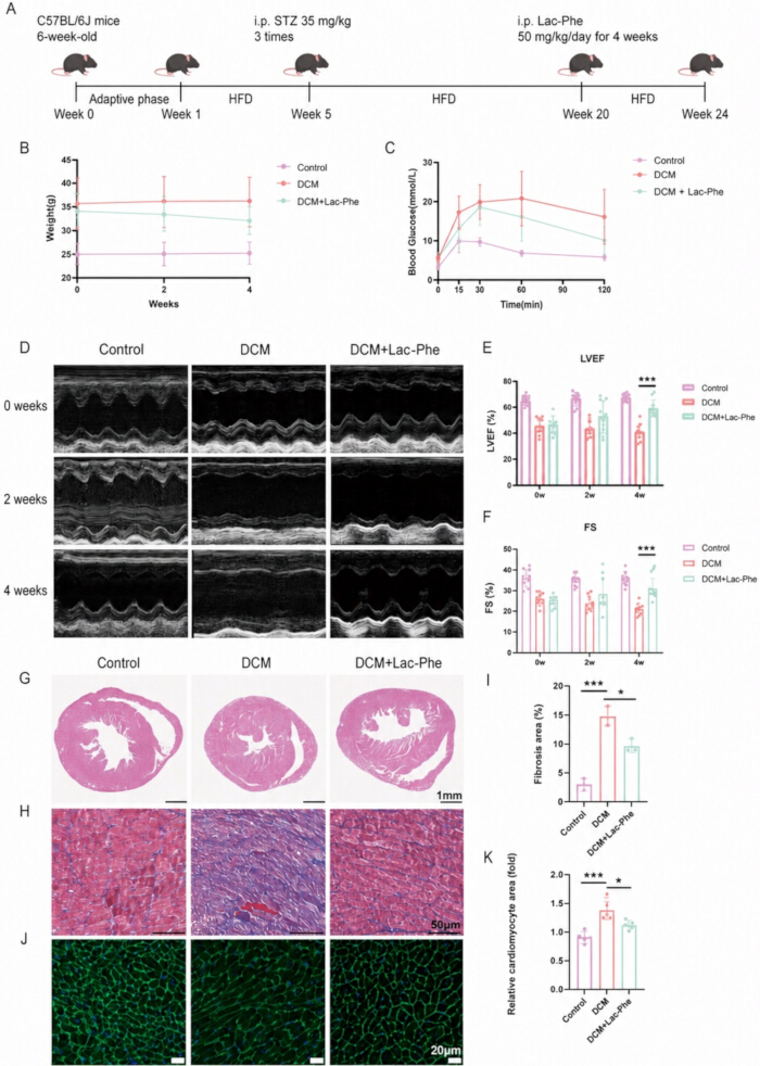
Figure 1. Lac-Phe improves cardiac function and pathological remodeling in mice with diabetic cardiomyopathy. (A) Schematic of the HFD/STZ-induced DCM mouse model and Lac-Phe treatment protocol. (B) Body weights of mice in the Control, DCM, and DCM + Lac-Phe groups during the 4-week treatment period; n = 10 per group. (C) Oral glucose tolerance test (OGTT) in the indicated groups; n = 10 per group. (D) Representative echocardiographic images at 0, 2, and 4 weeks of treatment. (E,F) Quantification of left ventricular ejection fraction (LVEF) (E) and fractional shortening (FS) (F); n = 10 per group. (G) Representative hematoxylin and eosin (H&E)-stained heart sections. Scale bar = 1 mm. (H,I) Representative Masson’s trichrome staining of heart sections (H) and quantification of fibrotic area (I); n = 3 per group. Scale bar = 50 μm. (J,K) Representative wheat germ agglutinin (WGA) staining of heart sections (J) and quantification of relative cardiomyocyte area (K); n = 3 per group. Scale bar = 20 μm. Data are presented as the mean ± standard deviation. P < 0.05, **P < 0.001. Please click here to view a larger version of this figure.
Cardiac function
Following anesthesia with isoflurane inhalation, mice were secured in a supine position on a temperature-controlled platform (37°C). Left ventricular dimensions and mitral valve blood flow velocity during diastole and systole were measured using an echocardiography machine equipped with a 40 MHz transducer, from which LVEF and shortening fraction (FS) were calculated. Three consecutive cardiac cycles were collected from each mouse.
Cell treatment
H9c2 cells were cultured in Dulbecco’s modified Eagle’s medium supplemented with 10% fetal bovine serum and 1% penicillin–streptomycin at 37°C in a humidified atmosphere containing 5% CO₂. Cells were passaged every 2–3 days upon reaching 70%–80% confluence. For experiments, cells were seeded in 10 cm dishes and then divided into three groups: Control, high glucose/palmitic acid (HG/PA), and HG/PA + Lac-Phe. Cells in the HG/PA and HG/PA + Lac-Phe groups were first exposed to 0.25 mM palmitic acid and 50 mM glucose for 24 h. Thereafter, cells in the HG/PA + Lac-Phe group were treated with 10 μg/mL Lac-Phe for an additional 24 h while HG/PA exposure was maintained; cells in the HG/PA group received an equivalent volume of PBS under the same HG/PA conditions. In the p-AMPK inhibition experiments, 10 μM AMPK-IN-3 (dissolved in DMSO) was added simultaneously with Lac-Phe for 24 h, and the corresponding control groups received an equal volume of DMSO. For the atorvastatin experiments, H9c2 cells under HG/PA conditions were treated with 10 μM atorvastatin for 24 h, either alone or in combination with 10 μg/mL Lac-Phe.
Histological staining
After 28 days of Lac-Phe administration, mouse hearts were harvested and immersion-fixed in 4% paraformaldehyde (pH 7.4) for 24 h. Fixed cardiac tissues were paraffin-embedded and sectioned into 6-μm-thick slices. Tissue sections were stained with hematoxylin and eosin, wheat germ agglutinin, and Masson’s trichrome. All staining procedures were performed strictly following the manufacturers’ protocols. Whole-slide imaging was performed at 20× magnification using a high-resolution brightfield microscope equipped with a digital camera system.
Western blot analysis
Protein concentration was determined using the Bradford assay. Tissue lysates were resolved by SDS-PAGE on 10% (w/v) acrylamide gels and electrophoretically transferred to polyvinylidene difluoride membranes. Nonspecific binding sites were blocked with 5% bovine serum albumin for 2 h at room temperature. Membranes were then incubated overnight at 4°C with primary antibodies (diluted 1:1,000), followed by six washes with TBST (5 min each) on a shaker. The membranes were then incubated with horseradish peroxidase-conjugated secondary antibodies (diluted 1:5,000) for 2 h at room temperature, followed by another six washes with TBST (5 min each). Protein bands were visualized using an enhanced chemiluminescence reagent kit. Quantification of western blot results was performed by densitometric analysis using ImageJ software. All antibodies used in this study are listed in the Table of Materials.
Transcriptomics
Total RNA was isolated from cardiac tissue of C57BL/6J mice using TRIzol Reagent and RNeasy mini kits. RNA quality was evaluated using a 2100 bioanalyzer, demonstrating high RNA purity and integrity. RNA-seq libraries were prepared according to the VAHTS Universal V10 RNA-seq Library Prep Kit and sequenced using an Illumina NovaSeq 6000 System (single-read, 150 bp). Differentially expressed genes were identified using DESeq2, with an adjusted P value < 0.05 and an absolute log₂ fold change > 1 considered statistically significant. Gene Ontology (GO) enrichment analysis was conducted on differentially expressed genes, and the significance of enrichment of differentially expressed genes in GO terms was calculated using the hypergeometric distribution algorithm.
Oil Red O staining and LD540 lipid staining
Cardiac tissue was embedded in OCT compound and then cut into 10-μm-thick sections. The frozen sections were equilibrated at room temperature for 30 min and fixed with 4% paraformaldehyde for 10 min. After washing with PBS, the sections were placed in 60% isopropyl alcohol for 15 s and covered with Oil Red O working solution for 30 min. The sections were then treated with 60% isopropyl alcohol for 15 s and washed three times with PBS. For LD540 lipid staining, H9c2 cells were washed with PBS and fixed with 4% paraformaldehyde for 10 min at room temperature. The cells were then incubated with 10 μM LD540 for 30 min at room temperature.
Transmission electron microscopy (TEM)
Fresh mouse heart tissues were cut into 1–2 mm3 pieces, fixed in electron microscope fixative at 4°C for 4 h, then washed with double-distilled water and immersed in ethanol solutions of progressively increasing concentrations. The samples were embedded, sectioned, and then observed under a transmission electron microscope.
Total cholesterol (TC) content
Total cholesterol (TC) levels in serum and H9c2 cell samples were measured using the Amplex Red Cholesterol and Cholesteryl Ester Assay Kit according to the manufacturer’s instructions.
Oral Glucose Tolerance Test (OGTT)
Mice were fasted for 14 h (from 19:00 to 9:00) and gavaged with glucose (20% stock, 2 g/kg). Blood glucose levels were determined using blood collected from the tail vein at 0, 15, 30, 60, and 120 min after gavage.
Cell counting kit-8 (CCK-8) measurement
CCK-8 was used to measure cell viability. H9c2 cells were seeded into 96-well plates at an initial density of 3 × 10⁴ cells/well. After treatment with different concentrations of Lac-Phe under HG/PA conditions for 24 h, 10 μL of the kit reagent was added to 100 μL of culture medium per well, followed by incubation at 37°C for 4 h. The absorbance was measured at 450 nm using a multimode microplate reader to obtain the optical density (OD) value.
Molecular docking
The crystal structures of Lac-Phe and AMPKα1 were downloaded from PubChem26. The molecular docking process was carried out using CB-DOCK227.
Statistical analysis
All echocardiographic measurements, histological staining, and image quantifications were performed by investigators blinded to group assignment. All data analyses were performed using Prism and SPSS 20.0. Each experiment was repeated at least three times, and the data were presented as the mean ± standard deviation. Homogeneity of variance was assessed using Levene’s test. Normality was confirmed by graphical inspection of residuals. Comparisons among multiple groups were conducted using one-way analysis of variance, followed by Tukey's post hoc test for pairwise comparisons. For OGTT data, two-way repeated-measures ANOVA was used, with group as the between-subjects factor and time as the within-subjects factor, followed by Bonferroni's post hoc test. P < 0.05 was considered statistically significant.
Zugriff eingeschränkt. Bitte melden Sie sich an oder starten Sie eine Testversion, um diesen Inhalt anzuzeigen.
Ergebnisse
Lac-Phe improves cardiac function in diabetic cardiomyopathy mice
We induced DCM in C57BL/6J mice using an HFD combined with STZ injections (Figure 1A) and randomly assigned the successfully established DCM mice to an untreated group (DCM group) and a Lac-Phe treatment group, with control C57BL/6J mice serving as the negative control. According to the study by Li et al.21, we selected 50 mg/kg as the therapeutic dose of Lac...
Zugriff eingeschränkt. Bitte melden Sie sich an oder starten Sie eine Testversion, um diesen Inhalt anzuzeigen.
Diskussion
In dieser Studie liefern wir Hinweise auf eine therapeutische Wirkung von Lac-Phe bei DCM. Lac-Phe verbesserte die kardiale Remodelung und kardiale Dysfunktion bei DCM, während es die Cholesterinproduktion in Kardiomyozyten verringerte und das Ungleichgewicht des Lipidstoffwechsels verbesserte. Mechanistisch gesehen erhöhte Lac-Phe die Phosphorylierung von AMPKα1. Anschließend war die erhöhte Phosphorylierung von AMPKα1 mit einer erhöhten Phosphorylierung und Hemmung seines downstream liegenden Zielproteins HMGCR assozii...
Zugriff eingeschränkt. Bitte melden Sie sich an oder starten Sie eine Testversion, um diesen Inhalt anzuzeigen.
Offenlegungen
Interessenkonflikt:
Die Autoren haben nichts zu offenbaren.
Danksagungen
Wir möchten den Forschern und Mitarbeitern des Klinischen Kollegs des Nanjing Drum Tower Hospitals und des Anschlusskrankenhauses der Medizinischen Fakultät der Nanjing-Universität für ihre technische Unterstützung herzlich danken. Diese Forschung wurde durch Zuschüsse der Nationalen Naturwissenschaftlichen Stiftung Chinas (Förderkennzeichen 81870291 und 82300384), der Chinesischen Stiftung für postdoktorale Wissenschaft (Förderkennzeichen 2023M731625) sowie der Naturwissenschaftlichen Stiftung der Provinz Jiangsu (Förderkennzeichen BK20220175) finanziert. Wir danken außerdem dem Institut für Fortgeschrittene Synthese der Nanjing Tech University für die zur Verfügung gestellten Ressourcen und die kooperative Unterstützung.
Zugriff eingeschränkt. Bitte melden Sie sich an oder starten Sie eine Testversion, um diesen Inhalt anzuzeigen.
Materialien
| Name | Unternehmen | Katalognummer | Kommentare |
|---|---|---|---|
| AMPK-IN-3 (p-AMPK-Inhibitor) | MedChemExpress (MCE) | HY-132921 | Wird bei AMPK-Inhibitionsexperimenten in einer Konzentration von 10 μM verwendet, um die AMPK-Signalübertragung in mit Lac-Phe behandelten Zellen zu hemmen. |
| Amplex Red Cholesterin- und Cholesterylester-Assay-Kit | Beyotime | S0211S | Wird zur Bestimmung der Gesamtcholesterinwerte in Serum- und H9c2-Zellproben verwendet. |
| Anti-AMPKα1 | Cell Signaling Technology | 2532; RRID: AB_330331 | Primärantikörper für die Western-Blot-Analyse von AMPKα1. |
| Anti-GAPDH | Cell Signaling Technology | 5174; RRID: AB_10622025 | Primärantikörper für die Western-Blot-Analyse von GAPDH. |
| Anti-HMGCR | Abcam | ab174830; RRID: AB_2749818 | Primärantikörper für die Western-Blot-Analyse von HMGCR. |
| Anti-p-AMPKα1 (Thr172) | Cell Signaling Technology | 2535; RRID: AB_331250 | Primärantikörper für die Western-Blot-Analyse von phosphoryliertem AMPKα1. |
| Anti-p-HMGCR (Ser872) | Abcam | ab124350 | Primärantikörper für die Western-Blot-Analyse von phosphoryliertem HMGCR. |
| Anti-Kaninchen-IgG, HRP-gekoppelter Antikörper | Cell Signaling Technology | 7074; RRID: AB_2099233 | Wird für die Western-Blot-Detektion in einer Verdünnung von 1:5.000 verwendet. |
| Anti-β-Tubulin | Cell Signaling Technology | 2128; RRID: AB_823664 | Primärantikörper für die Western-Blot-Analyse von β-Tubulin. |
| Atorvastatin | MedChemExpress (MCE) | HY-17379 | Wird allein oder in Kombination mit Lac-Phe verwendet, um die Effekte auf Lipidakkumulation, Gesamtcholesterinspiegel und HMGCR-Phosphorylierung in mit HG/PA behandelten H9c2-Zellen zu untersuchen. |
| Rinderserumalbumin | Sigma-Aldrich | A7906 | Wird in einer Konzentration von 5 % verwendet, um unspezifische Bindungen während der Western-Blot-Analyse zu blockieren. |
| Bradford-Assay-Reagenz/Kit | Thermo Fisher Scientific | 23200 | Wird zur Bestimmung der Proteinkonzentration vor der Western-Blot-Analyse verwendet. |
| C57BL/6J-Mäuse (5 Wochen alt, männlich) | Model Animal Research Center of Nanjing University | N/V | Wurden verwendet, um das durch HFD/STZ induzierte Mausmodell der diabetischen Kardiomyopathie zu etablieren. |
| Cell Counting Kit-8 (CCK-8) | Sharebio | SB-CCK8S | Wird verwendet, um die Lebensfähigkeit von H9c2-Zellen nach Behandlung zu messen. |
| Citratpuffer | Eigenherstellung | N/V | Wird bei pH 4,5 hergestellt und dient zum Lösen von STZ sowie als entsprechendes Fahrzeugkontrollmittel. |
| Kontrollnahrung (10 % Fett, 20 % Protein, 70 % Kohlenhydrate) | Research Diets, Inc. | D12450J | Wird als Nahrung für Mäuse der Kontrollgruppe verwendet. |
| D-(+)-Glukose | Sigma-Aldrich | N/V | Wird für die HG/PA-Zellbehandlung und für den oralken Glukosetoleranztest verwendet. |
| Dimethylsulfoxid (DMSO) | Sigma-Aldrich | D2650 | Wird als Lösungsmittel für AMPK-IN-3 und als entsprechendes Fahrzeugkontrollmittel verwendet. |
| Dulbecco’s modifiziertes Eagle’s Medium | Gibco, Thermo Fisher Scientific | 11965092 | Wird als Basalkulturmedium für H9c2-Zellen verwendet. |
| Echokardiographiegerät mit 40-MHz-Wandler | FUJIFILM VisualSonics | Vevo 3100 LT; 40-MHz-Wandler | Wird verwendet, um die Dimensionen des linken Ventrikels und die Blutflussgeschwindigkeit an der Mitralklappe zu messen sowie LVEF und FS abzuleiten. |
| Elektronenmikroskopie-Fixiermittel | ServiceBio | G1102 | Wird verwendet, um frisches Herzmuskelgewebe von Mäusen für die Transmissionselektronenmikroskopie zu fixieren. |
| Äthanol | Sigma-Aldrich | E7023 | Wird in schrittweise steigenden Konzentrationen zur Dehydrierung während der Probenvorbereitung für die Transmissionselektronenmikroskopie verwendet. |
| Fötales Rinderserum | Gibco, Thermo Fisher Scientific | 10099141 | Wird in einer Konzentration von 10 % zur Supplementierung des Kulturmediums für H9c2-Zellen verwendet. |
| H9c2-Zelllinie | Sunncell | RRID: CVCL_0286 | Wird als in-vitro-Kardiomyozytenmodell für HG/PA- und Lac-Phe-Behandlungsversuche verwendet. |
| Hämatoxylin- und Eosin-Färbung | ServiceBio | G1005-1/2 | Wird für die histologische Beurteilung von Herzmuskelgewebe verwendet. |
| Diät mit hohem Fettgehalt (60 kcal % Fett) | Research Diets, Inc. | D12492 | Wird zusammen mit niedrig dosiertem STZ verwendet, um das Mausmodell der diabetischen Kardiomyopathie zu etablieren. |
| Hellfeldmikroskop mit hoher Auflösung | Leica | DM2500 | Wird für die Ganzpräparat-Aufnahme von gefärbtem Herzmuskelgewebe bei einer 20×-Vergrößerung verwendet. |
| ImageJ | National Institutes of Health | N/V | Wird für die dichtometrische Quantifizierung der Western-Blot-Ergebnisse verwendet. |
| Isfluran | Sigma-Aldrich | 792632 | Wird zur Anästhesie während der Echokardiographie bei Mäusen verwendet. |
| Isopropanol | Sigma-Aldrich | I9516 | Wird in einer Konzentration von 60 % während der Oil-Red-O-Färbung von gefrorenen Herzgewebeschnitten verwendet. |
| LD540-Lipidfarbstoff | Beyotime | C2050S-1 | Wird in einer Konzentration von 10 μM zur Färbung intrazellulärer Lipide verwendet. |
| Masson’s Trichrom-Färbung | ServiceBio | G1006 | Wird für die histologische Färbung von Herzmuskelgewebe verwendet. |
| N-Lactoyl-Phenylalanin (Lac-Phe) | MedChemExpress (MCE) | HY-146098 | Wird DCM-Mäusen verabreicht und zur Behandlung von H9c2-Zellen verwendet, die HG/PA ausgesetzt waren. |
| Oil-Red-O-Arbeitslösung | ServiceBio | G1015-100ML | Wird zur Färbung von Lipiden in gefrorenen Schnitten von Herzmuskelgewebe verwendet. |
| Omni-ECL-Effizientes Licht-Chemilumineszenz-Kit | EpiZyme | SQ203L | Wird zur chemilumineszenten Visualisierung von Proteinbanden nach dem Western Blot verwendet. |
| Palmitinsäure | Sigma-Aldrich | P0500 | Wird in einer Konzentration von 0,25 mM zusammen mit hohem Glukosegehalt verwendet, um das HG/PA-behandelte H9c2-Zellmodell zu etablieren. |
| Paraformaldehyd | Sigma-Aldrich | P6148 | Wird als 4 %ige Lösung hergestellt und dient zur Fixierung von Herzmuskelgewebe und Zellen vor histologischen oder Lipidfärbungen. |
| Penicillin-Streptomycin | Gibco, Thermo Fisher Scientific | 15140122 | Wird im H9c2-Zellkulturmedium in einer Konzentration von 1 % verwendet. |
| Phosphatgepufferte Kochsalzlösung (PBS) | Gibco, Thermo Fisher Scientific | 10010023 | Wird zum Waschen während Färbeprozeduren und als entsprechendes Fahrzeugkontrollmittel in Zellbehandlungsversuchen verwendet. |
| Prism | GraphPad Software | Version 8.0 | Wird für die statistische Analyse experimenteller Daten verwendet. |
| PVDF-Transfermembran, 0,45 μm | Thermo Fisher Scientific | 88518 | Wird für den elektrophoretischen Proteintransfer während der Western-Blot-Analyse verwendet. |
| RNeasy Mini Kit | QIAGEN | 74104 | Wird zur Isolierung/Reinigung von Gesamt-RNA aus Herzmuskelgewebe von Mäusen verwendet. |
| Natriumchloridlösung, 0,9 % (physiologische Kochsalzlösung) | Sigma-Aldrich | S8776 | Wird zum Lösen von Lac-Phe und als Fahrzeugkontrollmittel für Kontroll- und DCM-Mäuse verwendet. |
| SPSS | IBM | Version 20.0 | Wird für die statistische Analyse experimenteller Daten verwendet. |
| Streptozotocin (STZ) | Sigma-Aldrich | S0130 | Wird zusammen mit HFD-Fütterung verabreicht, um das Mausmodell der diabetischen Kardiomyopathie zu etablieren. |
| TBS-Tween-20-Puffer (20×) | Thermo Fisher Scientific | 28360 | Wird zum Waschen der Membranen nach Inkubation mit Primär- und Sekundärantikörpern während der Western-Blot-Analyse verwendet. |
| Tissue-Tek O.C.T. Compound | Sakura Finetek | 4583 | Wird zum Einbetten von Herzmuskelgewebe vor der Herstellung gefrorener Schnitte für die Oil-Red-O-Färbung verwendet. |
| Transmissionselektronenmikroskop | HITACHI | HT7800 | Wird verwendet, um ultrastrukturelle Veränderungen im Herzmuskelgewebe von Mäusen zu untersuchen. |
| TRIzol-Reagenz | Invitrogen | 15596026 | Wird zur Isolierung von Gesamt-RNA aus Herzmuskelgewebe von Mäusen verwendet. |
| VAHTS Universal V10 RNA-seq Library Prep Kit | Vazyme | NR616-01 | Wird zur Herstellung von RNA-seq-Bibliotheken für die transkriptomische Analyse verwendet. |
| Weizenkeimagglutinin (WGA) | ServiceBio | W7024 | Wird für die histologische Beurteilung von Herzmuskelgewebe verwendet. |
Referenzen
- Pan KL, Hsu YC, Chang ST, Chung CM, Lin CL. The role of cardiac fibrosis in diabetic cardiomyopathy: from pathophysiology to clinical diagnostic tools. Int J Mol Sci. 2023;24(10):8604.
- Saeedi P, et al. Global and regional diabetes prevalence estimates for 2019 and projections for 2030 and 2045: results from the International Diabetes Federation Diabetes Atlas, 9th edition. Diabetes Res Clin Pract. 2019;157:107843.
- Zou R, et al. PGAM5-mediated PHB2 dephosphorylation contributes to diabetic cardiomyopathy by disrupting mitochondrial quality surveillance. Research. 2022;2022:0001.
- Jia G, DeMarco VG, Sowers JR. Insulin resistance and hyperinsulinaemia in diabetic cardiomyopathy. Nat Rev Endocrinol. 2016;12(3):144–53.
- Low Wang CC, Hess CN, Hiatt WR, Goldfine AB. Clinical update: cardiovascular disease in diabetes mellitus: atherosclerotic cardiovascular disease and heart failure in type 2 diabetes mellitus—mechanisms, management, and clinical considerations. Circulation. 2016;133(24):2459–502.
- Liu P, Zhang Z, Chen H, Chen Q. Pyroptosis: mechanisms and links with diabetic cardiomyopathy. Ageing Res Rev. 2024;94:102182.
- Park JJ. Epidemiology, pathophysiology, diagnosis and treatment of heart failure in diabetes. Diabetes Metab J. 2021;45(2):146–57.
- Gilca GE, et al. Diabetic cardiomyopathy: current approach and potential diagnostic and therapeutic targets. J Diabetes Res. 2017;2017:1310265.
- Nakamura K, et al. Pathophysiology and treatment of diabetic cardiomyopathy and heart failure in patients with diabetes mellitus. Int J Mol Sci. 2022;23(7):3587.
- Han X, et al. Cardiomyocyte OTUD1 drives diabetic cardiomyopathy via directly deubiquitinating AMPKα2 and inducing mitochondrial dysfunction. Nat Commun. 2025;16(1):666.
- Jia G, Hill MA, Sowers JR. Diabetic cardiomyopathy: an update of mechanisms contributing to this clinical entity. Circ Res. 2018;122(4):624–38.
- Yao Y, Yang B, Shi J. TiO(x)(OH)(4-2x) nanosheets with catalytic antioxidative activity alleviate oxidative injury in diabetic cardiomyopathy. J Am Chem Soc. 2025;147(4):3885–95.
- Seferovic PM, et al. Diabetic myocardial disorder: a clinical consensus statement of the Heart Failure Association of the ESC and the ESC Working Group on Myocardial & Pericardial Diseases. Eur J Heart Fail. 2024;26(9):1893–903.
- Wang H, et al. Inhibition of fatty acid uptake by TGR5 prevents diabetic cardiomyopathy. Nat Metab. 2024;6(6):1161–77.
- Zhao Y, et al. Ferroptosis: roles and molecular mechanisms in diabetic cardiomyopathy. Front Endocrinol. 2023;14:1140644.
- Nakamura M. Lipotoxicity as a therapeutic target in obesity and diabetic cardiomyopathy. J Pharm Pharm Sci. 2024;27:12568.
- Nakamura M, et al. Glycogen synthase kinase-3α promotes fatty acid uptake and lipotoxic cardiomyopathy. Cell Metab. 2019;29(5):1119–34.e12.
- Ritchie RH, Abel ED. Basic mechanisms of diabetic heart disease. Circ Res. 2020;126(11):1501–25.
- Thakur M, Tupe RS. Lipoxin and glycation in SREBP signaling: insight into diabetic cardiomyopathy and associated lipotoxicity. Prostaglandins Other Lipid Mediat. 2023;164:106698.
- Dhar A, et al. A comprehensive review of the novel therapeutic targets for the treatment of diabetic cardiomyopathy. Ther Adv Cardiovasc Dis. 2023;17:17539447231210170.
- Li VL, et al. An exercise-inducible metabolite that suppresses feeding and obesity. Nature. 2022;606(7915):785–90.
- Xiao S, et al. Lac-Phe mediates the effects of metformin on food intake and body weight. Nat Metab. 2024;6(4):659–69.
- Ying W, et al. N-lactoyl-phenylalanine modulates lipid metabolism in microglia/macrophage via the AMPK-PGC1α-PPARγ pathway to promote recovery in mice with spinal cord injury. J Neuroinflammation. 2025;22(1):167.
- Sha L, et al. N-Lac-Phe ameliorates myocardial cell death by improving mitochondrial dysfunction, ultimately improving TAC-induced heart failure. Cardiovasc Drugs Ther. 2026;40:1391–401.
- Ke J, Pan J, Lin H, Gu J. Diabetic cardiomyopathy: a brief summary on lipid toxicity. ESC Heart Fail. 2023;10(2):776–90.
- PubChem. PubChem. Available from: https://pubchem.ncbi.nlm.nih.gov/. Accessed August 15, 2026.
- Liu Y, Yang X, Gan J, et al. CB-Dock2: improved protein-ligand blind docking by integrating cavity detection, docking and homologous template fitting. Nucleic Acids Res. 2022;50(W1):W159–W164.
- GeneCards. GeneCards: the Human Gene Database. Version 6.1, June 17, 2026.
- Daina A, Michielin O, Zoete V. SwissTargetPrediction: updated data and new features for efficient prediction of protein targets of small molecules. Nucleic Acids Res. 2019;47(W1):W357–W364.
- Jo Y, DeBose-Boyd RA. Post-translational regulation of HMG CoA reductase. Cold Spring Harb Perspect Biol. 2022;14(12):a041253.
- Steinberg GR, Hardie DG. New insights into activation and function of the AMPK. Nat Rev Mol Cell Biol. 2023;24(4):255–72.
- Istvan ES, Deisenhofer J. Structural mechanism for statin inhibition of HMG-CoA reductase. Science. 2001;292(5519):1160–4.
- Beaudry D, et al. Statin therapy in cardiovascular diseases other than atherosclerosis. Curr Atheroscler Rep. 2007;9(1):25–32.
- Koch CG. Statin therapy. Curr Pharm Des. 2012;18(38):6284–90.
- Dewanjee S, et al. Autophagy in the diabetic heart: a potential pharmacotherapeutic target in diabetic cardiomyopathy. Ageing Res Rev. 2021;68:101338.
- Madonna R, et al. Empagliflozin inhibits excessive autophagy through the AMPK/GSK3β signalling pathway in diabetic cardiomyopathy. Cardiovasc Res. 2023;119(5):1175–89.
- Madhavi YV, et al. Targeting AMPK in diabetes and diabetic complications: energy homeostasis, autophagy and mitochondrial health. Curr Med Chem. 2019;26(27):5207–29.
- Fang C, et al. The AMPK pathway in fatty liver disease. Front Physiol. 2022;13:970292.
- Liu S, et al. AICAR-induced activation of AMPK inhibits TSH/SREBP-2/HMGCR pathway in liver. PLoS One. 2015;10(5):e0124951.
- Zhou H, et al. Empagliflozin rescues diabetic myocardial microvascular injury via AMPK-mediated inhibition of mitochondrial fission. Redox Biol. 2018;15:335–46.
- Lu QB, et al. METRNL ameliorates diabetic cardiomyopathy via inactivation of cGAS/STING signaling dependent on LKB1/AMPK/ULK1-mediated autophagy. J Adv Res. 2023;51:161–79.
- Wang D, et al. FGF1(ΔHBS) prevents diabetic cardiomyopathy by maintaining mitochondrial homeostasis and reducing oxidative stress via AMPK/Nur77 suppression. Signal Transduct Target Ther. 2021;6(1):133.
- He L. Metformin and systemic metabolism. Trends Pharmacol Sci. 2020;41(11):868–81.
- Yang F, et al. Metformin inhibits the NLRP3 inflammasome via AMPK/mTOR-dependent effects in diabetic cardiomyopathy. Int J Biol Sci. 2019;15(5):1010–9.
- Regitz-Zagrosek V, Gebhard C. Gender medicine: effects of sex and gender on cardiovascular disease manifestation and outcomes. Nat Rev Cardiol. 2023;20(4):236–47.
- Evrony GD, Hinch AG, Luo C. Applications of single-cell DNA sequencing. Annu Rev Genomics Hum Genet. 2021;22:171–97.
- Verboom K, et al. Smarter single cell total RNA sequencing. Nucleic Acids Res. 2019;47(16):e93.
Zugriff eingeschränkt. Bitte melden Sie sich an oder starten Sie eine Testversion, um diesen Inhalt anzuzeigen.
Nachdrucke und Genehmigungen
Weitere Artikel entdecken
Dieser Artikel wurde veröffentlicht
Video demnächst verfügbar