This study demonstrates that adipose-derived stem cell-derived exosomes protect human melanocytes from H₂O₂-induced oxidative damage through activation of the Keap1–Nrf2–ARE pathway, supporting further investigation of their therapeutic potential for vitiligo.
Forschungsartikel
Amelioration of Oxidative Stress in Melanocytes by Adipose-Derived Stem Cell Exosomes via the Keap1–Nrf2 Axis
* These authors contributed equally
In diesem Artikel
Zusammenfassung
Zusammenfassung
Oxidative stress, particularly H₂O₂ accumulation, is a key pathogenic factor in vitiligo, yet therapies that directly protect melanocytes remain limited. Adipose-derived stem cell-derived exosomes (ADSC-Exos) are cell-free vesicles with reported antioxidant effects, but whether they act through the Kelch-like ECH-associated protein 1 (Keap1)-nuclear factor erythroid 2-related factor 2 (Nrf2) pathway is unknown. This study aimed to investigate the protective effects of ADSC-Exos on hydrogen peroxide (H₂O₂)-induced oxidative damage in human melanocytes and to elucidate the underlying molecular mechanisms, focusing on the Keap1–Nrf2 signaling pathway. ADSCs and their exosomes were isolated and characterized using flow cytometry, transmission electron microscopy (TEM), nanoparticle tracking analysis (NTA), and western blot. An oxidative stress model was established by treating PIG1 human melanocytes with H₂O₂. The therapeutic effects of ADSC-Exos were assessed by measuring cell viability, migration, intracellular ROS levels, DNA damage, and antioxidant enzyme activities. The involvement of the Keap1–Nrf2 pathway was examined by western blot and immunofluorescence. H₂O₂ treatment significantly induced oxidative stress in PIG1 cells, evidenced by reduced cell viability and migration, increased ROS accumulation, and elevated DNA damage. Treatment with ADSC-Exos markedly restored cellular functions. Mechanistically, ADSC-Exos downregulated the expression of Keap1, facilitated the nuclear translocation of Nrf2, and upregulated the expression of downstream antioxidant enzymes, including heme oxygenase 1 (HO-1) and glutathione peroxidase 4 (GPX4). The findings demonstrate that ADSC-Exos effectively ameliorate H₂O₂-induced oxidative damage in melanocytes. The cytoprotection is mediated through the activation of the Keap1–Nrf2–ARE signaling pathway. These findings support further investigation of ADSC-Exos as a potential therapeutic strategy for vitiligo.
Einleitung
Vitiligo is a common acquired depigmenting skin disorder characterized by the progressive loss of functional melanocytes, which results in disfiguring white patches1,2. Although vitiligo can develop at any age, its onset most commonly occurs by 30 years of age3. The condition has a global prevalence of 0.5–2.0%4, which varies geographically, and comparable rates are seen in adult, pediatric, and adolescent populations5. Beyond its visible symptoms, the disease imposes a significant psychological burden and markedly impairs patients' quality of life6. The precise etiology of vitiligo is multifactorial, involving a convergence of genetic predisposition, autoimmunity, environmental triggers, and—critically—oxidative stress in its pathogenesis7. Among these factors, oxidative stress is increasingly viewed not merely as a secondary consequence but as a potential primary driver in the disease process8,9.
The central role of oxidative stress in vitiligo is well established10. The epidermal microenvironment in affected patients exhibits a redox imbalance, marked by elevated reactive oxygen species (ROS)—particularly hydrogen peroxide (H₂O₂)—and a weakened antioxidant defense system. This pro-oxidant environment directly damages melanocytes, leading to apoptosis11. Moreover, oxidative stress can expose or create neoantigens, initiating and amplifying an autoimmune response against melanocytes that perpetuates and extends depigmentation12,13. Moreover, the autoimmune response can exacerbate melanocyte oxidative damage, creating a self-amplifying deleterious cycle that contributes to disease initiation and progression14. Consequently, developing approaches to enhance melanocyte resistance to oxidative stress constitutes a pivotal therapeutic strategy15.
In this context, adipose-derived stem cell-derived exosomes (ADSC-Exos) have emerged as a potential therapeutic approach16. These nanoscale extracellular vesicles encapsulate a rich cargo of bioactive molecules mirroring the reparative and regenerative functions of their parent cells17. Previous studies have reported that ADSC-Exos reduce oxidative stress in diabetic wounds and ischemic injuries17,18. Their ability to affect multiple pathological pathways may differ from that of single-target agents19. However, despite this promise, the specific application of ADSC-Exos in vitiligo20, particularly their direct protective effects on melanocytes and the underlying molecular mechanisms, remains incompletely understood21.
This study aimed to evaluate the cytoprotective effects of ADSC-Exos against H₂O₂-induced oxidative damage in human melanocytes and to determine whether these effects involve modulation of the Keap1–Nrf2 signaling pathway.
Protokoll
Ethics approval
Human adipose tissue was obtained from 5 healthy surgical donors (aged 18–30 years; with a body mass index ranging from 22 to 26 kg/m2; all of whom tested negative for metabolic diseases) undergoing abdominal surgery, who had provided written informed consent. All procedures were approved by the Medical Ethics Review Committee of the First People’s Hospital of Yinchua. The study was approved on August 20, 2021, under approval number 2021–013.
The materials and equipment used in this study are listed in the Table of Materials.
Isolation of ADSCs
ADSCs were isolated as previously described with minor modifications. Briefly, the fresh adipose tissue was washed extensively with phosphate-buffered saline (PBS) containing 1% penicillin-streptomycin. After meticulous removal of connective tissues using scissors and forceps, the tissue was minced into a fine paste and digested with 0.1% type IV collagenase solution (2:1 volume/tissue volume) for 1 h at 37 °C with constant shaking. The digestion was terminated by adding an equal volume of Dulbecco’s modified Eagle medium (DMEM) supplemented with 10% fetal bovine serum (FBS). The digest was centrifuged at 300 x g for 5 min to pellet the stromal vascular fraction. The pellet was resuspended, passed through a 100 µm cell strainer, and centrifuged again. The collected cells were then cultured in a human ADSC growth medium. The culture medium was replaced after 24 h and subsequently every 3–4 days until the cells reached 80–90% confluence.
Characterization of ADSCs
Multilineage differentiation potential
For adipogenic differentiation, passage 3–5 ADSCs at approximately 60% confluence were induced with adipogenic induction medium for 2 weeks. Cells were then fixed and stained with Oil Red O to visualize lipid droplets. For osteogenic differentiation, ADSCs at 90–100% confluence were cultured in osteogenic induction medium for 3 weeks. The formation of calcium nodules was confirmed by Alizarin Red S staining.
Flow cytometry
Passage 3–5 ADSCs in the logarithmic growth phase were harvested and resuspended in PBS. Approximately 1 x 10⁶ cells per tube were incubated with fluorescently labeled antibodies against human CD29, CD44, CD73, CD90 (positive markers), and CD34, CD45 (negative markers), or corresponding isotype controls for 30 min at 4 °C in the dark. After washing, cells were analyzed using a flow cytometer.
Extraction and characterization of ADSC-derived exosomes (ADSC-Exos)
Exosome isolation
Passage 3–5 ADSCs were cultured in medium supplemented with exosome-depleted FBS (prepared by ultracentrifuging FBS at 120,000 x g for 18 h). After 48 h, the conditioned medium was collected and subjected to differential ultracentrifugation. The medium was sequentially centrifuged at 300 x g for 10 min to remove cells, 2,000 x g for 10 min to remove dead cells and debris, and 10,000 x g for 30 min to remove large vesicles. The resulting supernatant was collected and stored at 4 °C. The supernatants from the different ADSC cultures were pooled and ultracentrifuged at 100,000 x g for 70 min at 4 °C using a fixed-angle rotor with a k factor of 133. The resulting exosome pellet was washed in PBS and centrifuged again at 100,000 x g for 70 min. The final pellet was resuspended in a small volume of PBS, aliquoted after protein quantification, and stored at −80 °C.
Exosome characterization
TEM: 15 µL of exosome suspension was adsorbed onto a copper grid for 2 min, negatively stained with 2% uranyl acetate for 1 min, and air-dried. Images were acquired using a TEM.
NTA: Exosomes were diluted 1:50 in PBS and analyzed using a nanoparticle tracking analyzer to determine particle size distribution and concentration.
Western blot analysis: Exosomal proteins were separated by SDS-PAGE on a 12% gel, transferred to a polyvinylidene fluoride (PVDF) membrane, and probed with antibodies against heat shock protein 70 (HSP70) (1:1,000), CD63 (1:1,000), ALG-2-interacting protein X (ALIX) (1:1,000), tumor susceptibility gene 101 (TSG101) (1:1,000), and β-actin (1:10,000).
Exosome labeling
Purified exosome pellets were reconstituted in the diluent supplied with the PKH26 labeling kit to a protein concentration of approximately 0.5–1 µg/µL. The exosome suspension and the dye preparation were combined at a 1:1 volume ratio and mixed by gentle pipetting. The labeling reaction proceeded for 30 min. To quench unreacted dye, an equal volume of FBS was introduced into the mixture. Labeled exosomes were then recovered by ultracentrifugation at 100,000 x g for 90 min at 4 °C, washed once with sterile PBS, and pelleted again under identical centrifugal conditions. The final PKH26-positive exosome preparation was resuspended in PBS and added to PIG1 cells for 12 h. Fluorescent signals were captured using a confocal laser scanning microscope. PKH26 fluorescence was excited at 551 nm and collected at 567 nm, while 4′,6-diamidino-2-phenylindole (DAPI) was excited at 358 nm and detected at 461 nm.
Cell culture and experimental groups
Human epidermal PIG1 melanocytes were cultured in RPMI-1640 medium supplemented with 10% FBS. To establish an oxidative stress model, PIG1 cells were treated with various concentrations of H₂O₂ (0–500 µM) for 24 h as reported previously22. Based on preliminary results, 300 µM H₂O₂ was selected for subsequent experiments. Cells were divided into three groups: (1) Control group (normal culture conditions), (2) H₂O₂ group (treated with 300 µM H₂O₂), and (3) H₂O₂ + ADSC-Exos group (treated with 300 µM H₂O₂ and 50 µg/mL ADSC-Exos simultaneously)22,23. For Nrf2 inhibition, 5 µM ML385 was added into PIG1 cells before H₂O₂ and ADSC-Exos treatment.
Cell viability assay
Cell viability was assessed using the CCK-8 kit. PIG1 cells were seeded in 96-well plates. After respective treatments, the medium was replaced with serum-free DMEM containing 10% CCK-8 reagent and incubated for 2 h at 37 °C. The absorbance was measured at 450 nm using a microplate reader. The experiment was repeated three times.
Cell migration assay
Cell migration was evaluated using Transwell chambers with 8 µm pores. PIG1 cells from different treatment groups were harvested, resuspended in serum-free medium, and seeded into the upper chamber. The lower chamber was filled with medium containing 10% FBS as a chemoattractant. After 24 h of incubation, non-migrated cells on the upper surface were removed with a cotton swab. Migrated cells on the lower surface were fixed with 4% paraformaldehyde, stained with 0.1% crystal violet, photographed, and counted under a microscope.
Immunofluorescence staining
After treatments, PIG1 cells grown on coverslips were fixed with 4% paraformaldehyde, permeabilized with 0.1% Triton X-100, and blocked with 5% bovine serum albumin. Cells were then incubated overnight at 4 °C with primary antibodies against Ki67 (proliferation marker) and 8-OHdG (DNA oxidation marker), followed by incubation with appropriate fluorescent secondary antibodies. Nuclei were counterstained with DAPI. Images were captured using a fluorescence or confocal microscope.
Measurement of intracellular reactive oxygen species
Intracellular ROS levels were detected using the fluorescent probe 2′,7′-dichlorodihydrofluorescein diacetate (DCFH-DA). After treatments, cells were incubated with 10 µM DCFH-DA in serum-free medium at 37 °C for 20 min. After washing, fluorescence was visualized by confocal microscopy and quantified by flow cytometry.
Assessment of antioxidant enzyme activities
Superoxide dismutase (SOD) activity
Cell lysates were prepared using cold lysis buffer. SOD activity was measured using a WST-8-based assay kit according to the supplied protocol. The absorbance was measured at 450 nm.
Catalase (CAT) activity
CAT activity was determined by measuring the decomposition of H₂O₂. A standard curve was generated using known concentrations of H₂O₂. Cell lysates were incubated with 250 mM H₂O₂, and the reaction was stopped after 5 min. The remaining H₂O₂ was quantified using a colorimetric method, and CAT activity was calculated based on the standard curve.
Western blot analysis
Protein lysates from cytosolic and nuclear fractions were prepared. Equal amounts of protein (50 µg per lane) were separated by SDS-PAGE and transferred to PVDF membranes. After blocking with 5% nonfat milk, the membranes were incubated overnight at 4 °C with primary antibodies against Keap1 (1:1,000), Nrf2 (1:1,000), HO-1 (1:1,000), GPX4 (1:1,000), and β-actin (1:10,000), followed by incubation with a horseradish peroxidase-conjugated secondary antibody. Protein bands were visualized using an enhanced chemiluminescence detection system.
Reverse transcription quantitative polymerase chain reaction (RT-qPCR)
Cells were lysed directly in a phenol-guanidinium-based ribonucleic acid (RNA) extraction reagent, and total RNA was recovered through chloroform phase separation followed by isopropanol precipitation. RNA pellets were washed with 75% ethanol, air-dried, and redissolved in RNase-free water. RNA integrity and concentration were checked spectrophotometrically (A260/A280). For each sample, 1,000 ng of total RNA was reverse-transcribed using a reverse transcription reagent kit in a 20 µL reaction containing random hexamers and oligo(dT) primers. The reverse transcription program was 37 °C for 15 min and 85 °C for 5 s. Quantitative PCR was performed using a real-time PCR system and an intercalating dye-based master mix. Each 20 µL reaction contained 2 µL complementary DNA (cDNA), 10 µL master mix, 0.4 µL of each primer (10 µM), and 7.2 µL nuclease-free water. Cycling conditions were 95 °C for 30 s, followed by 40 cycles of 95 °C for 5 s and 60 °C for 30 s. A melting curve (65–95 °C) was included to confirm amplicon specificity. Relative gene expression was calculated using the 2−ΔΔCt method, with glyceraldehyde-3-phosphate dehydrogenase (GAPDH) as the reference gene. All reactions were run in triplicate. Primer sequences:
Microphthalmia-associated transcription factor M isoform (MITF-M): TCTACCGTCTCTCACTGGATTGG (forward (5′→3′)), GCTTTACCTGCTGCCGTTGG (reverse (5′→3′));
Tyrosinase (TYR): CATTCTTCTCCTCTTGGCAGA (forward (5′→3′)), CCGCTATCCCAGTAAGTGGA (reverse (5′→3′));
Tyrosinase-related protein 1 (TYRP1): GCTTTTCTCACATGGCACAG (forward (5′→3′)), GGCTCTTGCAACATTTCCTG (reverse (5′→3′));
Dopachrome tautomerase (DCT): CGACTCTGATTAGTCGGAACTCA (forward (5′→3′)), GGTGGTTGTAGTCATCCAAGC (reverse (5′→3′));
GAPDH: ACGGGAAGCTTGTCATCAAT (forward (5′→3′)), TGGACTCCACGACGTACTCA (reverse (5′→3′)).
Tyrosinase activity
Dopachrome formation was used to assess tyrosinase activity24,25,26. Treated cells were lysed in 50 mM phosphate buffer (pH 6.8) containing 1% Triton X-100. After centrifugation (12,000 x g, 15 min, 4 °C), equal protein amounts were mixed with L-3,4-dihydroxyphenylalanine (L-DOPA) (2 mg/mL) and kept at 37 °C in the dark. Absorbance at 475 nm was recorded every 10 min for 1 h. Activity was normalized to the control. Each sample was assayed in triplicate.
Statistical analysis
Data were analyzed using statistical analysis software and are presented as the mean ± standard deviation (SD). Three independent biological replicates were performed for each experiment. Before parametric testing, the normality of all datasets was assessed using the Shapiro-Wilk test, and the assumption of normality was satisfied. Homogeneity of variance was confirmed by Levene’s test. For single-factor comparisons, a one-way analysis of variance (ANOVA) was employed, followed by least significant difference post hoc testing. Statistical significance was defined as *p < 0.05, **p < 0.01, and ***p < 0.001.
Ergebnisse
Establishment of an H₂O₂-induced oxidative stress model in melanocytes
To simulate the oxidative stress microenvironment of vitiligo, human epidermal melanocytes (PIG1) were treated with various concentrations of H₂O₂ (0–500 µM) for 24 h. The CCK-8 assay demonstrated that H₂O₂ inhibited cell viability in a concentration-dependent manner. A significant reduction in cell survival was observed at 300 µM (p < 0.001, Figure 1A). Transwell migration assays revealed that 300 µM H₂O₂ markedly impaired the migratory capacity of PIG1 cells (p < 0.01, Figure 1B). Consistent with these findings, immunofluorescence staining showed a significant decrease in both the percentage of Ki67-positive cells and Ki67 fluorescence intensity following H₂O₂ treatment (Figure 1C,E), indicating suppressed proliferation. Conversely, H₂O₂ treatment led to a pronounced increase in 8-OHdG positivity and fluorescence intensity (Figure 1D,E), suggesting elevated DNA oxidative damage. Furthermore, intracellular ROS levels were significantly elevated as detected by the DCFH-DA probe (Figure 1F). These results demonstrated that 300 µM H₂O₂ induced oxidative stress in melanocytes, and this concentration was therefore selected for subsequent experiments.
Characterization of ADSCs and ADSC-Exos
ADSCs were successfully isolated and cultured. The primary ADSCs exhibited a spindle-shaped morphology and formed whorl-like arrangements upon reaching confluence (Figure 2A). To verify their multilineage differentiation potential, ADSCs were subjected to adipogenic and osteogenic induction. Oil Red O staining after 14 days of adipogenic induction revealed abundant lipid droplet formation (Figure 2A), and Alizarin Red staining after 21 days of osteogenic induction confirmed the presence of calcium nodules (Figure 2A). Flow cytometric analysis confirmed that the ADSCs were highly positive for the classic mesenchymal stem cell markers CD29, CD44, CD73, and CD90, while being negative for the hematopoietic markers CD34 and CD45 (<1% expression), meeting the criteria set by the International Society for Cell Therapy (Figure 2B). Exosomes were isolated from ADSC-conditioned media via ultracentrifugation. TEM revealed the characteristic cup-shaped, bilayered membrane structure of the extracted vesicles (Figure 2C). NTA indicated that the particle size distribution peaked at approximately 110 nm, with the majority of particles falling within the 80–150 nm range (Figure 2D). Western blot analysis detected the exosomal marker proteins CD63, ALIX, TSG101, and HSP70, with β-actin included as the loading control (Figure 2E). To confirm cellular uptake, exosomes were labeled with the red fluorescent dye PKH26 and co-cultured with PIG1 cells. Immunofluorescence imaging showed that the PKH26-labeled exosomes were effectively internalized by PIG1 cells and primarily localized in the cytoplasm (Figure 2F). These data demonstrate the successful isolation and characterization of ADSC-Exos.
ADSC-Exos ameliorate H₂O₂-induced impairment of melanocyte proliferation and migration
The protective effects of ADSC-Exos on PIG1 cells under H₂O₂-induced oxidative stress were subsequently investigated. Treatment with ADSC-Exos (50 µg/mL, 89.26 ± 1.48, n = 3) significantly restored the viability of melanocytes injured by 300 µM H₂O₂ (67.50 ± 2.86, n = 3, p < 0.0001) (Figure 3A). Similarly, the Transwell assay showed that the impaired migratory capacity of PIG1 cells caused by H₂O₂ was markedly rescued by exosome treatment (p < 0.01, Figure 3B). Furthermore, immunofluorescence staining revealed that the internalization of PKH26-labeled ADSC-Exos by PIG1 cells was associated with a significant increase in both the percentage of Ki67-positive cells and the Ki67 fluorescence intensity compared to the H₂O₂-only group (Figure 3C). Melanogenesis-related indices were also evaluated in the differently treated PIG1 cells. As shown in Figure 3D, H₂O₂ significantly decreased tyrosinase activity, whereas ADSC-Exos increased it. RT-qPCR analysis indicated that H₂O₂ decreased the expression of melanin-biosynthesis genes, whereas ADSC-Exos increased their expression (Figure 3E). Taken together, these results indicate that ADSC-Exos can effectively mitigate the H₂O₂-induced suppression of proliferation, migration, and melanin biosynthesis in melanocytes.
ADSC-Exos attenuate H₂O₂-induced oxidative stress damage in melanocytes
After establishing the functional recovery, the underlying mechanisms were investigated by evaluating oxidative stress parameters. DCFH-DA staining demonstrated that ADSC-Exos treatment significantly attenuated the H₂O₂-induced accumulation of intracellular ROS (41,156 ± 4,519 vs 157,510 ± 9,055, n = 3, p < 0.0001, Figure 4A,B). Assessment of antioxidant capacity showed that H₂O₂ treatment severely compromised the cellular antioxidant defense system, which was substantially restored by ADSC-Exos (Figure 4B). When treated with H2O2, glutathione (GSH) content in PIG1 cells was 1.94 ± 0.14, whereas ADSC-Exos treatment significantly boosted the content of GSH to 4.17 ± 0.12 (n = 3, p < 0.0001, Figure 4B). Similarly, the SOD activity increased from 0.85 ± 0.06 to 1.30 ± 0.05 (n = 3, p < 0.0001, Figure 4B), and CAT activity increased from 3.65 ± 0.27 to 6.04 ± 0.09 (n = 3, p < 0.0001, Figure 4B). Moreover, the H₂O₂-induced increase in 8-OHdG fluorescence, indicative of DNA damage, was significantly reduced upon exosome treatment (Figure 4C). These findings suggest that the cytoprotective effects of ADSC-Exos are mediated, at least in part, by reducing ROS accumulation and enhancing the antioxidant capacity, thereby alleviating oxidative DNA damage.
ADSC-Exos enhance melanocyte antioxidant capacity by activating the Keap1–Nrf2 pathway
To explore the molecular mechanism by which ADSC-Exos enhance antioxidant defenses, the Keap1–Nrf2 signaling pathway was examined. Western blot analysis showed that H₂O₂ treatment alone slightly downregulated Keap1 protein expression in the cytoplasm and led to a modest increase in nuclear Nrf2 levels, suggesting a mild activation of the endogenous antioxidant response (Figure 5A). Treatment with ADSC-Exos resulted in a more pronounced downregulation of Keap1 and a significant upregulation of nuclear Nrf2 (Figure 5A). Consequently, the protein expression levels of key Nrf2 downstream antioxidant enzymes, HO-1 and GPX4, were significantly increased by exosome treatment. Immunofluorescence analysis further showed increased nuclear localization of Nrf2 following ADSC-Exos treatment (Figure 5B). To determine whether the protective effects of ADSC-Exos depended on Nrf2 regulation, Nrf2 was subsequently inhibited using ML385. As shown in Figure 5C, inhibition of Nrf2 by ML385 significantly reduced the protective effects of ADSC-Exos on cell viability, ROS accumulation, melanin content, and tyrosinase activity. These results indicate that ADSC-Exos activate the Nrf2–ARE signaling pathway by inhibiting Keap1-mediated degradation of Nrf2, thereby enhancing the endogenous antioxidant defense system in melanocytes.
DATA AVAILABILITY:
The raw data supporting the findings of this study are provided in the Supplementary File. Please click here to download this file.
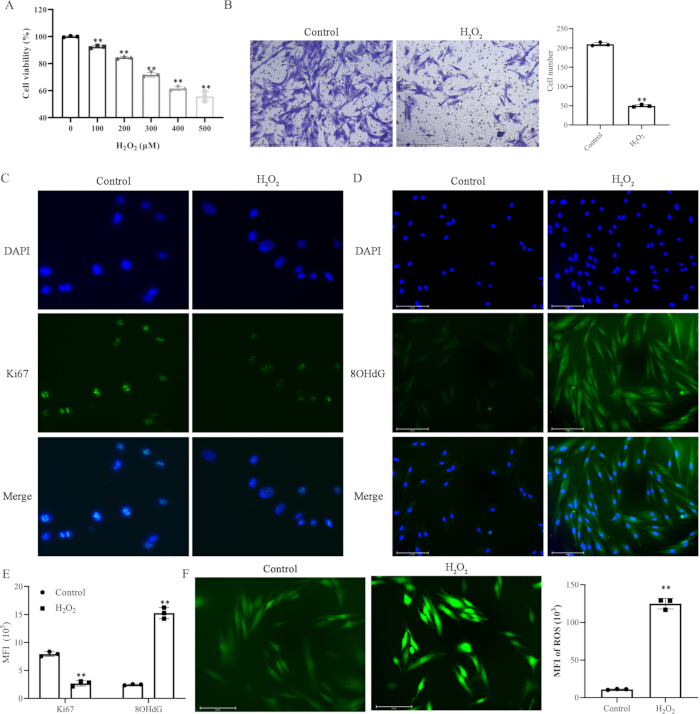
Figure 1: Establishment of an H₂O₂-induced oxidative stress model in melanocytes. (A) Viability of PIG1 cells treated with the indicated concentrations of H₂O₂ for 24 h, measured using the Cell Counting Kit-8 assay. (B) Representative Transwell images and quantification of migrated cells in the control and H₂O₂-treated groups. (C) Representative immunofluorescence images of Ki67 expression. (D) Representative immunofluorescence images of 8-OHdG expression. (E) Quantification of Ki67 and 8-OHdG MFI. (F) Representative fluorescence images and quantification of intracellular ROS. Scale bars = 50 µm. Data are presented as the mean ± SD (n = 3 independent experiments). **p < 0.01. Abbreviations: DAPI = 4′,6-diamidino-2-phenylindole; MFI = mean fluorescence intensity; ROS = reactive oxygen species; SD = standard deviation; 8-OHdG = 8-hydroxy-2′-deoxyguanosine. Please click here to view a larger version of this figure.
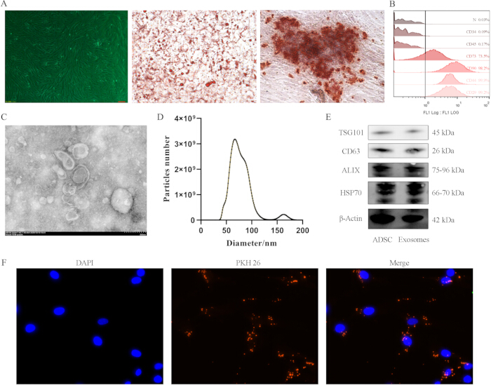
Figure 2: Characterization of ADSCs and ADSC-Exos. (A) Representative images showing ADSC morphology, adipogenic differentiation assessed using Oil Red O staining, and osteogenic differentiation assessed using Alizarin Red S staining. (B) Flow cytometric analysis of the ADSC surface markers CD29, CD44, CD73, CD90, CD34, and CD45. (C) Transmission electron microscopy image of ADSC-Exos. Scale bar = 200 nm. (D) Particle size distribution of ADSC-Exos determined using nanoparticle tracking analysis. (E) Western blot analysis of the exosomal markers TSG101, CD63, ALIX, and HSP70, with β-actin as the loading control. (F) Representative fluorescence images showing the association or uptake of PKH26-labeled ADSC-Exos by PIG1 cells. Blue = DAPI; red = PKH26-labeled ADSC-Exos. Abbreviations: ADSC = adipose-derived stem cell; ADSC-Exos = adipose-derived stem cell exosomes; ALIX = ALG-2-interacting protein X; DAPI = 4′,6-diamidino-2-phenylindole; HSP70 = heat shock protein 70; TSG101 = tumor susceptibility gene 101. Please click here to view a larger version of this figure.
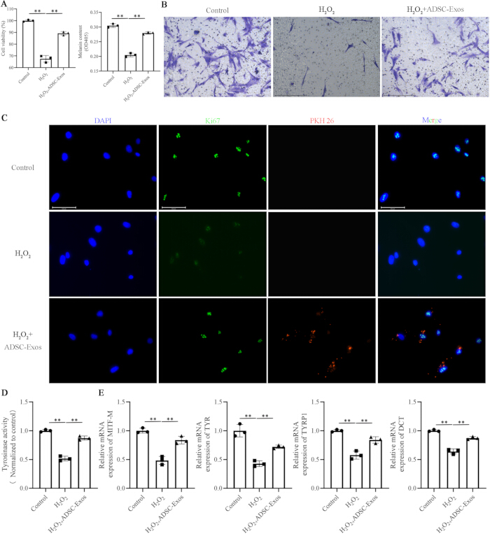
Figure 3: ADSC-Exos ameliorate H₂O₂-induced impairment of melanocyte function. (A) Viability and melanin content of PIG1 cells following the indicated treatments. (B) Representative Transwell images showing PIG1 cell migration. (C) Representative immunofluorescence images showing Ki67 expression and the association or uptake of PKH26-labeled ADSC-Exos. Blue = DAPI; green = Ki67; red = PKH26-labeled ADSC-Exos. (D) Tyrosinase activity in PIG1 cells following the indicated treatments. (E) Relative mRNA expression of MITF-M, TYR, TYRP1, and DCT. Data are presented as the mean ± SD (n = 3 independent experiments). **p < 0.01. Abbreviations: ADSC-Exos = adipose-derived stem cell exosomes; DAPI = 4′,6-diamidino-2-phenylindole; DCT = dopachrome tautomerase; MITF-M = microphthalmia-associated transcription factor M isoform; SD = standard deviation; TYR = tyrosinase; TYRP1 = tyrosinase-related protein 1. Please click here to view a larger version of this figure.
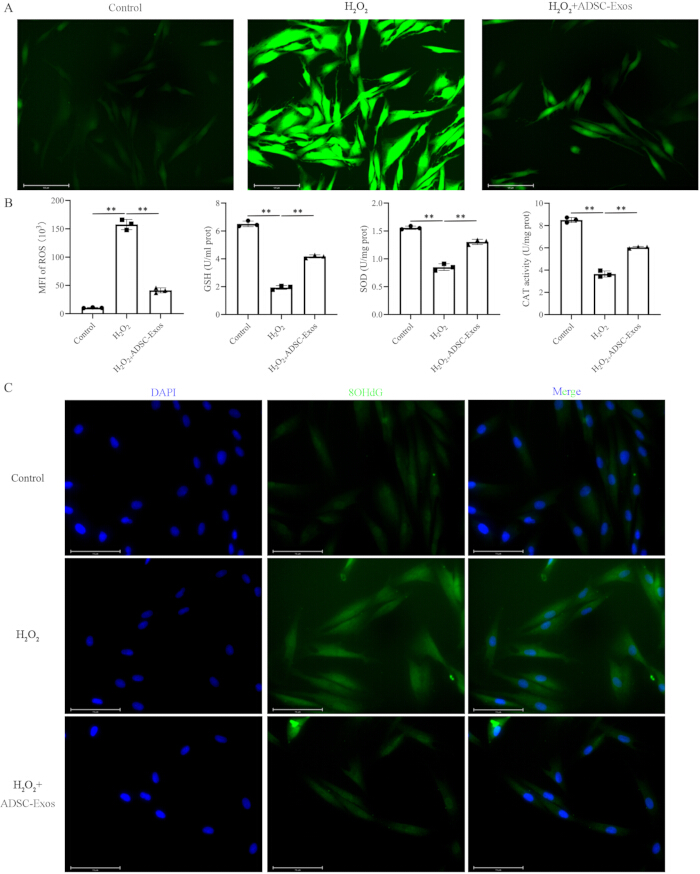
Figure 4: ADSC-Exos attenuate H₂O₂-induced oxidative stress in melanocytes. (A) Representative fluorescence images showing intracellular ROS in PIG1 cells following the indicated treatments. (B) Quantification of ROS MFI, GSH content, SOD activity, and CAT activity. SOD and CAT activities are expressed as U/mL protein. (C) Representative immunofluorescence images showing 8-OHdG expression. Blue = DAPI; green = 8-OHdG. Scale bars = 50 µm. Data are presented as the mean ± SD (n = 3 independent experiments). **p < 0.01. Abbreviations: ADSC-Exos = adipose-derived stem cell exosomes; CAT = catalase; DAPI = 4′,6-diamidino-2-phenylindole; GSH = glutathione; MFI = mean fluorescence intensity; ROS = reactive oxygen species; SD = standard deviation; SOD = superoxide dismutase; 8-OHdG = 8-hydroxy-2′-deoxyguanosine. Please click here to view a larger version of this figure.
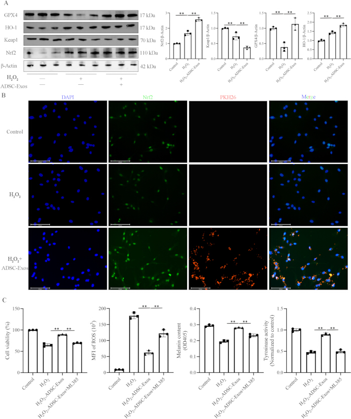
Figure 5: ADSC-Exos enhance melanocyte antioxidant capacity through the Keap1–Nrf2 pathway. (A) Representative western blots and densitometric quantification of Nrf2, Keap1, GPX4, and HO-1 expression in PIG1 cells following the indicated treatments. β-actin was used as the loading control. (B) Representative immunofluorescence images showing Nrf2 expression and the association or uptake of PKH26-labeled ADSC-Exos. Blue = DAPI; green = Nrf2; red = PKH26-labeled ADSC-Exos. (C) Cell viability, ROS MFI, melanin content, and tyrosinase activity following treatment with H₂O₂, ADSC-Exos, and the Nrf2 inhibitor ML385. Data are presented as the mean ± SD (n = 3 independent experiments). **p < 0.01. Abbreviations: ADSC-Exos = adipose-derived stem cell exosomes; DAPI = 4′,6-diamidino-2-phenylindole; GPX4 = glutathione peroxidase 4; HO-1 = heme oxygenase 1; Keap1 = Kelch-like ECH-associated protein 1; MFI = mean fluorescence intensity; Nrf2 = nuclear factor erythroid 2-related factor 2; ROS = reactive oxygen species; SD = standard deviation. Please click here to view a larger version of this figure.
Diskussion
Like many other diseases, the pathogenesis of vitiligo involves complex interactions between immune dysregulation and oxidative stress27,28,29,30. This study demonstrated that ADSC-Exos protect melanocytes from H₂O₂-induced injury primarily through activation of the Keap1–Nrf2 axis, as evidenced by restored viability, migration, and antioxidant enzyme expression. These findings position oxidative stress as an actionable target and suggest that exosome-based modulation of Nrf2 signaling may offer a therapeutic strategy for vitiligo. The pathogenesis of vitiligo is multifaceted, involving an intricate interplay of immune, inflammatory, and oxidative stress pathways. Among these, oxidative stress is increasingly recognized not only as an initiating factor but also as a key driver of disease progression31. Oxidative stress occurs when the production of ROS overwhelms the cellular antioxidant capacity, leading to a state of redox imbalance and subsequent tissue damage32. In vitiligo, endogenous and exogenous stimuli can lead to the accumulation of H₂O₂ in the epidermis, which generates highly damaging hydroxyl radicals, induces lipid peroxidation, and freely permeates cellular and nuclear membranes, causing extensive damage33,34. The findings are consistent with this paradigm, demonstrating that H₂O₂ treatment induces a concentration-dependent increase in intracellular ROS, escalates DNA damage, and ultimately impairs the proliferative and migratory capacities of human melanocytes (PIG1 cells).
ADSC-Exos have been widely reported to facilitate tissue repair in various injury models by promoting the proliferation and migration of resident cells while modulating excessive inflammatory responses and ROS levels35. For instance, in diabetic wounds—a condition also characterized by debilitating oxidative stress—ADSC-Exos have been shown to enhance the antioxidant capacity of fibroblasts and endothelial cells, thereby restoring their function36,37. Stem cell-derived exosomes protect melanocytes in vitiligo models. For example, bone marrow mesenchymal stem cell-derived exosomes deliver hydroxyacyl-CoA dehydrogenase to activate Nrf2/HO-1 and inhibit pyroptosis38, while three-dimensional-cultured human umbilical cord mesenchymal stem cell-derived exosomes enriched with miR-125b-5p and miR-132-3p ameliorate vitiligo by reducing oxidative stress and enhancing regulatory T-cell responses27. The present study demonstrated that ADSC-Exos effectively rescue the H₂O₂-induced functional impairments in melanocytes, significantly restoring their viability and migration. This underscores the feasibility of leveraging the multifaceted regenerative properties of ADSC-Exos to counteract the adverse microenvironment in vitiligo.
The pivotal balance of oxidative stress is governed by a sophisticated antioxidant system, with the Keap1–Nrf2 signaling pathway serving as a master regulator39. Under homeostatic conditions, Nrf2 is constitutively bound by its repressor, Keap1, and targeted for proteasomal degradation40. Under oxidative stress, this interaction is disrupted, allowing Nrf2 to stabilize, translocate to the nucleus, and initiate the transcription of a battery of cytoprotective genes, including antioxidant enzymes41. Previous studies have suggested that the Nrf2-regulated oxidative stress response is a potential therapeutic target for vitiligo42,43. The Keap1–Nrf2 pathway was therefore examined to elucidate the molecular mechanism underlying the protective effects of ADSC-Exos. The results indicate that ADSC-Exos mitigated oxidative stress in association with modulation of the Keap1–Nrf2 axis. Treatment with ADSC-Exos decreased Keap1 expression, facilitated the nuclear accumulation of Nrf2, and consequently upregulated the expression of downstream effector enzymes such as HO-1 and GPX4. This orchestrated activation of the endogenous antioxidant defense system represents a potential mechanism through which ADSC-Exos protect melanocytes from oxidative demise.
Beyond cytoprotection, whether ADSC-Exos directly modulate melanogenesis remains an unresolved question. The data demonstrate that ADSC-Exos restore melanin content and the expression of microphthalmia-associated transcription factor (MITF), tyrosinase, TYRP1, and DCT in H₂O₂-challenged melanocytes. However, the translational interpretation of these findings remains limited. The observed recovery may be secondary to improved melanocyte survival rather than direct regulation of melanogenic pathways. However, this distinction may be less absolute than it appears, as emerging evidence indicates that exosomal miRNAs can directly target melanogenic regulators: exosomal miR-330-5p directly inhibits MITF protein expression, leading to suppression of TYR, TYRP1, and TYRP2, while exosomal miR-25-5p from ultraviolet B (UVB)-induced fibroblasts inhibits melanin synthesis-related proteins TYR and MITF. These findings raise the possibility that ADSC-Exos may carry specific miRNAs capable of directly engaging melanogenic transcription factors, a hypothesis that warrants systematic miRNA profiling of ADSC-Exos in future studies. The mechanistic basis for the observed antioxidant effects may also involve specific exosomal miRNA cargo. A growing body of evidence demonstrates that exosomal miRNAs can directly target KEAP1 mRNA, thereby activating Nrf2 signaling. Fibroblast-derived exosomes deliver miR-29a-3p, which targets the 3′ untranslated region (3′UTR) of Keap1 mRNA and activates the KEAP1/Nrf2 antioxidant pathway44. Similarly, exosomal miR-141-3p from periodontal ligament stem cells decreases KEAP1 expression and activates the Nrf2 antioxidant pathway45, and miR-200a-3p in human umbilical cord MSC-derived exosomes attenuates UVB-induced oxidative stress by targeting Keap146. Most relevant to the present study, ADSC-derived exosomes deliver miR-125b-5p to inhibit Keap1 expression, increase Nrf2 nuclear translocation, and alleviate ferroptosis via the Keap1–Nrf2–GPX4 axis36. These findings strongly suggest that the observed Keap1 downregulation and Nrf2 activation in ADSC-sEV-treated melanocytes may be mediated by specific exosomal miRNAs, rather than solely by direct protein cargo. The studies cited above provide a roadmap for this endeavor—miRNA sequencing of ADSC-Exos followed by target prediction and functional validation (e.g., miRNA mimic/inhibitor experiments, luciferase reporter assays for Keap1 3′UTR) would be required to establish whether specific exosomal miRNAs directly mediate the observed Keap1–Nrf2 activation.
Furthermore, ADSC-Exos may interact with this neuroimmunoendocrine network. ADSC-Exos have been shown to exert potent immunomodulatory effects—including shifting T-cell responses toward anti-inflammatory phenotypes and modulating cytokine profiles. They may attenuate the neurogenic inflammation that sustains local oxidative stress and immune activation in the vitiligo milieu47. The observed Keap1–Nrf2 activation may not only counteract oxidative damage directly but could also intersect with neuroendocrine signaling, as Nrf2 has been reported to modulate inflammatory cytokine production and stress responses that are themselves regulated by neuropeptides and stress hormones. Moreover, MITF activity is regulated by multiple upstream signaling cascades, including the cyclic adenosine monophosphate (cAMP)/protein kinase A (PKA) pathway, which is a central driver of melanogenic gene transcription. However, the precise crosstalk between ADSC-Exos and this network remains unexplored.
Although the in vitro data support a cytoprotective mechanism, the translational interpretation of these findings is limited by the experimental model. The clinical microenvironment of vitiligo is more complex than a monolayer of stressed melanocytes and includes interactions among keratinocytes, dermal fibroblasts, tissue-resident immune cells, and peripheral nerve endings47. The H₂O₂-challenged PIG1 model, albeit methodologically standardized, does not reproduce the autoimmune and inflammatory signals present in the lesional microenvironment of patients with vitiligo. However, melanosome formation and transfer were not evaluated in this study. Furthermore, the utilization of an immortalized melanocyte lineage, while enabling mechanistic dissection, cannot fully capture the phenotypic heterogeneity or the donor-dependent redox setpoints characteristic of primary human melanocytes, which may substantially influence the therapeutic threshold. Moreover, the specific exosomal miRNAs or proteins responsible for the observed effects have not been identified. Consequently, further investigation using more physiologically relevant models, including reconstructed epidermal equivalents and ex vivo patient-derived skin explants, is necessary to evaluate the effects of ADSC-Exos in the complex human disease microenvironment.
In conclusion, this study demonstrates that ADSC-Exos effectively ameliorate H₂O₂-induced oxidative damage in human melanocytes by restoring intracellular redox homeostasis and enhancing cellular antioxidant defenses. The core mechanistic insight is the activation of the Keap1–Nrf2–ARE signaling pathway. These findings not only provide mechanistic support for the use of ADSC-Exos for vitiligo but also support further investigation of a potential therapeutic approach targeting the oxidative stress component.
Offenlegungen
The authors declare no competing interests. No artificial intelligence tools were used in the preparation of the manuscript or figures.
AUTHOR CONTRIBUTIONS:
Xiaobing Fang and Ziyong Ma conceptualized the study. Xiaobing Fang and Chao Yang conducted the investigation. Yafang Wang performed the validation, and Xiaodong Yang curated the data. Ziyong Ma prepared the original draft and contributed to visualization and supervision. Xiaobing Fang reviewed and edited the manuscript, administered the project, and acquired funding. All authors read and approved the final manuscript
Danksagungen
This work was supported by the Ningxia Natural Science Foundation (No. 2022AAC03735).
Materialien
| Name | Unternehmen | Katalognummer | Kommentare |
|---|---|---|---|
| Anti-8-OHdG antibody | ABCAM | ab62623 | Immunofluorescence – DNA oxidative damage marker |
| Anti-ALIX antibody | Proteintech | 12422-1-AP | Exosomal marker for western blot |
| Anti-CD63 antibody | Proteintech | 25682-1-AP | Exosomal marker for western blot |
| Anti-GPX4 antibody | Selleck | F1580 | Western blot – downstream antioxidant enzyme |
| Anti-HO-1 antibody | Proteintech | 10701-1-AP | Western blot – downstream antioxidant enzyme |
| Anti-HSP70 antibody | Proteintech | 10995-1-AP | Exosomal marker for western blot |
| Anti-Keap1 antibody | Abmart | TA5266 | Western blot – Keap1 protein |
| Anti-Ki67 antibody | Servicebio | GB151499-100 | Immunofluorescence – proliferation marker |
| Anti-Nrf2 antibody | Abmart | T55136 | Western blot and immunofluorescence – Nrf2 protein |
| Anti-TSG101 antibody | Proteintech | 67381-1-Ig | Exosomal marker for western blot |
| Anti-β-Actin antibody | Proteintech | 81115-1-RR | Western blot – loading control |
| Catalase (CAT) activity assay kit | Sangon | D799597 | Measures CAT activity by H2O2 decomposition |
| Cell Counting Kit-8 (CCK-8) | Boster | AR1199 | Cell viability assay (absorbance at 450 nm) |
| DCFH-DA probe | Beyotime | S1105 | Intracellular ROS detection (fluorescent probe) |
| FITC mouse anti-human CD29 antibody | Becton Dickinson | 569943 | Positive MSC marker for flow cytometry |
| FITC mouse anti-human CD34 antibody | Becton Dickinson | 560942 | Hematopoietic negative marker for flow cytometry |
| FITC mouse anti-human CD44 antibody | Becton Dickinson | 560977 | Positive MSC marker for flow cytometry |
| FITC mouse anti-human CD45 antibody | Becton Dickinson | 561865 | Hematopoietic negative marker for flow cytometry |
| FITC mouse anti-human CD73 antibody | Becton Dickinson | 561254 | Positive MSC marker for flow cytometry |
| FITC mouse anti-human CD90 antibody | Becton Dickinson | 561969 | Positive MSC marker for flow cytometry |
| Fluorescent secondary antibodies | Proteintech | SA00003-2 | For immunofluorescence staining |
| HRP-conjugated secondary antibodies | Proteintech | SA00001-2 | For western blot detection |
| Human adipose-derived mesenchymal stem cell adipogenic differentiation kit | Cyagen | HUXMD-90031 | Staining of lipid droplets after adipogenic differentiation |
| Human adipose-derived mesenchymal stem cell culture kit | Cyagen | HUXMD-9001 | Human adipose-derived mesenchymal stem cell culture |
| Human adipose-derived mesenchymal stem cell osteogenic differentiation kit | Cyagen | HUXMD-90021 | Staining of calcium nodules after osteogenic differentiation |
| Isotype control antibodies | Becton Dickinson | 556658 | Corresponding isotype controls for flow cytometry |
| ML385 | Targetmol | T4360 | Nrf2 inhibition |
| Nanoparticle tracking analyzer | Malvern Panalytical | NS300 | Particle size distribution and concentration of exosomes |
| PKH26 Red Fluorescent Dye | Sigma-Aldrich | PKH26GL | Labeling of exosomes for cellular uptake assay |
| PrimeScript RT | TaKaRa | RR037Q | Reverse transcription for quantitative PCR |
| Statistical analysis software | GraphPad | 8 | Statistical analysis |
| Superoxide dismutase (SOD) activity assay kit (WST-8-based) | Sangon | D799606 | Measures SOD activity in cell lysates |
| Transwell chambers (8-μm pore size) | Corning | 3428 | Migration assay (24-well format) |
| TRIzol | TaKaRa | 9108 | Total RNA extraction |
| Type IV collagenase | Sigma-Aldrich | 17104019 | Digestion of adipose tissue (1 h at 37 °C) |
| Tyrosinase activity assay kit | Solarbio | BC4055 | Tyrosinase activity assay |
Referenzen
- Seneschal J et al. Worldwide expert recommendations for the diagnosis and management of vitiligo: Position statement from the international Vitiligo Task Force-Part 2: Specific treatment recommendations. J Eur Acad Dermatol Venereol. 2023;37(11):2185-2195.
- Böhm M et al. S1 Guideline: Diagnosis and therapy of vitiligo. J Dtsch Dermatol Ges. 2022;20(3):365-378.
- LeWitt TM, Kundu RV. Vitiligo. JAMA Dermatol. 2021;157(9):1136.
- Bergqvist C, Ezzedine K. Vitiligo: A focus on pathogenesis and its therapeutic implications. J Dermatol. 2021;48(3):252-270.
- Ezzedine K et al. Psychosocial Effects of Vitiligo: A Systematic Literature Review. Am J Clin Dermatol. 2021;22(6):757-774.
- Kussainova A et al. Vitiligo and anxiety: A systematic review and meta-analysis. PLoS One. 2020;15(11):e0241445.
- Matarrese P et al. An Overview of the Biological Complexity of Vitiligo. Oxid Med Cell Longev. 2024;2024:3193670.
- Białczyk A, Wełniak A, Kamińska B, Czajkowski R. Oxidative Stress and Potential Antioxidant Therapies in Vitiligo: A Narrative Review. Mol Diagn Ther. 2023;27(6):723-739.
- Seneschal J, Boniface K, D'Arino A, Picardo M. An update on Vitiligo pathogenesis. Pigment Cell Melanoma Res. 2021;34(2):236-243.
- Chen J et al. Oxidative stress-induced hypermethylation and low expression of ANXA2R: Novel insights into the dysfunction of melanocytes in vitiligo. J Dermatol Sci. 2024;114(3):115-123.
- Jaganjac M, Milkovic L, Zarkovic N, Zarkovic K. Oxidative stress and regeneration. Free Radic Biol Med. 2022;181:154-165.
- Zhang J et al. Research Progress on Targeted Antioxidant Therapy and Vitiligo. Oxid Med Cell Longev. 2022;2022:1821780.
- Wang J et al. Damage-associated molecular patterns in vitiligo: igniter fuse from oxidative stress to melanocyte loss. Redox Rep. 2022;27(1):193-199.
- Ng CY et al. Targeting the elevated IFN-γ in vitiligo patients by human anti- IFN-γ monoclonal antibody hampers direct cytotoxicity in melanocyte. J Dermatol Sci. 2023;110(3):78-88.
- Guo S, Zhang Q. Paeonol protects melanocytes against hydrogen peroxide-induced oxidative stress through activation of Nrf2 signaling pathway. Drug Dev Res. 2021;82(6):861-869.
- Papadopoulos KS, Piperi C, Korkolopoulou P. Clinical Applications of Adipose-Derived Stem Cell (ADSC) Exosomes in Tissue Regeneration. Int J Mol Sci. 2024;25(11):5916.
- Tian C et al. ADSC exosomes improve high glucose induced fibroblast oxidative stress injury and accelerate DFU wound healing via regulating Keap1/Nrf2 axis. Cell Signal. 2025;134:111936.
- Liu Z et al. ADSC-derived exosomes mitigate radiation-induced skin injury by reducing oxidative stress, inflammation and cell death. Front Public Health. 2025;13:1603431.
- Ren H et al. Adipose mesenchymal stem cell-derived exosomes promote skin wound healing in diabetic mice by regulating epidermal autophagy. Burns Trauma. 2024;12:tkae001.
- Zhang M et al. Vitiligo: An immune disease and its emerging mesenchymal stem cell therapy paradigm. Transpl Immunol. 2023;76:101766.
- Yang RL et al. The immunological pathogenesis of vitiligo and the potential role of mesenchymal stromal/stem cells in its treatment. J Transl Med. 2025;23(1):707.
- Ma J et al. Exosomes enriched with miR-31-3p from keratinocytes under oxidative stress promote vitiligo progression by destructing melanocytes and activating CD8+ T cells. Int J Biol Macromol. 2025;310(Pt 1):143070.
- Kemaloğlu CA et al. The Optimal Effective Dose of Adipose-Derived Stem Cell Exosomes in Wound Healing. Ann Plast Surg. 2024;93(2):253-260.
- Slominski RM et al. Melanoma, Melanin, and Melanogenesis: The Yin and Yang Relationship. Front Oncol. 2022;12:842496.
- Slominski A, Tobin DJ, Shibahara S, Wortsman J. Melanin pigmentation in mammalian skin and its hormonal regulation. Physiol Rev. 2004;84(4):1155-1228.
- Slominski A, Zmijewski MA, Pawelek J. L-tyrosine and L-dihydroxyphenylalanine as hormone-like regulators of melanocyte functions. Pigment Cell Melanoma Res. 2012;25(1):14-27.
- Wang Q et al. 3D-hUMSCs Exosomes Ameliorate Vitiligo by Simultaneously Potentiating Treg Cells-Mediated Immunosuppression and Suppressing Oxidative Stress-Induced Melanocyte Damage. Adv Sci (Weinh). 2024;11(31):e2404064.
- Iwanowski T, Kołkowski K, Nowicki RJ, Sokołowska-Wojdyło M. Etiopathogenesis and Emerging Methods for Treatment of Vitiligo. Int J Mol Sci. 2023;24(11):9749.
- Al-Karawi AS, Al-Budairi AS, Atoom AM. Serum soluble CD25, Galectin-9, and CXCL9 as immunological markers differentiating complete remission from refractory acute myeloid leukemia. Clin Immunol. 2026;287:110750.
- Al-Karawi AS, Kadhim AS. Correlation of autoimmune response and immune system components in the progression of IgA nephropathy: A comparative study. Hum Immunol. 2024;85(6):111181.
- Chen J, Li S, Li C. Mechanisms of melanocyte death in vitiligo. Med Res Rev. 2021;41(2):1138-1166.
- Mathachan SR et al. Does oxidative stress correlate with disease activity and severity in vitiligo? An analytical study. J Cosmet Dermatol. 2021;20(1):352-359.
- Lin Y et al. The underestimated role of mitochondria in vitiligo: From oxidative stress to inflammation and cell death. Exp Dermatol. 2024;33(1):e14856.
- Diotallevi F et al. Vitiligo, from Pathogenesis to Therapeutic Advances: State of the Art. Int J Mol Sci. 2023;24(5):4910.
- Shen K et al. Exosomes from adipose-derived stem cells alleviate the inflammation and oxidative stress via regulating Nrf2/HO-1 axis in macrophages. Free Radic Biol Med. 2021;165:54-66.
- Shen K et al. miR-125b-5p in adipose derived stem cells exosome alleviates pulmonary microvascular endothelial cells ferroptosis via Keap1/Nrf2/GPX4 in sepsis lung injury. Redox Biol. 2023;62:102655.
- Yang C et al. Exosomes from adipose-derived stem cells restore fibroblast function and accelerate diabetic wound healing. Heliyon. 2024;10(1):e22802.
- Tang S, Li X, Xi J. Bone marrow mesenchymal stem cell-derived exosomes HADH alleviate vitiligo by activating the Nrf2/HO-1 pathway. Exp Cell Res. 2025;453(2):114798.
- Zhang DD. Thirty years of NRF2: advances and therapeutic challenges. Nat Rev Drug Discov. 2025;24(6):421-444.
- George M, Reddy AP, Reddy PH, Kshirsagar S. Unraveling the NRF2 confusion: Distinguishing nuclear respiratory factor 2 from nuclear erythroid factor 2. Ageing Res Rev. 2024;98:102353.
- Dong Y et al. Nrf2 activators for the treatment of rare iron overload diseases: From bench to bedside. Redox Biol. 2025;81:103551.
- Lin X, Meng X, Song Z, Lin J. Nuclear factor erythroid 2-related factor 2 (Nrf2) as a potential therapeutic target for vitiligo. Arch Biochem Biophys. 2020;696:108670.
- Panieri E, Telkoparan-Akillilar P, Saso L. NRF2, a crucial modulator of skin cells protection against vitiligo, psoriasis, and cancer. Biofactors. 2023;49(2):228-250.
- Yan L et al. Fibroblast exosomes promote wound healing and improve the quality of healed skin via miR-29a-3p-mediated KEAP1/Nrf2 pathway activation. Burns Trauma. 2025;13:tkaf035.
- Liu M et al. Exosomal miR-141-3p from PDLSCs Alleviates High Glucose-Induced Senescence of PDLSCs by Activating the KEAP1-NRF2 Signaling Pathway. Stem Cells Int. 2023;2023:7136819.
- Gui Q et al. miR-200a-3p in Human Umbilical Cord Mesenchymal Stem Cell-Derived Exosomes Attenuates UVB-Induced Skin Inflammatory Response and Oxidative Stress via Keap1-Nrf2 Pathway. Stem Cells Int. 2025;2025:7831890.
- Slominski RM, Raman C, Jetten AM, Slominski AT. Neuro-immuno-endocrinology of the skin: how environment regulates body homeostasis. Nat Rev Endocrinol. 2025;21(8):495-509.