This study integrates Global Burden of Disease data and bibliometric analysis to characterize the global burden of colorectal cancer and the research landscape of exosomes in colorectal cancer. Key research domains and emerging frontiers, including biomarker applications, drug delivery, and immunotherapy, were identified to inform future research.
Forschungsartikel
Mapping the Spatiotemporal Evolution and Knowledge Landscape of Exosomes in Colorectal Cancer
* These authors contributed equally
In diesem Artikel
Zusammenfassung
Zusammenfassung
Many studies have investigated the roles of exosomes in colorectal cancer (CRC), but the global research landscape and future directions in this field remain insufficiently characterized. This study aimed to integrate epidemiological evidence with bibliometric analysis to map the research landscape of exosome research in CRC. Global Burden of Disease (GBD) data were analyzed to assess the geographic and socioeconomic distribution of CRC incidence and mortality, and a bibliometric analysis was conducted to systematically evaluate publication trends, research clusters, and emerging themes. Network-based community detection was conducted using the Walktrap algorithm. GBD analysis revealed pronounced geographic and socioeconomic disparities in CRC incidence and mortality, indicating a heterogeneous global disease burden. Knowledge mapping demonstrated a rapid and sustained increase in publications on exosomes in CRC. Clustering analyses identified four major research domains: exosome biogenesis and molecular characteristics; tumor microenvironment and therapy resistance; exosomal non-coding RNAs in tumor progression and metastasis; and exosomal biomarkers for diagnosis and prognosis. The Walktrap analysis further identified biomarkers, diagnosis, liquid biopsy, drug delivery, and immunotherapy as highly relevant but relatively underdeveloped research directions. Overall, this integrated epidemiological and knowledge-mapping study provides a comprehensive overview of the evolution and structure of exosome research in CRC and highlights key research hotspots and emerging frontiers that may guide future investigations into precision diagnostics and therapeutic innovation.
Einleitung
Colorectal cancer (CRC) represents one of the most serious global public health challenges, ranking third in cancer incidence and second in mortality worldwide, with approximately 1.93 million new cases and over 900,000 deaths in 20221. The burden of CRC is projected to rise to 3.2 million new cases and 1.6 million deaths by 20402. A recent systematic review of over 3.8 million CRC patients from 36 countries reported that the proportion of distant metastasis at diagnosis ranged from 16.2% to 28.2%, with a median of 23.7%3. Metastasis is the critical cause of cancer-related death in CRC4, with a 5-year survival rate of approximately 14%5. Despite improvements in surgical techniques and targeted therapies that have prolonged survival, the prognosis for CRC patients with metastasis remains poor. This highlights the urgent need for innovative biomarkers with high sensitivity and specificity to enhance both diagnosis and prognosis6.
CRC metastasis is a complex biological process that involves the metastatic cascade, including invasion of surrounding tissues, entry into the bloodstream, survival in circulation, adhesion to vessel walls, extravasation into distant parenchyma, micrometastasis, and colonization4,7. Growing evidence demonstrates the pivotal role of the tumor microenvironment (TME) in CRC metastasis. The TME mainly consists of tumor cells, stromal cells such as fibroblasts and endothelial cells, immune cells, and other noncellular components8. Tumor cells and their microenvironment interact to promote tumor initiation, development, and metastasis through complex intercellular signaling9, in which exosomes play a central role.
Initially, exosomes were regarded as garbage bins for clearing unwanted cellular components10. However, extensive research has revealed the pivotal role of exosomes in mediating intercellular communication through investigations into their biological origin, secretion, structure, and function11,12. Exosomes, nanosized extracellular vesicles ranging from 30 to 150 nm, are secreted by nearly all cell types into bodily fluids such as blood, urine, saliva, and cerebrospinal fluid. They encapsulate and carry bioactive molecules such as proteins, lipids, and nucleic acids, including DNA, mRNA, and noncoding RNAs13. Exosomes drive various biological processes, including cell signaling, immune responses, and cancer progression14. Moreover, their presence in biological fluids makes them an attractive minimally invasive liquid biopsy approach for cancer diagnosis, prognosis, and surveillance13. In the CRC field, exosomes play dual roles: on one hand, exosome-mediated intercellular communication promotes tumor growth, invasion, migration, and immune evasion, alters vascular permeability, and facilitates pre-metastatic niche formation15. On the other hand, they serve as promising biomarkers for early diagnosis and therapeutic targeting16. With advances in precision medicine, identifying and validating reliable cancer biomarkers is essential for early detection, accurate diagnosis, and effective treatment. Among emerging candidates, exosomes have attracted considerable interest as potential carriers of critical information in cancer studies17.
In recent years, studies on exosomes in CRC have expanded rapidly, leading to the accumulation of a large amount of unstructured knowledge. Although numerous studies have highlighted the roles of exosomes in tumor progression, metastasis, and therapeutic resistance, the overall research landscape, developmental trends, and future directions of this field remain insufficiently characterized. By analyzing and visualizing research information, bibliometric analysis and knowledge mapping can evaluate the scale, distribution, structure, and evolution of scientific research, thereby providing evidence to guide future strategies18.
Previous bibliometric analyses have mainly focused on publication trends and research hotspots, providing an overview of the development of this research field. However, these literature-based analyses do not reflect the geographical and socioeconomic heterogeneity of the CRC burden, which may provide important context for interpreting the relevance of emerging research directions across different regions.
Accordingly, our study primarily used visualization analysis to characterize the research landscape of exosomes in CRC, identify critical research clusters, and explore emerging and promising research directions. Global Burden of Disease (GBD) data were additionally analyzed to characterize the global burden of CRC and its geographical and socioeconomic distribution, thereby providing an epidemiological context for interpreting the research patterns.
Protokoll
Information source
The Web of Science Core Collection was chosen as the data source for this study due to its comprehensive coverage and reliable records18. Although multiple databases could increase the number of retrieved articles, they often contain overlapping records and may introduce confounding factors that affect data quality. Selecting a single reliable and authoritative database is a common approach in bibliometric analysis. Publications published between 2003 and 2025 were identified through a literature search conducted in the Web of Science Core Collection on 1 January 2026. The retrieved records were independently screened by two researchers according to the predefined eligibility criteria. The titles and abstracts of all retrieved records were independently reviewed to assess relevance to the research topic, and irrelevant records were excluded. Full-text screening and formal assessment of inter-reviewer agreement were not performed. Any discrepancies between the two researchers were resolved through discussion and consensus. If a consensus could not be reached, a third, senior researcher was consulted to make the final decision. Full records and cited references for relevant literature were downloaded in plain-text format using predefined search terms. The flowchart for data retrieval, screening, and inclusion is shown in Figure 1.
Data cleaning and standardization
The retrieved records underwent data cleaning and standardization to minimize inconsistencies in the information. Author names were standardized to address variations in initials and name presentation, while institutional and country/region names were harmonized to reduce inconsistencies in affiliation information. For keyword analysis, synonyms, abbreviations, spelling variants, and singular/plural forms were manually standardized. Terms with distinct scientific meanings were retained separately. The cleaned and standardized dataset was used for subsequent analysis and visualization.
Clustering analysis
Clustering analysis was performed using VOSviewer to identify thematic clusters based on keyword co-occurrence relationships18. A co-occurrence analysis of all keywords was conducted using full counting, and keywords occurring in at least 30 publications were included. Association strength was used to normalize the strength of links between keywords. Before network construction, keywords were manually reviewed and cleaned to remove terms unrelated to the research topic and to merge synonyms, abbreviations, spelling variants, and other equivalent terms. The resulting network was clustered using the VOSviewer clustering technique with a resolution of 1.00 and a minimum cluster size of 1, with small clusters merged. The total link strength of each keyword was calculated as the sum of its link strengths with all other keywords. Node size in the network visualization was weighted by total link strength. The resulting clusters were used to characterize the major thematic domains of exosome research in CRC.
Walktrap algorithm and community map analysis
Community detection can be performed using algorithms such as Walktrap, Girvan-Newman, Louvain, Label Propagation, Spectral Clustering, and Infomap, each suited to different types of networks. For instance, Girvan-Newman is more appropriate for small networks, Louvain works efficiently on large networks, and Infomap is advantageous for multilayer structures. The Walktrap algorithm detects communities by simulating random walks to capture close relationships among nodes, which here represent research topics19. Each node is initially treated as an independent community, and the similarity between topics is estimated using co-occurrence frequency. These values are then converted into a distance matrix, where communities with the shortest distances are progressively merged. The algorithm ultimately selects the partition that best reflects the community structure, characterized by strong internal connections and sparse links between communities20.
In this study, thematic mapping was performed using the Biblioshiny interface of the bibliometrix package in R to identify major research themes based on keyword co-occurrence. All Keywords were selected as the field for analysis. Considering the number of included publications and the overall number of keywords, the number of words and minimum cluster frequency were adjusted to obtain 10 thematic clusters, facilitating the identification and interpretation of major research themes. Walktrap was selected as the clustering algorithm, using the default random-walk length of 4 steps in the underlying igraph implementation, with a community repulsion value of 0.5. Three labels were displayed for each cluster, with a label size of 0.4. The resulting thematic clusters were characterized using centrality and density measures. Centrality reflects the strength of connections between a thematic cluster and other clusters, whereas density reflects the strength of internal connections among keywords within the same cluster. The thematic map was divided into four quadrants according to the relative centrality and density of the identified themes, reflecting their relevance to the overall research field and their degree of internal development, respectively.
The global burden of CRC
The GBD Study is an ongoing international project that assesses the global impact of diseases, injuries, and risk factors. Drawing on data from registries, surveys, vital statistics, and literature, it applies unified statistical models to produce comparable health metrics21. The GBD 2021 (https://vizhub.healthdata.org/gbd-results/) was utilized as the source for analyzing the burden of CRC.
Data were extracted in July 2025, with colon and rectum cancer as the cause, deaths and incidence as the measures, and number, percent, and rate as the metrics. Estimates were obtained for all ages and both sexes from 1990 to 2021, covering the global level, 21 GBD regions, and 204 countries and territories. All-age counts of incident cases and deaths were used to describe the absolute CRC burden, whereas age-standardized rates (ASRs) were obtained directly from the GBD Results Tool to facilitate comparisons across locations and over time by minimizing the influence of differences in population age structure. Specifically, the age-standardized incidence rate (ASIR) represents the number of new CRC cases per 100,000 population per year after age standardization, whereas the age-standardized mortality rate (ASMR) represents the number of CRC-related deaths per 100,000 population per year after age standardization.
The Socio-demographic Index (SDI) is a composite indicator widely used in GBD studies to assess the socioeconomic development of regions and countries. It integrates per capita income, educational levels, and fertility rates into an index ranging from 0 to 122. The associations of SDI with the ASIR and ASMR of CRC were assessed using Pearson correlation analysis. Pearson correlation coefficients (r) and p-values were calculated to assess the strength and direction of the associations, with statistical significance set at p < 0.05.
Software applications
The visualization and statistical analysis were performed using various software tools: Microsoft Excel; the “maps”, “bibliometrix”, and “ggplot2” packages in R; the “plotly” and “pandas” packages in Python; VOSviewer; and CiteSpace. The tools used for different visualizations are displayed in Figure 1.
Ergebnisse
The global burden of CRC
Global level
Using data from the GBD database, we assessed CRC burden from 1990 to 2021. Globally, the ASIR increased by 6.5%, while the ASMR decreased by 20.3% (Table 1). This divergent pattern indicates that the global incidence and mortality burden of CRC have evolved differently over time.
Regional level
Substantial regional variation was observed. Mortality declined sharply in high-income regions such as North America (by 37.1%) and Australasia (by 41.2%), which is likely attributable to widespread screening programs and access to advanced therapies. In contrast, several low- and middle-income regions experienced pronounced increases in both incidence and mortality. Central Latin America showed the largest increases in incidence (90.3%) and mortality (29.0%), while Southeast Asia and Tropical Latin America recorded incidence increases of 56.9% and 56.1%, with mortality increases of 26.1% and 21.2%, respectively. These trends reflect differences in healthcare infrastructure, early detection, and treatment availability across regions.
National level
At the national level, the Netherlands reported the highest ASIR (69.8 per 100,000), followed by Monaco (68.3) and Bermuda (61.8), whereas the Gambia, Papua New Guinea, and Mozambique had the lowest ASIRs (3.3–4.1 per 100,000). Uruguay had the highest ASMR (27.5 per 100,000), followed by Hungary (26.0) and Bulgaria (25.7), while the Gambia, Papua New Guinea, and Bangladesh had the lowest ASMRs (3.1–3.6 per 100,000) (Figure 2A,B). Correlation analysis revealed a strong positive relationship between the ASIR and the SDI (r = 0.821, p < 0.001) (Figure 2C), suggesting that higher levels of social development are associated with greater CRC incidence. The ASMR demonstrated a moderate-to-strong positive association with SDI (r = 0.629, p < 0.001) (Figure 2D). Visual inspection of Figure 2D showed that ASMR tended to increase with SDI across countries with an SDI of approximately 0.25–0.75, whereas a declining pattern was observed at higher SDI levels. These findings indicate substantial cross-national differences in CRC burden and suggest variation in the association between social development and CRC mortality across SDI levels. Consequently, low- to middle-SDI countries should focus on strengthening cancer prevention and control strategies, while high-SDI countries should continue to optimize screening and therapeutic approaches to further reduce the disease burden.
Visualization study of publication trends, productive authors, journal impact, and disciplinary evolution
The annual publication output from 2005 to 2025 reveals three stages in the growth pattern (Figure 3A). The embryonic stage (2005–2009) was characterized by minimal activity, with annual publications consistently below 5. This was followed by an initial development stage (2010–2015), during which outputs rose to approximately 30. The field then entered a rapid development stage (2016–2025), peaking at over 180 publications in 2021. Thereafter, publication output remained relatively high, with more than 120 publications per year, reflecting sustained academic interest in this field. In bibliometric analysis, publications and citations are standard metrics for evaluating academic performance, productivity, and impact. Li J had the highest number of publications, followed by Zhang Y and Li Y (Figure 3B), indicating strong productivity in this research domain. In contrast, citation analysis revealed that Simpson RJ received the highest total citations, followed by Ji H and Greening DW (Figure 3C), reflecting substantial academic influence. Beyond these conventional measures, the h-index, m-index, and g-index were further applied to provide a more comprehensive evaluation of author impact (Figure 3D–F). The h-index indicates that a scholar has h articles each cited at least h times, thus reflecting both productivity and citation impact. Li J achieved the highest h-index, followed closely by Zhang Y and Simpson RJ. However, since the h-index may not fully represent authors with fewer but highly influential papers or early-career researchers, the g-index and m-index were also considered. The m-index, calculated by dividing the h-index by the number of active publication years, and the g-index, defined as the squared number of citations accumulated by an author who has published at least g articles23, both placed Li J and Zhang Y in the top three. Collectively, these metrics demonstrate that Li J has made the most balanced and influential contributions, while Simpson RJ and Zhang Y also play pivotal roles in advancing this research field.
The journal distribution and citation landscape in this research field were analyzed to identify major publication sources and knowledge flow patterns. The top three journals by publication count were Frontiers in Oncology, International Journal of Molecular Sciences, and Scientific Reports (Figure 3G), indicating that these journals serve as the primary platforms for related studies. In terms of citations, Cancer Research ranked first, followed by Molecular Cancer (Figure 3H), suggesting that high-impact journals in oncology and molecular biology play a key role in shaping the field’s foundational knowledge. Furthermore, the dual-map overlay of journals generated by CiteSpace, using thresholds of ≥10 citing articles and ≥100 cited times, illustrates the overall distribution and citation relationships among journals (Figure 3I). The citing journals are located on the left, representing the sources of new research findings, while the cited journals on the right reflect the established knowledge bases. Two primary citation pathways are identified. This indicates that studies published in Molecular/Biology/Immunology and Medicine/Medical/Clinical journals commonly cited those from Molecular/Biology/Genetics journals, suggesting that studies in this domain are primarily rooted in molecular and clinical research foundations. The width of the citation paths reflects close knowledge flow between these fields, highlighting strong interdisciplinary connections that facilitate the integration of molecular mechanisms and clinical applications in cancer research.
Spatial distribution of the countries
The worldwide distribution of publications and citations suggests that China and the USA are firmly in the top two for these two metrics (Figure 4A–D). Single Country Publications (SCP) refer to publications involving authors from a single country, whereas Multiple Country Publications (MCP) involve authors from multiple countries. Further examination of collaboration patterns through the SCP and MCP indicators uncovered distinct national strategies. China, despite its vast output, conducted the majority of its research domestically, with an MCP ratio of only 8.0% (Figure 4E). This stands in stark contrast to several other productive nations, such as Australia (45.5%), the USA (44.6%), and Italy (32.5%), which all demonstrated a strong propensity for international collaboration, producing a substantial share of their research through partnerships. This marked disparity suggests a clear need for China to strengthen international exchange and collaboration in this field to further synergize its productivity with global influence.
Spatial and temporal networks of research hotspots
Using VOSviewer-based clustering, research hotspots on exosomes in CRC were categorized into four dominant clusters (Figure 5A). Cluster 1 focuses on the biogenesis and molecular characteristics of exosomes, particularly their protein-related components, as well as the mechanisms underlying exosome formation and secretion. Cluster 2 highlights the tumor microenvironment and therapy resistance, emphasizing the roles of cellular and microenvironmental interactions in chemoresistance and therapeutic responses in CRC. Cluster 3 centers on tumor progression and metastasis associated with exosomal non-coding RNAs, which are linked to CRC proliferation, migration, invasion, and other malignant behaviors. Cluster 4 is associated with the identification and application of exosomal biomarkers, particularly for liquid biopsy, diagnosis, and prognosis based on plasma samples. Collectively, these clusters capture the diverse molecular, cellular, and translational aspects of exosome research in CRC. Temporal analysis further indicates that cluster 2 represents an emerging research focus (Figure 5B). Spatial density networks based on total link strength and occurrence frequency provide an intuitive visualization of these research hotspots (Figure 5C,D).
Exploring themes worthy of further study
The community discovery analysis was performed on keywords using the Walktrap algorithm. In the motor theme community (quadrant I), exosomes, CRC, and expression, which reflect the molecular characteristics and expression patterns in this field, are identified as highly relevant and well-developed topics. Notably, the community map (quadrant IV) highlights biomarker, diagnosis, liquid biopsy, drug delivery, and immunotherapy as highly relevant, application-oriented themes that remain relatively underdeveloped, representing promising directions for future research (Figure 6).
Data Availability:
The datasets used in this study were obtained from the Web of Science (https://webofscience.clarivate.cn/wos/alldb/basic-search) and the GBD 2021 (https://vizhub.healthdata.org/gbd-results/). Additional raw data and supporting materials are available from the corresponding author upon reasonable request.
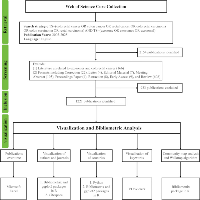
Figure 1: Flowchart of the bibliometric analysis. Please click here to view a larger version of this figure.
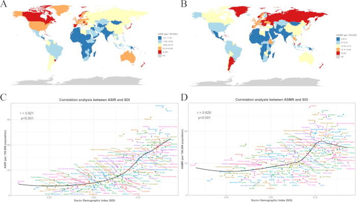
Figure 2: The national and territorial burden of colorectal cancer in 2021. (A) ASIR in 204 countries and territories. (B) ASMR in 204 countries and territories. (C) Relationship between SDI and ASIR of CRC by nation. (D) Relationship between SDI and ASMR of CRC by nation, with ASMR increasing at SDI values of approximately 0.25–0.75 but declining above 0.75. Please click here to view a larger version of this figure.
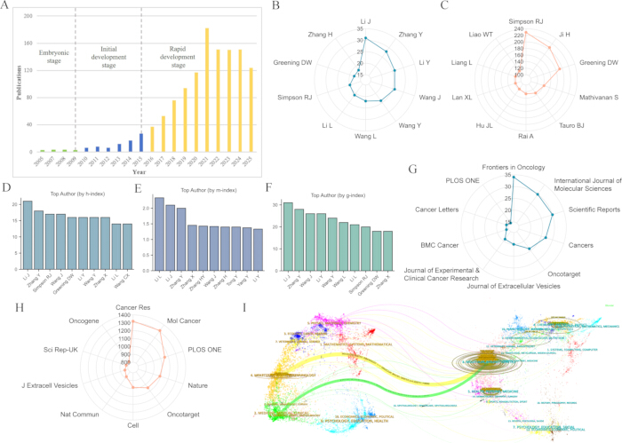
Figure 3: Visualization study of publication trends, productive authors, journal impact, and disciplinary evolution. (A) The timeline for the number of publications. (B) The top ten authors of publications. (C) The top ten authors of citations. (D) The top ten authors by h-index. (E) The top ten authors by m-index. (F) The top ten authors by g-index. (G) The top ten journals of publications. (H) The top ten journals of citations. (I) The dual-map overlay of journals in this field. Please click here to view a larger version of this figure.
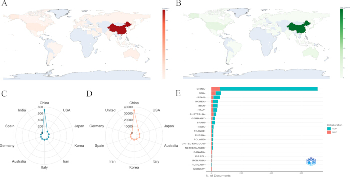
Figure 4: Spatial distribution of the countries. (A) The world map of publications by countries. (B) The world map of citations by countries. (C) The top ten countries of publications. (D) The top ten countries of citations. (E) Collaborations by countries of corresponding authors. SCP: Single country publications; MCP: Multiple country publications. Please click here to view a larger version of this figure.
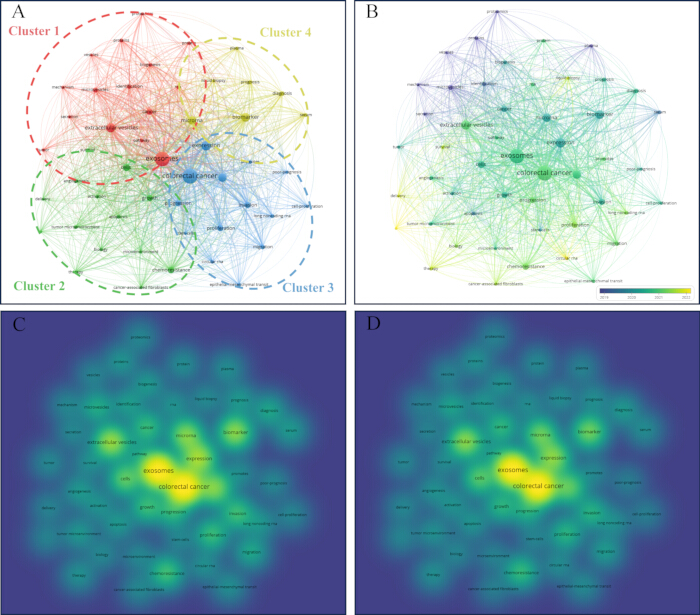
Figure 5: Spatial and temporal networks of research hotspots of exosomes in colorectal cancer. (A) Clustering analysis of research hotspots. The color indicates the cluster type; the node size indicates the total link strength. (B) Time series analysis of research hotspots. The color mapping range was based on the distribution of the average publication year across all research topics, with purple representing earlier topics and yellow representing more recent topics. (C) Spatial density network based on total link strength. (D) Spatial density network based on occurrence frequency. Please click here to view a larger version of this figure.
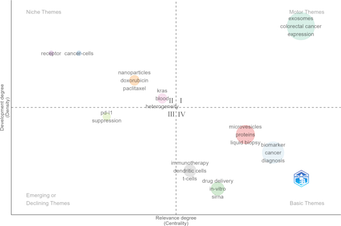
Figure 6: The research prospect discovery for the themes. The horizontal coordinate indicates the degree of relevance to the field. The vertical coordinate reflects its current level of development. Themes in quadrant IV are highly relevant to the field but remain relatively underdeveloped, indicating potential directions for future research. Please click here to view a larger version of this figure.
Table 1: Global and regional colorectal cancer burden in 2021. The table presents colorectal cancer incidence and deaths, corresponding age-standardized rates (ASRs) per 100,000 population, 95% uncertainty intervals (UIs), and percentage changes in ASRs from 1990 to 2021 across Global Burden of Disease regions. Abbreviations: 95% UI = 95% uncertainty intervals; ASRs = age-standardized rates. Please click here to download this table.
Diskussion
The global burden of CRC
Our analysis highlights a clear global shift in the burden of CRC over the past three decades. While the overall ASIR has increased modestly globally, improvements in diagnosis and therapeutic interventions have contributed to a substantial decline in the ASMR. However, these benefits have been unevenly distributed across regions and countries. High-income regions have experienced notable reductions in mortality, likely reflecting the impact of widespread screening programs, early detection, and access to advanced treatment. In contrast, many low- and middle-income regions show marked increases in both incidence and mortality, underscoring emerging public health challenges and inequities in healthcare infrastructure. At the national level, cross-country disparities are pronounced: higher CRC incidence is generally associated with greater social development, as measured by the SDI, whereas mortality patterns show complex relationships with SDI and healthcare access.
Importantly, these epidemiological disparities also provide a context for interpreting and prioritizing future exosome research in CRC. The unmet clinical needs across different SDI settings are not necessarily identical; therefore, the translational priorities of exosome-based technologies may need to be context-specific. In low- and middle-SDI regions, where CRC incidence and mortality are increasing amid limited screening capacity and unequal access to advanced diagnostic and therapeutic resources, exosome research may be particularly valuable for developing scalable, minimally invasive approaches, including liquid biopsy, early detection, and prognostic biomarkers. Such applications could potentially complement conventional screening and facilitate earlier identification of high-risk patients. In contrast, in high-SDI regions, where improvements in screening and treatment have contributed to declining CRC mortality, exosome research may increasingly focus on precision-oriented applications, including prediction of treatment response and resistance, targeted drug delivery, and immunotherapy, to address residual mortality and treatment-refractory disease.
Thus, the GBD analysis and bibliometric analysis provide complementary perspectives. The GBD analysis identifies the geographical and socioeconomic distribution of unmet CRC needs, whereas the bibliometric analysis identifies emerging scientific and technological opportunities within exosome research. Integrating these perspectives suggests that emerging themes identified by the bibliometric analysis may have different levels of translational relevance across SDI settings. This burden-oriented framework may therefore help align future exosome research with region-specific clinical needs and promote more targeted and equitable development of exosome-based diagnostic and therapeutic strategies.
Global research trends and scientific landscapes of exosomes in CRC
This comprehensive visualization analysis delineates the evolution and research landscape of exosome research in CRC. The field has progressed from an early exploration phase to a period of rapid growth, supported by influential authors and journals and characterized by strong interdisciplinary links between molecular biology, genetics, and clinical medicine. Spatially, the global contribution is markedly asymmetric, with China demonstrating overwhelming productivity yet a notably low rate of international collaboration compared to other major contributors such as the USA and Australia. This disparity may reflect not only differences in international collaboration practices but also the strong domestic research networks, national and regional funding schemes, institutional incentives, and research policies that shape patterns of scientific collaboration. Therefore, the relatively low MCP ratio should not be regarded as a direct indicator of insufficient international cooperation or research capacity, but rather as a reflection of the predominantly domestic collaboration structure of research in this field. Nevertheless, expanding strategic international partnerships could facilitate knowledge exchange, access to complementary expertise, and broader dissemination of research findings, thereby potentially enhancing the international visibility and global academic influence of China’s research on exosomes in CRC.
Using VOSviewer-based clustering, the analysis identified four major research domains, ranging from fundamental molecular mechanisms to biomarker applications. The Walktrap analysis further highlighted emerging application-oriented themes, pointing to potential priorities for future research on exosomes in CRC. To further examine the robustness of these thematic findings, the major patterns identified using VOSviewer were compared with the communities generated by Walktrap. Although the two approaches produced different levels of thematic organization, several findings showed correspondence. The domain of exosomal biomarkers with diagnostic applications was consistent with the directions of diagnosis and liquid biopsy, while the domain related to the tumor microenvironment and therapy resistance was conceptually connected with emerging directions such as immunotherapy and drug delivery. Although the two approaches were not expected to generate identical cluster boundaries, their complementary analyses converged on several important research themes and emerging directions, supporting the overall interpretability and robustness of the thematic landscape identified in this study.
Application of exosomes in the diagnosis and treatment
Research on the role of exosomes in disease is still developing, but studies exploring their diagnostic and therapeutic potential have grown rapidly. In general, the sensitivity and specificity of blood-based tumor markers are low, and no marker has 100% tumor specificity. By contrast, exosomes exhibit higher sensitivity and specificity than conventional markers such as carcinoembryonic antigen (CEA) and carbohydrate antigen 19-9 (CA19-9)24. Moreover, because exosomes are stable and widely present in body fluids, they enable convenient and non-invasive detection compared with endoscopic biopsy25. In recent years, the idea of liquid biopsy as an alternative to tissue biopsy has attracted considerable attention26. As emerging liquid biopsy modalities, exosomes hold great promise not only as diagnostic and prognostic biomarkers but also as therapeutic vehicles, bridging experimental research and clinical applications.
A biomarker is a biological molecule that can indicate physiological or pathological changes. In CRC, exosomal cargoes such as long non-coding RNAs (lncRNAs), microRNAs (miRNAs), and circular RNAs (circRNAs) can be used as potential biomarkers. Using exosomal contents offers certain benefits, as the vesicles shield their cargo from RNase degradation, thereby preserving both their integrity and biological activity27. Exosomal lncRNA CRNDE-h is upregulated in CRC patients and significantly correlates with metastasis and reduced overall survival, highlighting its value as a non-invasive biomarker for both diagnosis and prognosis28. Exosomal lncRNA MIR4713HG, which can be packaged into CRC-derived exosomes, promotes CRC cell proliferation and invasion, and its knockdown impairs tumor growth in vivo, highlighting its potential as a liquid biopsy biomarker and a therapeutic target for RNA nanotherapeutics29.
Similar to lncRNAs, certain exosomal miRNAs have been recognized as potential biomarkers for CRC diagnosis and prognosis. For instance, exosomal miR-6803-5p is significantly elevated in the serum of CRC patients, especially in those with advanced stage and metastasis, and is closely associated with poor overall and disease-free survival, highlighting its potential as a diagnostic and prognostic biomarker30. Clinically, increased plasma exosomal miR-106a-5p is associated with liver metastasis and unfavorable prognosis31. Exosomal miR-188-3p is significantly elevated in CRC patients with liver metastasis, correlates with tumor size and advanced stage, and serves as an independent predictor of poor disease-free and overall survival32.
Moreover, CRC patients exhibited high levels of circRNAs in serum-derived exosomes, which were absent in healthy controls27,33. In addition to RNA cargoes, exosomal proteins on the vesicle surface and within vesicles have emerged as potential biomarkers for cancer detection. Exosomes are present in all body fluids, but plasma is generally preferred for detecting most solid tumors34. Plasma-derived exosomal proteins GPC1 and CPNE3 were significantly upregulated in CRC patients, whereas the QSOX1 protein was significantly downregulated, indicating that dysregulated exosomal proteins can serve as promising biomarkers for non-invasive diagnosis in CRC35,36,37. Moreover, plasma exosomal proteins PLG and SERPINA1 have been reported to exhibit superior diagnostic performance compared with CEA and CA19-9 in CRC, with expression levels associated with TNM stage and coagulation abnormalities, indicating their potential as non-invasive biomarkers for early detection38. Furthermore, ADAM17 derived from circulating exosomes of patients with CRC could serve as a blood-based biomarker for predicting metastasis39.
Furthermore, exosomes can also be considered potential therapeutic targets; for example, exosomal lncRNA BANCR derived from SW620 and HCT-15 cells, which promotes CRC metastasis by inducing M2 macrophage polarization, might represent a novel therapeutic target40. Tumor-derived exosomal miR-208b promotes Treg expansion by targeting PDCD4, thereby facilitating tumor growth and chemoresistance in CRC, highlighting its potential as both a predictive biomarker for oxaliplatin-based therapy response and a novel target for immunotherapy41.
Challenges and considerations
The emerging directions identified in our bibliometric analysis, including liquid biopsy, drug delivery, and immunotherapy, should be interpreted as research trends derived from keyword network analysis rather than as evidence of established clinical efficacy or translational maturity. Their identification as emerging or underexplored themes reflects growing research attention and thematic connectivity but does not necessarily mean these approaches are ready for clinical implementation. The transition from promising research signals to clinically applicable technologies requires rigorous validation. The clinical translation of exosome-based therapies faces several critical challenges. First, heterogeneity in origin, size, cargo composition, and isolation procedures results in batch-to-batch variation and raises safety concerns42. Second, exosome yield remains limited and is influenced by donor cell type and the activity of the endosomal sorting complex, highlighting the need to better understand exosome biogenesis and secretion. Third, the lack of standardized, efficient isolation and purification methods hampers reproducibility, as current techniques cannot achieve an ideal balance among high yield, purity, scalability, and cost-effectiveness. Fourth, drug-loading efficiency varies across methods, necessitating further optimization with exosomes from diverse cellular sources. Fifthly, large-scale production of good manufacturing practice (GMP) grade exosomes remains insufficient, and novel technologies are required to ensure scalability, reproducibility, safety, and consistent therapeutic potency for clinical applications43. Beyond these technical and regulatory safeguards, ethical issues also demand attention: donor permission, traceability, and long-term risk are particularly relevant when exosomes originate from human tissues such as stem cells or malignant cells, highlighting the need for clear and transparent consent procedures to ensure that donors fully understand how their biological material will be used; moreover, as exosomes may carry sensitive biological information, strict governance and data protection measures are required, and the early involvement of institutional review boards is essential, especially in first-in-human studies with engineered or tumor-derived exosomes34. Collectively, integrating these technical, regulatory, and ethical measures into development pathways is necessary to minimize risk and to enable the responsible clinical translation of exosome-based interventions.
Future direction
As research on exosomes in CRC continues to evolve, multiple promising avenues for clinical translation are emerging. Among these, liquid biopsy represents one of the most attractive directions. Tissue biopsy has long been regarded as the gold standard for cancer diagnosis. However, its invasiveness poses potential risks, and due to tumor heterogeneity, it provides only site-specific information rather than a comprehensive assessment of the disease. Compared with tissue biopsy, liquid biopsy offers key advantages, including minimally invasive procedures, early diagnosis, and dynamic monitoring44. Circulating tumor cells (CTCs), circulating tumor DNA (ctDNA), and exosomes are three major categories of liquid biopsy for cancer45. Compared with CTCs and ctDNA, exosome-based liquid biopsy offers distinct advantages. Exosomes deliver diverse nucleic acids and proteins that provide comprehensive biological information, occur in higher abundance in clinical samples, and their lipid bilayer structure protects cargo from degradation, ensuring reliable downstream analysis46. Recent advances in biomedical engineering and materials science have provided a solid foundation for the development of novel technologies. Various exosome isolation systems, such as microfluidic devices and automated technology, have emerged in recent years47. As a result, exosome-based liquid biopsy strategies are increasingly regarded as a promising direction for future research and clinical application. Furthermore, a deeper understanding of exosome biology may not only reshape cancer research strategies but also open new avenues for therapies that target the complex interplay between tumor cells and their surrounding microenvironment.
Exosome-mediated drug delivery has been proposed as a promising strategy for cancer treatment. Exosomes have already been successfully engineered to deliver conventional chemotherapeutics such as doxorubicin and paclitaxel48,49, as well as gene-silencing agents such as PD-L1 small interfering RNA (siRNA) and CTLA-4 siRNA, to exert anticancer effects in CRC50. Compared with artificial nanocarriers, exosomes offer advantages including high biocompatibility, low immunogenicity, ability to cross biological barriers, minimal toxicity, long-distance transport, and selective accumulation in target tissues, suggesting that exosome-based delivery systems may become valuable tools in future clinical applications51. Moreover, by serving as versatile carriers of both chemotherapeutics and genetic materials, exosomes have the potential to enhance therapeutic efficacy while minimizing side effects, and, when integrated with personalized medicine, they are expected to further refine cancer treatment toward more efficient, individualized strategies.
Beyond drug delivery, exosome-based vaccines have emerged as a promising immunotherapeutic strategy, as both tumor-derived and immune cell-derived exosomes can stimulate antitumor immune responses by presenting major histocompatibility complex molecules, costimulatory proteins, and other bioactive cargos52. For example, exosomes derived from embryonic stem cells engineered to produce granulocyte-macrophage colony-stimulating factor (GM-CSF) have been shown to act as effective prophylactic vaccines, eliciting robust antitumor immune responses and significantly suppressing tumor growth in preclinical models53. Early clinical trials using dendritic cell-derived exosomes have shown favorable safety profiles and the capacity to induce tumor-specific immune responses54. Relative to earlier treatment approaches, dendritic cell-derived exosome immunotherapy provides a more targeted and precise antitumor immune response. In addition, it shows advantages in cost-effectiveness, stability, and accessibility42. Although current clinical evidence remains limited and further research is needed to optimize large-scale production and enhance biocompatibility, exosome-based vaccines hold significant promise for future CRC immunotherapy.
Collectively, advancing exosome research in CRC will require close collaboration among basic scientists, clinicians, bioengineers, and industry partners. Standardized methodologies, reference materials, and shared data platforms are essential for improving reproducibility, accelerating translation, and facilitating the development of safe, effective, and clinically implementable exosome-based diagnostics and therapies. Continued multidisciplinary efforts will be key to unlocking the full potential of exosomes for early detection, precise treatment, and personalized immunotherapeutic strategies in CRC.
Strengths and limitations
This study provides a comprehensive overview of the research landscape on exosomes in CRC, identifying hotspots and highlighting emerging trends. These findings contribute to a clearer understanding of current progress and potential directions in the field. Nonetheless, some limitations should be acknowledged. First, only English-language publications from the Web of Science Core Collection were included, which may introduce language and database selection biases and potentially underrepresent relevant research. Second, review articles were excluded to maintain a relatively homogeneous dataset focused on original research and to minimize potential distortion in citation-based analyses. However, this may have resulted in the omission of some influential conceptual contributions. Third, as a bibliometric study, this work maps the existing published knowledge landscape but cannot independently validate the biological functions or clinical efficacy of exosomes in CRC. Despite these limitations, this study offers valuable insights into the status, trends, and future directions of exosome research in CRC.
Conclusion
This study mapped the global research landscape on exosomes in CRC, revealing both well-established findings and emerging areas with high translational potential. The results underscore the growing significance of exosomes in the development of non-invasive diagnostics and the advancement of therapeutic strategies. Continued multidisciplinary efforts in this field are likely to accelerate the translation of exosome research into clinical applications, ultimately improving precision medicine and patient outcomes in CRC.
Offenlegungen
The authors have no conflicts of interest to declare.
Danksagungen
This work was supported by the National Natural Science Foundation of China (82500640 and 82260807).
Materialien
| Name | Unternehmen | Katalognummer | Kommentare |
|---|---|---|---|
| CiteSpace | Chaomei Chen | Version 6.3.R1 | Bibliometric and knowledge mapping analysis |
| Microsoft Excel | Microsoft Corporation, USA | Microsoft Office 2019 | Data analysis and visualization |
| Python | Python Software Foundation | Version 3.12.4 | Data analysis and visualization |
| R | R Core Team | Version 4.5.1 | Data analysis and visualization |
| VOSviewer | Centre for Science and Technology Studies, Leiden University | Version 1.6.20 | Bibliometric and knowledge mapping analysis |
Referenzen
- Bray F, et al. Global cancer statistics 2022: GLOBOCAN estimates of incidence and mortality worldwide for 36 cancers in 185 countries. CA Cancer J Clin. 2024;74(3):229-63. doi:10.3322/caac.21834.
- Morgan E, et al. Global burden of colorectal cancer in 2020 and 2040: incidence and mortality estimates from GLOBOCAN. Gut. 2023;72(2):338-44. doi:10.1136/gutjnl-2022-327736.
- Guo L, et al. Global distribution of colorectal cancer staging at diagnosis: an evidence synthesis. Clin Gastroenterol Hepatol. 2025;23(12):2088-101. doi:10.1016/j.cgh.2024.11.019.
- Zhou H, et al. Colorectal liver metastasis: molecular mechanism and interventional therapy. Signal Transduct Target Ther. 2022;7(1):70. doi:10.1038/s41392-022-00922-2.
- Shin AE, Giancotti FG, Rustgi AK. Metastatic colorectal cancer: mechanisms and emerging therapeutics. Trends Pharmacol Sci. 2023;44(4):222-36. doi:10.1016/j.tips.2023.01.003.
- Ghofrani-Shahpar M, et al. Cancer-associated fibroblasts drive colorectal cancer cell progression through exosomal miR-20a-5p-mediated targeting of PTEN and stimulating interleukin-6 production. BMC Cancer. 2024;24(1):400. doi:10.1186/s12885-024-12190-0.
- Abdul Pari AA, Singhal M, Augustin HG. Emerging paradigms in metastasis research. J Exp Med. 2021;218(1):e20190218. doi:10.1084/jem.20190218.
- Cheng B, Yu Q, Wang W. Intimate communications within the tumor microenvironment: stromal factors function as an orchestra. J Biomed Sci. 2023;30(1):1. doi:10.1186/s12929-022-00894-z.
- Yue M, et al. Extracellular vesicles remodel tumor environment for cancer immunotherapy. Mol Cancer. 2023;22(1):203. doi:10.1186/s12943-023-01898-5.
- Liu Y, Wang Y, Lv Q, Li X. Exosomes: from garbage bins to translational medicine. Int J Pharm. 2020;583:119333. doi:10.1016/j.ijpharm.2020.119333.
- Kalluri R, LeBleu VS. The biology, function, and biomedical applications of exosomes. Science. 2020;367(6478):eaau6977. doi:10.1126/science.aau6977.
- van Niel G, D'Angelo G, Raposo G. Shedding light on the cell biology of extracellular vesicles. Nat Rev Mol Cell Biol. 2018;19(4):213-28. doi:10.1038/nrm.2017.125.
- Zhu L, et al. Isolation and characterization of exosomes for cancer research. J Hematol Oncol. 2020;13(1):152. doi:10.1186/s13045-020-00987-y.
- Miron RJ, Zhang Y. Understanding exosomes: part 1-characterization, quantification and isolation techniques. Periodontol 2000. 2024;94(1):231-56. doi:10.1111/prd.12520.
- Yimin E, et al. Function and mechanism of exosomes derived from different cells as communication mediators in colorectal cancer metastasis. iScience. 2024;27(4):109350. doi:10.1016/j.isci.2024.109350.
- Zhang Y, et al. Exosomes in tumor-stroma crosstalk: shaping the immune microenvironment in colorectal cancer. FASEB J. 2024;38(6):e23548. doi:10.1096/fj.202302297R.
- Ghosh S, et al. Harnessing exosomes as cancer biomarkers in clinical oncology. Cancer Cell Int. 2024;24(1):278. doi:10.1186/s12935-024-03464-5.
- Guo SB, et al. Artificial intelligence AlphaFold model for molecular biology and drug discovery: a machine-learning-driven informatics investigation. Mol Cancer. 2024;23(1):223. doi:10.1186/s12943-024-02140-6.
- Yu C, et al. Research trends, hotspots and future directions of tertiary lymphoid structures in cancer: a comprehensive informatics analysis and visualization study. Discov Oncol. 2024;15(1):665. doi:10.1007/s12672-024-01556-2.
- Guo SB, et al. Comparative investigation of neoadjuvant immunotherapy versus adjuvant immunotherapy in perioperative patients with cancer: a global-scale, cross-sectional, and large-sample informatics study. Int J Surg. 2024;110(8):4660-71. doi:10.1097/JS9.0000000000001479.
- GBD 2021 Risk Factors Collaborators. Global burden and strength of evidence for 88 risk factors in 204 countries and 811 subnational locations, 1990-2021: a systematic analysis for the Global Burden of Disease Study 2021. Lancet. 2024;403(10440):2162-203. doi:10.1016/S0140-6736(24)00933-4.
- GBD 2019 Diseases and Injuries Collaborators. Global burden of 369 diseases and injuries in 204 countries and territories, 1990-2019: a systematic analysis for the Global Burden of Disease Study 2019. Lancet. 2020;396(10258):1204-22. doi:10.1016/S0140-6736(20)30925-9.
- Guo SB, et al. Comprehensive scientometrics and visualization study profiles lymphoma metabolism and identifies its significant research signatures. Front Endocrinol (Lausanne). 2023;14:1266721. doi:10.3389/fendo.2023.1266721.
- Baassiri A, et al. Exosomal non coding RNA in liquid biopsies as a promising biomarker for colorectal cancer. Int J Mol Sci. 2020;21(4):1398. doi:10.3390/ijms21041398.
- Tang XH, et al. Exosome-derived noncoding RNAs in gastric cancer: functions and clinical applications. Mol Cancer. 2021;20(1):99. doi:10.1186/s12943-021-01396-6.
- De Rubis G, Rajeev Krishnan S, Bebawy M. Liquid biopsies in cancer diagnosis, monitoring, and prognosis. Trends Pharmacol Sci. 2019;40(3):172-86. doi:10.1016/j.tips.2019.01.006.
- Shakerian N, et al. Therapeutic and diagnostic applications of exosomes in colorectal cancer. Med Oncol. 2024;41(8):203. doi:10.1007/s12032-024-02440-3.
- Liu T, et al. Exosomal long noncoding RNA CRNDE-h as a novel serum-based biomarker for diagnosis and prognosis of colorectal cancer. Oncotarget. 2016;7(51):85551-63. doi:10.18632/oncotarget.13465.
- Zhou C, et al. Novel exosome-associated lncRNA model predicts colorectal cancer prognosis and drug response. Hereditas. 2025;162(1):79. doi:10.1186/s41065-025-00445-0.
- Yan S, et al. Exosomal miR-6803-5p as potential diagnostic and prognostic marker in colorectal cancer. J Cell Biochem. 2018;119(5):4113-9. doi:10.1002/jcb.26609.
- Liang Y, et al. Exosomal miR-106a-5p from highly metastatic colorectal cancer cells drives liver metastasis by inducing macrophage M2 polarization in the tumor microenvironment. J Exp Clin Cancer Res. 2024;43(1):281. doi:10.1186/s13046-024-03204-7.
- Li T, et al. Colorectal cancer cells-derived exosomal miR-188-3p promotes liver metastasis by creating a pre-metastatic niche via activation of hepatic stellate cells. J Transl Med. 2025;23(1):369. doi:10.1186/s12967-025-06334-4.
- Qiao XX, Shi HB, Xiao L. Serum exosomal hsa-circ-0004771 modulates the resistance of colorectal cancer to 5-fluorouracil via regulating miR-653/ZEB2 signaling pathway. Cancer Cell Int. 2023;23(1):243. doi:10.1186/s12935-023-03072-9.
- Javdani-Mallak A, Mowla SJ, Alibolandi M. Tumor-derived exosomes and their application in cancer treatment. J Transl Med. 2025;23(1):751. doi:10.1186/s12967-025-06814-7.
- Li J, et al. GPC1 exosome and its regulatory miRNAs are specific markers for the detection and target therapy of colorectal cancer. J Cell Mol Med. 2017;21(5):838-47. doi:10.1111/jcmm.12941.
- Sun B, et al. Circulating exosomal CPNE3 as a diagnostic and prognostic biomarker for colorectal cancer. J Cell Physiol. 2019;234(2):1416-25. doi:10.1002/jcp.26936.
- Ganig N, et al. Proteomic analyses of fibroblast- and serum-derived exosomes identify QSOX1 as a marker for non-invasive detection of colorectal cancer. Cancers (Basel). 2021;13(6):1351. doi:10.3390/cancers13061351.
- Li L, et al. Plasma exosomal protein PLG and SERPINA1 in colorectal cancer diagnosis and coagulation abnormalities. J Cancer Res Clin Oncol. 2023;149(11):8507-19. doi:10.1007/s00432-023-04776-1.
- Li K, et al. Tumor-derived exosomal ADAM17 promotes pre-metastatic niche formation by enhancing vascular permeability in colorectal cancer. J Exp Clin Cancer Res. 2024;43(1):59. doi:10.1186/s13046-024-02991-3.
- Ding AX, et al. lncRNA BANCR promotes the colorectal cancer metastasis through accelerating exosomes-mediated M2 macrophage polarization via regulating RhoA/ROCK signaling. Mol Cell Biochem. 2024;479(1):13-27. doi:10.1007/s12010-023-04709-z.
- Ning T, et al. Exosomal miR-208b related with oxaliplatin resistance promotes Treg expansion in colorectal cancer. Mol Ther. 2021;29(9):2723-36. doi:10.1016/j.ymthe.2021.04.028.
- Jangam TC, et al. Exosomes as therapeutic and diagnostic tools: advances, challenges, and future directions. Cell Biochem Biophys. 2025;83(3):2805-29. doi:10.1007/s12013-025-01730-5.
- Thakur A, Rai D. Global requirements for manufacturing and validation of clinical grade extracellular vesicles. J Liq Biopsy. 2024;6:100278. doi:10.1016/j.jlb.2024.100278.
- Heitzer E, Haque IS, Roberts CES, Speicher MR. Current and future perspectives of liquid biopsies in genomics-driven oncology. Nat Rev Genet. 2019;20(2):71-88. doi:10.1038/s41576-018-0071-5.
- Yu W, et al. Exosome-based liquid biopsies in cancer: opportunities and challenges. Ann Oncol. 2021;32(4):466-77. doi:10.1016/j.annonc.2021.01.074.
- Zhou B, et al. Application of exosomes as liquid biopsy in clinical diagnosis. Signal Transduct Target Ther. 2020;5(1):144. doi:10.1038/s41392-020-00258-9.
- Tang H, et al. The new advance of exosome-based liquid biopsy for cancer diagnosis. J Nanobiotechnology. 2024;22(1):610. doi:10.1186/s12951-024-02863-0.
- Hosseini NF, et al. AS1411 aptamer-functionalized exosomes in the targeted delivery of doxorubicin in fighting colorectal cancer. Biomed Pharmacother. 2022;155:113690. doi:10.1016/j.biopha.2022.113690.
- Wang X, et al. Enhanced therapeutic potential of hybrid exosomes loaded with paclitaxel for cancer therapy. Int J Mol Sci. 2024;25(7):3645. doi:10.3390/ijms25073645.
- Li J, et al. Exosomes-delivered PD-L1 siRNA and CTLA-4 siRNA protect against growth and tumor immune escape in colorectal cancer. Genomics. 2023;115(4):110646. doi:10.1016/j.ygeno.2023.110646.
- Li J, et al. A comprehensive review on the composition, biogenesis, purification, and multifunctional role of exosome as delivery vehicles for cancer therapy. Biomed Pharmacother. 2023;165:115087. doi:10.1016/j.biopha.2023.115087.
- Xu Z, Zeng S, Gong Z, Yan Y. Exosome-based immunotherapy: a promising approach for cancer treatment. Mol Cancer. 2020;19(1):160. doi:10.1186/s12943-020-01278-3.
- Yaddanapudi K, et al. Exosomes from GM-CSF expressing embryonic stem cells are an effective prophylactic vaccine for cancer prevention. Oncoimmunology. 2019;8(3):1561119. doi:10.1080/2162402X.2018.1561119.
- Pitt JM, et al. Dendritic cell-derived exosomes for cancer therapy. J Clin Invest. 2016;126(4):1224-32. doi:10.1172/JCI81137.