This video demonstrates preparation of a bacteriophage lysate containing transducing particles. A donor strain is infected, plaques form, and the soft agar overlay is processed with chloroform to yield a clarified lysate for downstream analysis.
Se requiere una suscripción a JoVE para ver este contenido. Inicie sesión o comience su prueba gratuita.
Artículo de método
Preparation of Bacteriophage Lysate for Generalized Transduction
176 visualizaciones
⸱
31 de julio de 2026
En este artículo
Resumen
Protocolo
- Infection of the donor strain
- Grow the donor strain JW4197 in 5 mL of lysogeny broth (LB) medium supplemented with 10 mM CaCl2 and optionally with Kan (25 µg/mL) to an optical density at 600 nm (OD600) of ~1.0. Measure the OD600 value using a spectrophotometer.
- Make a dilution series of an existing P1 phage stock in the LB medium: recommended dilutions are between 10-3 to 10-7.
- Mix 200 μL of the bacterial suspension and 100 μL of a given phage dilution in a 15 mL centrifuge tube or equivalent. Prepare as many tubes as phage dilutions. Incubate the tubes for 20 min at 37 °C without shaking.
- Add ~3 mL of molten top agar (~50 °C) supplemented with 10 mM CaCl2 to the tubes, mix the contents thoroughly by vortexing the tubes shortly, and pour the mixtures onto prewarmed LB plates to make even layers.
- Incubate the plates overnight at 37 °C.
- Lysate preparation
- The following day choose a plate with a semi-confluent growth of phage plaques. On a semi-confluent plate, approximately half the surface area of the plate is clear (Figure 1).
- Scrape the top agar layer from such a plate using an inoculation loop or a similar tool and place the top agar in a centrifuge tube. Add 1–2 mL of LB and a drop of chloroform and vortex the tube vigorously for ~1 min. Add the chloroform in a fume hood.
- Centrifuge the tube for 15 min at 4,000 x g or faster to pellet the agar and bacterial cells.
- Move the supernatant to a fresh microcentrifuge tube, avoiding carrying over any debris from the pellet. Add 2 drops of chloroform and store the lysate at 4–10 °C. Add chloroform in a fume hood. Do not freeze the phage lysate as this will result in a significant reduction of the number of infectious particles.
Acceso restringido. Inicie sesión o comience una prueba gratuita para ver este contenido.
Resultados
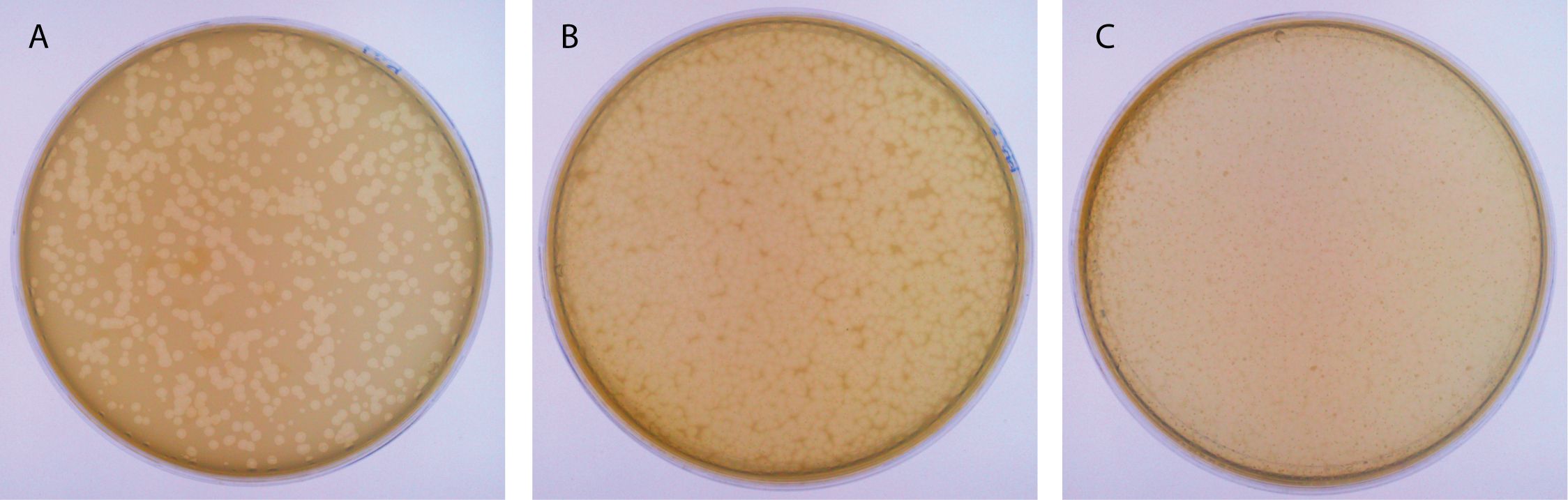
Figure 1: Examples of plates after a P1 infection. (A) This panel shows a plate with individual plaques. (B) This panel shows a semi-confluent plate. (C) This panel shows an over-infected plate...
Acceso restringido. Inicie sesión o comience una prueba gratuita para ver este contenido.
Materiales
| Nombre | Empresa | Número de catálogo | Comentarios |
|---|---|---|---|
| Strains | |||
| E. coli JW4179 | NIG | JW4179-KC | tamA deletion mutant |
| P1 vir | NIG | HR16 | Generally transducing bacteriophage |
| Agar | BD Bacto | 214010 | |
| Calcium chloride | Merck | 102382 | |
| Chloroform | Merck | 102445 |
