Se requiere una suscripción a JoVE para ver este contenido. Inicie sesión o comience su prueba gratuita.

Artículo de método

Immunofluorescence Assay to Detect Hepatitis C Virus Replication Complexes in Infected Cells

156 visualizaciones

31 de julio de 2026

En este artículo

Resumen

Source: Ren, S., et al. A Protocol for Analyzing Hepatitis C Virus Replication. J. Vis. Exp. (2014).

This video demonstrates an immunofluorescence-based method to detect Hepatitis C virus replication in infected hepatoma cells. The protocol involves cell fixation, antibody staining for viral protein and double-stranded RNA, and visualization using fluorophore-tagged secondary antibodies. Dual signal detection under a fluorescence microscope confirms active viral replication within the host cells.
Protocolo

1. Immunofluorescence Assay (IFA)

  1. Fix the Hepatitis C virus (HCV)-infected and transfected cells using methanol for 30 min at -20 ºC for the immunofluorescence assay.

  2. Wash the cell with phosphate-buffered saline (PBS) three times and block with IFA blocking buffer.

  3. Use rabbit polyclonal anti-NS5A primary antibody or mouse monoclonal anti-double-stranded RNA (dsRNA) antibody J2 at a dilution of 1:200 (1 μg/ml) and incubate for 5 hr to overnight at 4 ºC.

  4. Wash the cells with PBS three times after the primary antibody.

  5. Add goat anti-rabbit Immunoglobulin G (IgG)-488 polyclonal secondary antibody or goat anti-mouse IgG-594 polyclonal secondary antibody at a 1:1,000 dilution (1 μg/ml) and incubate for 1 hr at room temperature.

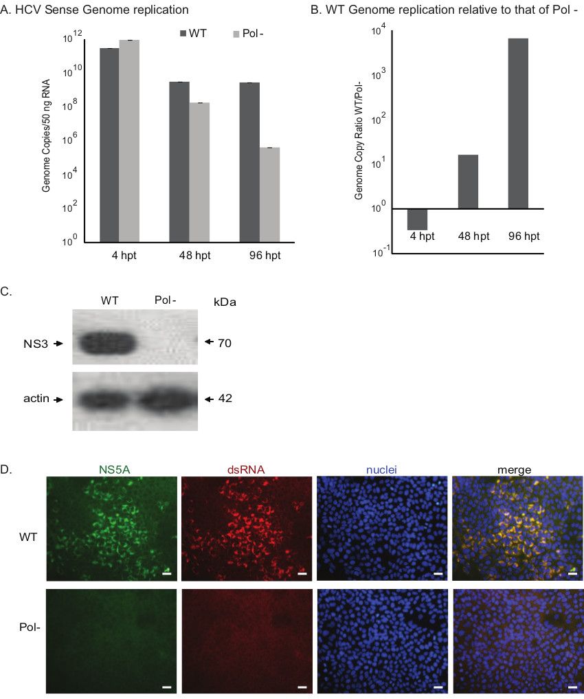
  6. Wash cells with PBS three times and stain nuclei using Hoechst dye and view using a fluorescence microscope (Figure 1D).

Acceso restringido. Inicie sesión o comience una prueba gratuita para ver este contenido.

Resultados

figure-results-1

Figure 1. Assessing the growth phenotypes of HCV constructs. A) Evaluating the genome replication kinetics of wild-type (WT) and polymerase null (Pol-) viruses. The genome copy numbers of the sense RNA strand assessed by RT-qPCR are present...

Acceso restringido. Inicie sesión o comience una prueba gratuita para ver este contenido.

Materiales

Lista de materiales utilizados en este artículo
NombreEmpresaNúmero de catálogoComentarios
Dulbecco’s modified Eagle’s medium (DMEM)Fisher Scientific10-017-CV 
Non essential amino acidFisher ScientificMT25025CI 
HEPESLife Technologies15630080 
GlutamaxLife Technologies35050061 
Opti-MEM Reduced Serum Medium,no Phenol RedLife Technologies11058-021 
Huh-7.5.1The Scripps Research Institute The cell line was kindly provided by Dr. Francis Chisari to Dr. Arumugaswami under executed MTA between The Scripps Research Institute and Cedars-Sinai Medical Center
mouse monoclonal anti-dsRNA antibody J2English & Scientific Consulting Kft.10010200 
Goat anti-rabbit IgG Alexa Fluor 488Life TechnologiesA11008 
Goat anti-rabbit IgG Alexa Fluor 594Life TechnologiesA11020 
Blotting Grade Blocker Non Fat Dry MilkBio-Rad170-6404XTU 
Tween-20Bio-Rad170-6531XTU 
Anti-Hepatitis C Virus NS3 antibody [8 G-2]Abcamab65407 
Anti-Hepatitis C Virus NS3 antibody [H23]Abcamab13830 

Etiquetas

Replicación del VHCcélulas de hepatoma infectadasARN de doble cadenatinción con anticuerposmicroscopía de fluorescenciadetección de proteínas viralesfijación celularanticuerpos secundarios