This video demonstrates the assessment of viral infectivity using reporter epithelial cells engineered with a β-galactosidase gene under a viral protein-sensitive promoter. Following infection with different viral dilutions, the infected host cells express β-galactosidase, which is detected using a chromogenic substrate. The resulting stained cells are then counted to quantify infective virion particles across varying virus concentrations.
Artículo de método
Assessing Viral Infectivity Using a β-Galactosidase-Based Assay
11 visualizaciones
⸱
29 de octubre de 2026
En este artículo
Resumen
Protocolo
- Quantitation of virus concentration and virion infectivity
- Count and seed 0.1 x 10⁶ TZM-bl cells in a 24-well plate using 500 µL of complete Dulbecco's Modified Eagle Medium (DMEM) for each well and keep them in a cell culture incubator for 10-12 h.
- Make sure the cells are 50%-60% confluent before beginning with infection for virus quantitation. Change the existing media with 250 µL of fresh complete DMEM to each well.
NOTE: Keep a positive control well to rule out any experimental fault. - Calculate the amount of stock virus needed for infections of 1 ng, 0.1 ng, and 0.01 ng in duplicates. Add the appropriate amount of virus to the wells and keep the plate in the incubator at 37 °C for 4 h.
- Wash the cells twice with 500 µL of incomplete DMEM and then add 500 µL of complete DMEM.
Proceed with the β-gal staining procedure after 36 h of media change.
- Discard the existing media and wash the cells twice with 1 mL of Phosphate-Buffered Saline (PBS). Fix the cells by adding 500 µL of the fixing solution (0.25% Glutaraldehyde in PBS) to each well and incubate at room temperature (RT) inside the biosafety cabinet for 5-7 min.
- Prepare the staining solution as given in Table 1. Keep it away from light, as X-gal is light-sensitive.
- Wash the cells once with 1 mL of PBS. Overlay the cells with 500 µL of freshly prepared staining solution and incubate at 37 °C in the dark for 2-18 h.
- To stop the reaction afterward, rinse the cells with 500 µL of 3% dimethyl sulfoxide (DMSO) in PBS twice and then keep the cells in 500 µL of fresh PBS.
- Count the blue infected cells (as shown in Figure 1A) under the microscope at 10x magnification for 5 random fields. Take an average count of the 5 fields and multiply it by the field factor and dilution factor. This quantifies the infective virion particles in the virus stock.
NOTE: For a 24-well plate, the field factor is 75.
Table 1: List of components required for β-gal staining in TZM-bl cells.
Components Preparation Required volume PBS Prepare 1x PBS from a stock of 10x PBS 14 mL Potassium Ferri-ferrocyanide Dissolve 0.82 g of Potassium Ferricyanide and 1.06 g of Potassium Ferrocyanide in 25 mL of 1x PBS 0.75 mL Magnesium chloride 1 M in 1 mL of 1x PBS 15 µL X-gal Dissolve 30 mg in 0.6 mL of N, N-dimethyl formamide (in dark) 0.3 mL Total = 15 mL
Resultados
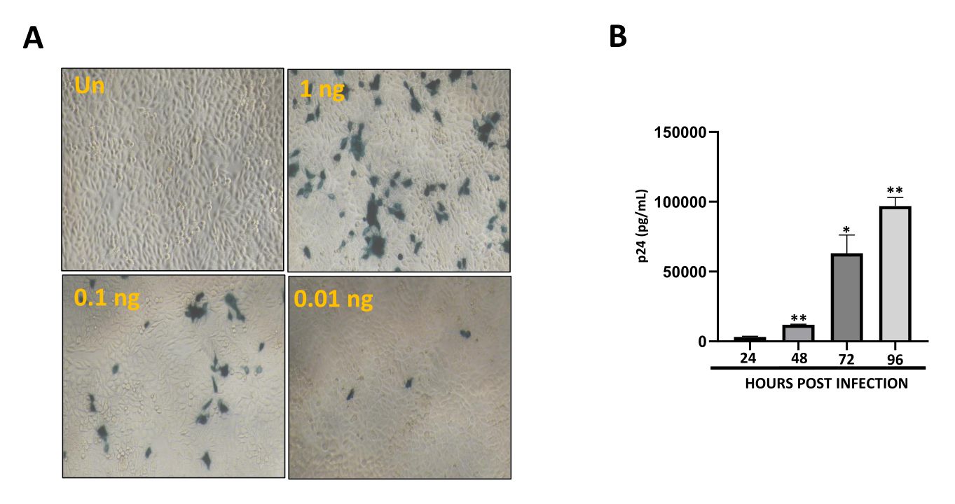
Figure 1: Quantification of virion infectivity and analysis of infection progression. (A) Quantitation of virion infectivity by TZM-bl based β-gal assay. The infectivity of the virus was determined by adding 1 ng, 0.1 ng, and 0.01 ng of the virus to the TZM-bl cells, followed by β-gal staining and counting the number of blue cells. Magnification: 10x. (B) Analysis of infection progression in 0.5 MOI HIV-1 infected CEM-GFP cells based on p24 antigen capture ELISA of the culture supernatants. CEM-GFP cells were infected with 0.5 MOI of HIV-1, and the supernatants were processed for p24 ELISA. The error bars are displayed as mean ± S.E values. Asterisks indicate the significance level, which is indicated in the figures as ns = p ≥ 0.05, * = p ≤ 0.05, ** = p ≤ 0.01, *** = p ≤ 0.001 and ****=p ≤ 0.0001 as analyzed by Student's t-test.
Materiales
| Nombre | Empresa | Número de catálogo | Comentarios |
|---|---|---|---|
| DMSO | Sigma-Aldrich, USA | D2650 | |
| Fetal Bovine Serum | Invitrogen, USA | 16000044 | |
| Glutaraldehyde 25% | Sigma-Aldrich, USA | G6257 | Infectivity assay |
| HEK-293T | NCCS, India | ||
| HIV-1 infectious Molecular Clone pNL4-3 | NIH, AIDS Repository, USA | 114 | |
| Inverted microscope | Nikon, Japan | Model: Eclipse Ti2 | |
| Magnesium chloride | Sigma-Aldrich, USA | M8266 | Infectivity assay |
| Penicillin/Streptomycin | Invitrogen, USA | 151140122 | |
| Potassium ferricyanide | Sigma-Aldrich, USA | 244023 | Infectivity assay |
| Potassium ferrocyanide | Sigma-Aldrich, USA | P3289 | Infectivity assay |
| UltraPure X-gal | Invitrogen, USA | 15520-018 | Infectivity assay |
