Method Article

Individual Lobe Extraction from Prostate: A Technique to Harvest Individual Prostate Lobes from Mouse Prostate

4.4K views

⸱

April 30th, 2023

In This Article

Abstract

Source: Nath, D. et al. Identification, Histological Characterization, and Dissection of Mouse Prostate Lobes for In Vitro 3D Spheroid Culture Models. J. Vis. Exp. (2018)

This video describes a technique to harvest individual lobes from the prostate gland of a mouse model. Individual lobes can then be used for histological analysis and cancer research.

Protocol

All procedures involving animals have been reviewed by the local institutional animal care committee and the JoVE veterinary review board.

1. Gross Anatomy of the Prostate and Individual Lobe Microdissection

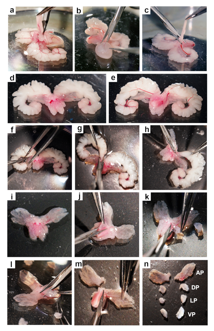
NOTE: This is depicted in Figures 1(i-n) and Figure 2

  1. Flip the tissue with a pair of blunt forceps so that the dorsal side faces up, showing the dorsal lobes, which resemble a butterfly’s wings.
  2. Collect the dorsal lobes by holding the lobe with forceps and snipping at the base with scissors (Figure 1j).
  3. Flip over the remaining tissue to the ventral side.
  4. Collect the lateral lobes, which are small and usually wrap the urethra on the side, which are wedged between the anterior, ventral, and dorsal lobes (Figure 1k).
  5. Next, collect the ventral lobes, which are larger than the lateral lobes and lie on the urethra ventrally (Figure 1l).
  6. Last, harvest the anterior lobes, the largest of the four, by cutting and discarding the urethra (Figure 1m).
  7. Process the tissue pieces according to experimental needs.

Results

Brain dissection process, step-by-step method using tweezers for anatomical study, labeled sections.
Figure 1: Dissection of the mouse prostate. Step-by-step images for the dissection of the mouse prostate from the UGS: (a) removing fat, (b) removing the urinary bladder, (c) removing the vas deferens, (d) ventral view of the prostate with the urethra, (e) dorsal view of the prostate with the urethra, (f-h) removing the seminal vesicles, (i) ventral view of the prostate, (j) dissecting a dorsal lobe, (k) dissecting a lateral lobe, (l) dissecting a ventral lobe, (m) dissecting an anterior lobe, and (n) the dissected prostate lobes: anterior = AP, dorsal = DP, lateral = LP and ventral =VP.
Dorsal and ventral brain anatomy, labeled regions, comparative diagram, educational use.
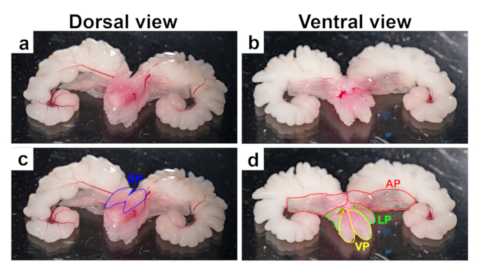
Figure 2: Anatomy of the mouse prostate lobes. Representative images from 3-month-old mouse UGS after removal of the fat, bladder, and vas deferens. Images show prostate lobes with seminal vesicles and urethra, with dorsal (a and c) and ventral (b and d) views. Top panels (a and b) show untouched images, while bottom panels (c and d) show the lobes outlined in blue (dorsal), red (anterior), yellow (ventral), and green (lateral).

Disclosures

No conflicts of interest declared.

Materials

List of materials used in this article
NameCompanyCatalog NumberComments
Mouse surgical instruments (Mouse Dissecting kit)World Precision InstrumentsMOUSEKIT
Dissection microscope
RPMI mediumThermofisher Scientific11875093
Dissection medium (DMEM + 10%FBS)Thermofisher Scientific11965-084
Fetal Bovine SerumThermofisher Scientific10438018
PBS (Phosphate buffered saline)Thermofisher Scientific10010031
CollagenaseThermofisher Scientific17018029Make 10x stock (10mg/ml) in RPMI, filter sterilize, aliquot and store at -20 °C
Syringes and NeedlesFisher Scientific

Explore More Articles

Dissecting MicroscopeTissue ExcisionVentral LobesDorsal LobesLateral LobesAnterior LobesUrogenital System