This video demonstrates a co-culture model using multi-species oral biofilms and human oral epithelium. It highlights epithelial immune activation and enables the downstream analysis of host-pathogen interaction.
Un abonnement JoVE est requis pour visualiser ce contenu. Connectez-vous ou commencez votre période d'essai gratuite.
Article de méthode
A Transwell Tissue Model for Studying Interactions Between Human Oral Biofilms and Epithelium
182 vues
⸱
1 juillet 2026
Dans cet article
Résumé
Protocole
The following protocol involves the preparation of a multi-species biofilm representative of gingivitis, containing a total of 7 species (spp.). Three Streptococcus spp., Streptococcus mitis (NCTC 12261), Streptococcus intermedius (DSM 20753), and Streptococcus oralis (NTCC 11427) are included to mimic oral health, acting as initial colonizers of the salivary pellicle. Four anaerobic microorganisms associated with the shift from oral health to disease are next added: Veillonella dispar (NCTC 11831), Actinomyces naeslundii (DSM 17233), and two Fusobacterium spp. Fusobacterium nucleatum (ATCC 10953) and Fusobacterium nucleatum subspecies (subspp.) vincentii (DSM 19507). All steps involved are conducted aseptically either at the flame or in a class II safety cabinet. All media and phosphate-buffered saline (PBS) used for microbiological preparations are autoclaved prior to use, and sterility is assessed at regular intervals during the protocol. The details of the reagents and the equipment used in this study are listed in the Table of Materials.
1. Preparation of microbial communities for co-culture
NOTE: This protocol depicts the generation of a multi-species biofilm representative of consortia associated with inflammation of the gum tissue, also known as gingivitis.
- Revive all three Streptococcus species on blood agar plates (Columbia blood agar base containing 5% sterile defibrinated horse blood) from frozen stocks of porous beads (commercially obtained) containing the microorganisms stored at -80°C. This is achieved by using an inoculating loop and the streak-plate technique.
- Incubate for 24 h at 37 °C, 5% CO2, then isolate 3-4 colonies for propagation into 10 mL of Tryptone Soya Broth medium. Culture broths for 16-18 h at 37 °C, 5% CO2.
- For the intermediate pathogens, revive V. dispar, A. naeslundii, F. nucleatum, and F. nucleatum subspp. vincentii anaerobically on fastidious anaerobic agar base containing 5% sterile defibrinated horse blood for 48 h at 37 °C, prior to culture in Schaedler's broth for an additional 24-48 h under the same conditions.
- After growth, pellet cell suspensions by centrifugation for 5 min, 20 °C at 3000 x g, then wash pellets in 10 mL of sterile PBS (pH 7.2-7.6). Pellet cell suspensions again via centrifugation for 5 min, 20 °C at 3000 x g, then repeat wash steps for a second time. Resuspend washed cells in 10 mL of sterile PBS for standardization.
- Standardize all three Streptococcus spp. individually using a spectrophotometer at 550 nm. Absorbance values of 0.50 (range from 0.45-0.55, acceptable) are indicative of a cell count of ~1 x 10⁸ cells/mL, as previously determined using the Miles and Misra cell count technique. To achieve this absorbance reading, further dilute 10 mL of washed cell suspensions in sterile PBS.
- Following standardization, dilute all Streptococcus spp. 1:10 to 1 x 10⁷ cells/mL in a 1:1 mix of Todd Hewitt broth (THB) and Roswell Park Memorial Institute (RPMI) medium. Add 500 μL of cell suspension by pipetting to a 24-well microtiter tissue culture plate containing a 13 mm diameter hydroxyapatite disc. Leave biofilms to mature for 24 h at 37 °C, 5% CO2.
NOTE: Multi-species biofilms can be grown on different oral-relevant substrates such as enamel, dentin, and poly(methyl methacrylate) denture surfaces. Alternative media can also be used for these models, such as artificial or synthetic saliva, although careful consideration should be made depending on the consortia of microorganisms used: studies have shown that growth medium selection has important implications for mixed community biofilm growth. - The next day, standardize the four anaerobic microorganisms in a similar manner to the above (steps 1.2-1.4). Pellet cell suspensions, wash twice, then standardize V. dispar to 0.50 absorbance (range from 0.45-0.55) and the three remaining microorganisms to 0.20 absorbance (range from 0.18-0.22). Once standardized, further dilute all suspensions 1:10 to 1 x 10⁷ cells/mL in a 1:1 mix of THB and RPMI.
- Carefully remove non-adhered cells and spent media, and discard them from the Streptococcus biofilms by pipetting. Replace microtiter plate wells with 500 μL of standardized 1 x 10⁷ cells/mL suspensions of the four anaerobes. Culture biofilms for 24 h under anaerobic conditions at 37 °C.
- After 24 h, remove non-adhered cells and spent media from the 7-species biofilms and replace with 500 µL of sterile 1:1 mix of THB: RPMI media. Biofilms are left to mature anaerobically at 37 °C for 4 days, with media removed and replenished on a daily basis (four media changes in total).
- On day 7, the multi-species biofilm is fully mature and ready for downstream experiments. Wash biofilms twice with 500 μL of sterile PBS for use in co-culture.
NOTE: Biofilms can be profiled using a range of biological methodologies such as qPCR (for compositional assessment) and microscopic profiling with confocal or electron microscopy.
2. Organotypic tissue handling and experimental setup
NOTE: The experimental setup described below involves human oral epithelium (HOE) tissue composed of TR146 cells cultivated on an inert polycarbonate membrane filter. Other models exist, including epidermis models, bladder, oesophageal, corneal, gingival, and vaginal epithelium. All models are handled and prepared in a manner similar to the one described below for investigating host-pathogen interactions.
- Upon arrival, unbox and transfer the HOE tissue and media to a class II safety cabinet. Add a total of 1 mL of maintenance media supplied with the tissue to 12-well plates.
- Remove polycarbonate inserts containing the HOE with sterile tweezers from the 24-well plates and nutrient agar used for shipping and transfer them to the 12-well plates containing the media, ensuring no air bubbles remain underneath the tissue. Ensure any excess agar attached to the sides or bottom of the inserts is carefully removed using an additional pair of tweezers or tissue paper.
- Incubate tissue models for 24 h at 37 °C, 5% CO2, prior to experimental setup to acclimatize to laboratory conditions following shipment. It is noteworthy that additional maintenance media or growth media are available for longer maintenance or further maturation of the tissue models.
- Co-culture experiments can now be conducted. For the example provided here, remove the 7-species biofilms created as above (steps 1.1-1.9) from their substrates by sonication. To achieve this, remove HA discs containing biofilms from the bottom of 24-well plates using a 19 G needle and tweezers, then transfer a bijoux containing 1 mL of sterile Dulbecco's PBS. Sonicate at 35 kHz for 10 min in a sonication water bath.
- Carefully remove inserts containing the tissue models using tweezers from the overnight acclimatization, and add 100 µL of biofilm sonicate suspension directly to the tissue by pipetting. Use unstimulated control tissues for comparative purposes. For these control tissue inserts, add 100 µL of sterile Dulbecco's PBS without the biofilm suspension.
NOTE: Planktonic cells, spent biofilm supernatants containing dispersed cells, or whole biofilms can be utilized for the co-culture model in place of biofilm sonicate. Different applications for these host-pathogen models using different microbial stimulants are schematized in Figure 1 as documented elsewhere. - Following the addition, transfer the inserts to another 12-well plate containing 1 mL of fresh maintenance media, again ensuring no air bubbles are present underneath the inserts. Incubate plates containing tissue models for 24 h at 37 °C, 5% CO2, prior to tissue processing for downstream applications.
NOTE: Tissue suppliers can provide additional media to support the continued culture of the tissue following exposure to sonicated aggregates or biofilms. To this end, several previous models investigating prolonged tissue-biofilm inoculation have been published. To achieve similar results using the current organotypic model, sonicate the tissue (as above, step 2.6) and leave the microorganisms to attach for 24 h. Discard any remaining microbial suspension and continue culture at the air-liquid interface for the required experimental time course.
3. Tissue processing for experimental outputs
NOTE: Following co-culture, tissue models are processed for experimental outputs.
- Firstly, prepare 350 µL of RLT lysis buffer in 2.0 mL screw-cap O-Ring tubes containing 1% of β-mercaptoethanol and ~100 µL equivalent of 0.5 mm acid-washed glass beads.
- Remove inserts containing the tissue from the media using tweezers, and discard any remaining microbial suspension from the insert. Next, hold, inverted, at eye level for ease. Using a 19 G needle, carefully slice the tissue and the membrane from the bottom of the insert and transfer to the RLT buffer.
- Homogenize tissue at 30 s using a benchtop bead beater homogenizer.
Accès restreint. Veuillez vous connecter ou commencer un essai pour afficher ce contenu.
Résultats
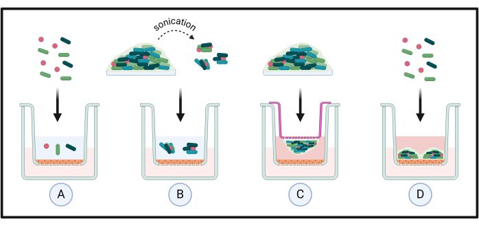
Figure 1: Different applications for organotypic tissue co-culture models. Standardized suspensions of microorganisms are directly applied to the tissue to investigate species-host interactions (A). These are often used for assessing mi...
Accès restreint. Veuillez vous connecter ou commencer un essai pour afficher ce contenu.
Matériaux
| Nom | Entreprise | Numéro de catalogue | Commentaires |
|---|---|---|---|
| 13 mm hydroxyapatite (HA) discs | Plasma Biotal Limited | n/a (made to order) | Substrate for oral biofilm formation, mimicking the tooth surface |
| 19 G needle (40 mm) | VWR | 613-2028 | Used to help with removal of HA discs from plates and to cut out tissue from inserts |
| 24 well TC-treated flat bottom microtitre plates with lids | Scientific Laboratory Supplies | 3524 (pack of 100) | Plates for culturing biofilms and tissue co-culture experiments |
| Acid washed glass beads (425-600 μm) | Merck: Sigma-Aldrich | G8772 | For tissue lysis to improve yield of RNA extraction |
| Actinomyces naeslundii DSM 17233 | Leibniz Institute DSMZ | n/a | One of the microorganisms used for the complex biofilm model |
| Bead Mill 24 Homogenizer (or equivalent) | Fisher Scientific | 15-340-163 | For tissue lysis to improve yield of RNA extraction |
| Bijouxes (7 mL) | Greiner Bio-One | 189170 (700 per bag) | Used for biofilm sonication |
| Columbia Blood Agar (CBA) base | Thermo Fisher Scientific | CM0331B (for 500 grams) | Used to prepare CBA plates for culturing aerobic microorganisms |
| Defibrinated horse blood | E&O Laboratories | DHB | Used to supplement CBA and FAA plates for culturing aerobic and anaerobic microorganisms |
| Dulbecco's phosphate buffered saline (dPBS: without CaCl₂ andMgCl₂) | Merck: Sigma-Aldrich | DB537 | dPBS for cell co-culture with biofilm sonicate |
| Fusobacterium nucleatum ATCC 10953 | American Type Culture Collection | n/a | One of the microorganisms used for the complex biofilm model |
| Fusobacterium nucleatum subspecies (subspp.) vincentii DSM 19507 | Leibniz Institute DSMZ | n/a | One of the microorganisms used for the complex biofilm model |
| GraphPad Prism (Version 10) | GraphPad Software, Inc | n/a | For graph creation and data analyses |
| Human oral epithelium (HOE) | Episkin | n/a (made to order) | The epithelial tissue utilised for the co-culture model system |
| Inoculating loops (disposable, 10 µL, or equivalent) | Fisher Scientific | 12870155 (pack of 1000) | For microbiological culture in solid and liquid media |
| Microbank beads | Pro-Lab Diagnostics | PL.170 | Storage and cyropreservation of microorganisms |
| Petri dishes (90 mm) | Fisher Scientific (Sterilin) | 11309283 (pack of 500) | Used for preparation of agar plates for microbial culture |
| Phosphate-buffered saline (PBS) | Thermo Fisher Scientific | 18912014 (pack of 100 tablets) | PBS for microorganism standardization and biofilm wash steps |
| Roswell Park Memorial Institute 1640 medium | Merck: Sigma-Aldrich | R7755-10L (for 10 L) | Media for biofilm culture |
| Schaedler anaerobe agar base | Merck: Sigma-Aldrich (Millipore) | 91019 (for 500 g) | Used to prepare fastidious anaerobic agar plates |
| Schaedler anaerobe broth | Thermo Fisher Scientific | CM0497B (for 500 g) | Liquid media for growth of anaerobic microorganisms |
| Screw-cap Beadbug O-Ring tube | Scientific Laboratory Supplies | Z763837-1000EA (pack of 1000) | For use in Bead Mill 24 homogenizer, to lyse tissue |
| Sonication bath | Fisher Scientific (Fisherbrand) | n/a | For sonication of biofilms from HA discs |
| Streptococcus intermedius DSM 20753 | Leibniz Institute DSMZ | n/a | One of the microorganisms used for the complex biofilm model |
| Streptococcus mitis NCTC 12261 | National Collection of Type Cultures | n/a | One of the microorganisms used for the complex biofilm model |
| Streptococcus oralis NTCC 11427 | National Collection of Type Cultures | n/a | One of the microorganisms used for the complex biofilm model |
| Todd Hewitt broth | Merck: Sigma-Aldrich | T1438 | Media for biofilm culture |
| Tryptone soya broth (TSB) medium | Merck: Sigma-Aldrich | 22092 (for 500 g) | Liquid media for growth of aerobic microorganisms |
| Tweezers (150 mm, Stainless Steel, Serrated) | RS-Pro | 545-187 | Used to move HA discs and tissue inserts/tissue extracts |
| Universal tubes (30 mL) | Greiner Bio-One | 201150 (400 per bag) | Vessels for culturing microorganisms in liquid culture |
| Veillonella dispar NCTC 11831 | National Collection of Type Cultures | n/a` | One of the microorganisms used for the complex biofilm model |
| β-metacapthoethanol | Merck: Sigma-Aldrich | M3148 | RNase deactivation in tissue samples |
