This video demonstrates the clonal isolation and PCR-based screening of recombinant herpesvirus following site-specific excision of a GFP marker. Fluorescence-activated cell sorting is used to isolate non-fluorescent infected cells, and junction PCR confirms retention of the target gene in GFP-negative clones.
Un abonnement JoVE est requis pour visualiser ce contenu. Connectez-vous ou commencez votre période d'essai gratuite.
Article de méthode
Clonal Isolation and PCR Screening of GFP-Excised Recombinant Herpesvirus Retaining the Target Gene
197 vues
⸱
1 juillet 2026
Dans cet article
Résumé
Protocole
Plaque Purification
- Fluorescence-activated cell sorting
- Seed two 96-well plates with 2 x 10⁴ Chick Embryo Fibroblast (CEF) cells per well the day before sorting.
- 72 h postinfection, prepare the infected cells for sorting. Sort the single nonfluorescence cells into 96-well plates seeded with CEFs.
- Passaging of the recombinant viruses and PCR confirmation
- Choose 5 - 10 single nonfluorescence plaques 5 d postsorting, trypsinize them with 50 µL of trypsin-EDTA at 38.5 °C for 3 min, and add 50 µL of culture medium to resuspend the cells.
NOTE: See Figure 1B for a representative GFP-negative plaque. - Pass half of the cells into each well of a 6-well plate preseeded with CEFs as the second generation.
- Centrifuge the remaining cells of each clone at 200 x g for 5 min, discard the supernatant, and resuspend the cells with 50 µL of squishing buffer for DNA extraction.
- Perform PCR with 5' junction primers using 1 µL of DNA template. For each sample, prepare the following 20 µL reaction mix on ice: 2x PCR Master Mix (10 µL), 10 µM upstream primer (0.5 µL), 10 µM downstream primer (0.5 µL), DNA template (1 µL), and nuclease-free water (8 µL). The amplification program is: 95 °C for 2 min; 95 °C for 30 s, 55 °C for 30 s, and 72 °C for 40 s for 35 cycles; 72 °C for 7 min. Load 2 µL of the amplification products to one well of 1% agarose gel for gel electrophoresis.
NOTE: See Figure 1B for a representative result of 5' junction PCR. - Based on the PCR results, choose three to five positive clones of recombinant HVT for further passages and verification.
- Choose 5 - 10 single nonfluorescence plaques 5 d postsorting, trypsinize them with 50 µL of trypsin-EDTA at 38.5 °C for 3 min, and add 50 µL of culture medium to resuspend the cells.
Accès restreint. Veuillez vous connecter ou commencer un essai pour afficher ce contenu.
Résultats
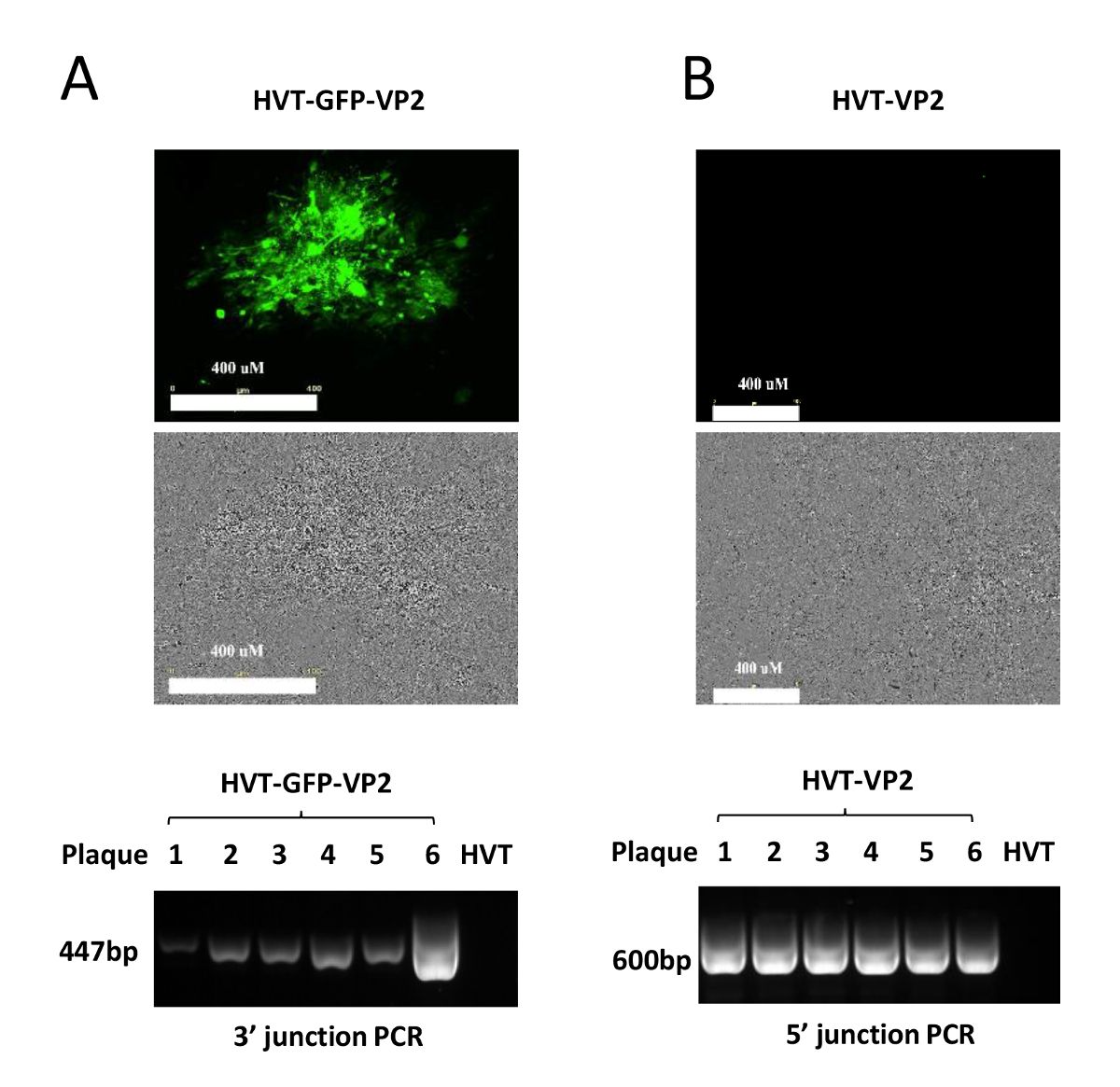
Figure 1: Verification of the recombinant HVT. (A) This panel shows a GFP-positive plaque (HVT-GFP-VP2) visualized under the fluorescence microscope (top panel) and the PCR verification of HVT-GFP-VP2 with primers VP2-F & UL46-R1 for the 3...
Accès restreint. Veuillez vous connecter ou commencer un essai pour afficher ce contenu.
Matériaux
| Nom | Entreprise | Numéro de catalogue | Commentaires |
|---|---|---|---|
| HVT Fc126 strain | Avian Disease and Oncology Laboratory | wildtype HVT virus | |
| GoTaq Master Mixes | Promega | M7123 | junction PCR amplification |
| Platinum Pfx DNA Polymerase | Thermofisher | 11708021 | PCR amplification of full insert |
| Confocal laser scanning microscope | Leica Microsystems | Leica TCS SP5 LASAF | immunoflourescence |
| FACS cell sorter | BD biosciences | BD FACSAria | single cell sorting |
