This video demonstrates the phenotyping of EBV-infected T and NK cell lines using flow cytometry, highlighting the preparation, antibody staining, and analysis steps to identify lineage-specific surface markers. It shows how marker profiles confirm distinct T-cell and NK-cell populations.
Un abonnement JoVE est requis pour visualiser ce contenu. Connectez-vous ou commencez votre période d'essai gratuite.
Article de méthode
Phenotyping EBV-Infected Cell Lines Using Flow Cytometry
316 vues
⸱
30 juin 2026
Dans cet article
Résumé
Protocole
All procedures involving sample collection have been performed in accordance with the institute's IRB guidelines.
1. Cell Phenotyping by Flow Cytometry
- When cells have expanded, collect 1×10⁶ cells to determine the phenotypes of cell lines. Centrifuge cells at 240 x g for 5 min at room temperature (NK/T cells will precipitate at the bottom of the tube).
- Discard the supernatant, wash the precipitation by adding 7 mL PBS. Centrifuge for 5 min at 240 x g at room temperature. Repeat once.
- Add 200 µL human serum to each tube, mix well and incubate for 10 min at room temperature.
- Centrifuge cells at 240 x g for 5 min at room temperature. Discard the supernatant and re-suspend the cells at 1×10⁶/mL with cold PBSA (PBS+0.2%BSA). Divide cells into 5 tubes, each tube containing 2×10⁵ cells.
- Centrifuge cells at 240 x g for 5 min at 4 °C. Discard the supernatant and re-suspend the cells with PBSA buffer or PBSA containing 10 µL PE (or PE-Cy7) and 10 µL FITC labeled antibodies to stain cell receptors of T or NK cells on ice as is indicated by Figure 1. Cells suspended with PBSA buffer are used as a negative control.
- Incubate the cells with antibodies for 20-30 min on ice in the dark.
- Wash the cells twice with cold PBSA, re-suspend the cells with 300 µL cold PBSA.
- Analyze the cell phenotype with a flow cytometer.
- Set up the Flow Cytometer in "Create worksheet" condition. Set up the experimental template with a dot plot that displays forward scatter (FSC) versus side scatter (SSC).
- Load the isotype control tube to optimize the FSC and SSC voltages, and optimize the FSC threshold value to eliminate debris without interfering with the cell population of interest. Delete all parameters except FSC, SSC, FITC, PE and PE-Cy7.
- Perform compensation using the isotype control and a single positive control in each 2-color analysis group.
- Load samples and create HLA-DR VS CD19, CD4 VS CD8, CD56 VS CD16 and CD3 VS CD16 dot plots showing different population of cells.
NOTE: PBMC were used to perform compensation using the negative/isotype control and the single positive control. 2-color immunofluorescence with flow cytometer was used routinely to analyze the expression of surface markers. The following antibodies are included: anti-HLA-DR, anti-CD4, anti-CD16 conjugated with fluorescein isothiocyanate (FITC), anti-CD8, anti-CD56, CD3 conjugated with phycoerythrin (PE), and anti-CD19 conjugated with PE-Cy7.
Accès restreint. Veuillez vous connecter ou commencer un essai pour afficher ce contenu.
Résultats
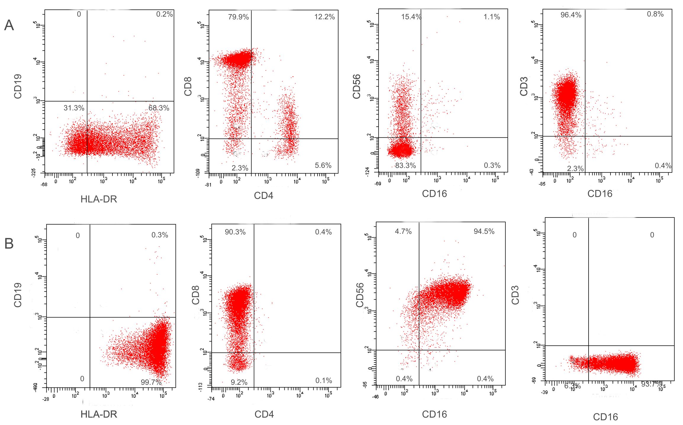
Figure 1: Representative gating strategy for flow cytometry analysis of cell lines L196 and M296. PBMC were used for adjusting FSC and SSC voltages. The cluster of live and single lymphocytes was gated for P1 in the FSC and SSC plot. The i...
Accès restreint. Veuillez vous connecter ou commencer un essai pour afficher ce contenu.
Matériaux
| Nom | Entreprise | Numéro de catalogue | Commentaires |
|---|---|---|---|
| Hu HLA-DR FITC | BD | 560944 | antibody for FACS |
| Hu CD4 FITC | BD | 555346 | antibody for FACS |
| Hu/NHP CD8 PE | BD | 557086 | antibody for FACS |
| Hu CD3 PE | BD | 555340 | antibody for FACS |
| Hu CD16 FITC 3G8 | BD | 555406 | antibody for FACS |
| Hu/NHP CD56 PE MY31 | BD | 556647 | antibody for FACS |
| hu/CD19 PE-Cy7 | BD | 560728 | antibody for FACS |
| human IL-2 | Roche | 11147528001 | |
| human serum | MRC | CCC118-125 | |
| Centrifuge | Techcomp | CT6T | Centrifugation |
| Automated Cell Counter | Countstar | IC 1000 | For cell counting |
| 24 well cell culture cluster | Corning Incorporated | 3524 | Polystyrene plates |
| 25cm2 cell culture flask | Nest | 707001 | Polystyrene |
| FBS | Gibco | 10270 | Component of cell medium |
| RPMI Medium 1640 | life | 22400089 | For cell medium |
| L-Glutamine | Amresco | 374 | Component of cell medium |
| 100 x streptomycin penicillin solution BioRoYeeBRY-2309 | BioRoYee | BRY-2309 | Component of cell medium |
| flow cytometer | BD | BD FACSAria II | |
| soft ware | BD | BD FACSDiva soft ware | FACS analysis |
