This protocol describes a standardized surgical method for inducing chronic constriction injury of the infraorbital nerve in mice, including nerve exposure, placement of a controlled constrictive ligature, wound closure, and postoperative care.
מאמר שיטה
A Surgical Protocol for Infraorbital Nerve Chronic Constriction Injury in Mice
* These authors contributed equally
במאמר זה
סיכום
תקציר
Trigeminal neuropathic pain arises from injury to sensory pathways of the trigeminal system and is associated with persistent changes in neuronal excitability, gene expression, and pain-related behaviors. Infraorbital nerve chronic constriction injury (IoN-CCI) is a widely used model for investigating the cellular and molecular mechanisms underlying chronic orofacial neuropathic pain. This video demonstrates a surgical approach for IoN-CCI in adult mice by placing a single controlled constrictive ligature around the infraorbital nerve while preserving the gross anatomical continuity of the nerve. The protocol includes preoperative preparation, anesthesia and analgesia, extraoral exposure of the infraorbital nerve, placement and tightening of the ligature, wound closure, perioperative care, and postoperative monitoring. The ligature is tightened under magnification until a mild indentation is visible without complete nerve occlusion, transection, or discoloration. Key procedural considerations include accurate nerve identification, consistent ligature placement and tension, and standardized postoperative care. Related IoN-CCI paradigms have been associated with long-lasting mechanical hypersensitivity and molecular changes within trigeminal ganglion neurons. This protocol provides a detailed method for studying mechanisms associated with trigeminal nerve injury and chronic orofacial neuropathic pain.
מבוא
Chronic neuropathic pain arises from injury to peripheral or central components of the somatosensory system and is characterized by persistent alterations in neuronal excitability and sensory processing. Rodent models of nerve injury have been widely used to investigate the mechanisms underlying neuropathic pain. The chronic constriction injury (CCI) model originally described by Bennett and Xie uses loose circumferential ligatures to produce a constrictive nerve injury that preserves the gross anatomical continuity of the nerve while generating persistent pain-like behaviors1. Adaptations of constrictive nerve-injury models to the trigeminal system, including infraorbital nerve chronic constriction injury (IoN-CCI), have enabled investigation of persistent orofacial pain after trigeminal nerve injury2,3,4,5. These models are associated with mechanical hypersensitivity and transcriptional remodeling within trigeminal ganglion neurons2,6,7.
Several infraorbital nerve-injury models have been developed, including proximal IoN-CCI, distal IoN-CCI, partial infraorbital nerve ligation, and trigeminal inflammatory compression (TIC), each of which differs in surgical location, injury mechanism, and expected outcome3,4,5,8,9,10,11. In partial infraorbital nerve ligation, a suture needle is passed under the lateral portion of the nerve and then through its middle so that approximately one-third to one-half of the nerve diameter is tightly ligated5. In contrast, the IoN-CCI procedure described here places a single circumferential 7-0 silk ligature around the intact nerve and tightens it only until a controlled indentation is produced without transection or complete occlusion. Both approaches preserve gross nerve continuity, but they differ in the mechanism used to produce the nerve injury. Behavioral assays such as mechanical stimulation of the whisker pad using von Frey filaments are commonly used to quantify hypersensitivity in these models12,13. However, variability in surgical technique, including the degree of nerve constriction and consistency of nerve exposure, can significantly impact experimental reproducibility and outcome measures3,8,14.
This protocol describes a standardized method for inducing neuropathic pain in mice through chronic constriction injury of the infraorbital nerve. Following surgical exposure of the infraorbital nerve, a single 7-0 silk ligature is placed around the nerve and gradually tightened until a mild, visible indentation is produced without blanching, discoloration, complete occlusion, or transection. This injury induces persistent changes in neuronal excitability and gene expression in trigeminal ganglion neurons, modeling chronic neuropathic pain2,6. Sham procedures involve nerve exposure without placement of the constrictive ligature and are typically performed on the contralateral side of the same animal to enable within-subject comparisons, although this design may not be appropriate for all behavioral assays unless they are conducted unilaterally. This method is well-suited for investigating cellular and molecular mechanisms underlying trigeminal neuropathic pain and for evaluating potential therapeutic strategies6,7.
פרוטוקול
All experiments involving animals were approved by the National Institute of Dental and Craniofacial Research Institutional Animal Care and Use Committee and were performed in accordance with institutional guidelines.
1. Subjects
- Use male and female C57BL/6J mice, 8–10 weeks of age. At this age, females typically weigh approximately 18–22 g and males approximately 23–29 g.
- House mice in standard cages under controlled environmental conditions (approximately 21–23 °C, 40%–60% humidity) with food and water available ad libitum.
- Maintain animals on a 14 h light/10 h dark cycle.
- Allow animals to acclimate to the animal facility housing environment for at least 72 h prior to surgery.
2. Surgery
- Preoperative Preparation
- Before beginning surgery, obtain approval from the appropriate institutional animal care and use committee. Ensure that the approved protocol includes the methods used for initial instrument sterilization and for cleaning, disinfection, and re-sterilization between animals.
- Prepare a clean, uncluttered surgical workspace and assemble sterilized surgical instruments.
- Begin each surgical session with sterilized instruments. Between animals, remove visible organic material from the instrument tips, disinfect them according to the approved institutional procedure, and place the clean, dry working tips in the preheated glass-bead sterilizer for approximately 15 s. Allow the instruments to cool completely before contacting tissue.
- Set a heating pad to 37 °C to maintain body temperature during surgery and post-surgical recovery.
- Prepare the ketamine/xylazine anesthetic working solution by combining 1 mL ketamine (100 mg/mL), 0.5 mL xylazine (20 mg/mL), and 8.5 mL sterile injectable 0.9% saline to yield final concentrations of 10 mg/mL ketamine and 1 mg/mL xylazine. Prepare the ketoprofen analgesic working solution (0.5 mg/mL) in sterile injectable 0.9% saline. Assemble the ophthalmic ointment and all required surgical supplies.
NOTE: Prepare a fresh ketamine/xylazine anesthetic working solution approximately once per month to reduce potential loss of ketamine potency and maintain consistent anesthetic efficacy.
- Anesthesia and Analgesia
- Weigh the mouse and anesthetize it using ketamine (100 mg/kg) and xylazine (10 mg/kg), administered intraperitoneally (IP) at a total injection volume of 10 mL/kg body weight (0.1 mL per 10 g).
CAUTION: Handle anesthetics according to institutional safety guidelines. - Confirm adequate anesthesia by absence of pedal withdrawal and palpebral reflexes before beginning surgery. Continue monitoring respiratory pattern and anesthetic depth throughout the procedure.
- If pedal withdrawal or palpebral reflexes return during surgery, administer one-half of the initial ketamine/xylazine dose intraperitoneally and confirm adequate anesthetic depth before proceeding. Supplemental anesthesia is rarely required when animals are weighed accurately before dosing.
NOTE: Sterile injectable 0.9% saline is used as the diluent for the ketamine/xylazine and ketoprofen working solutions; a separate perioperative saline injection is not routinely administered. If animals appear dehydrated, warmed sterile saline may be administered at the discretion of veterinary staff. The duration of adequate anesthesia may vary among animals; therefore, assess anesthetic depth continuously by monitoring pedal withdrawal and palpebral reflexes rather than relying solely on elapsed time. With accurate weight-based dosing, the approximately 20 min surgical procedure is typically completed without supplemental anesthesia, and recovery from ketamine/xylazine anesthesia generally occurs within approximately 1 h.
- Weigh the mouse and anesthetize it using ketamine (100 mg/kg) and xylazine (10 mg/kg), administered intraperitoneally (IP) at a total injection volume of 10 mL/kg body weight (0.1 mL per 10 g).
- Surgical Site Preparation
- Apply ophthalmic ointment to both eyes to prevent corneal desiccation.
- Remove fur over the whisker pad using depilatory cream (~30 s). Following depilatory cream application, thoroughly remove residual cream using moistened sterile gauze.
CAUTION: Avoid prolonged contact of depilatory cream with the skin, as excessive exposure may cause irritation or tissue damage. - Trim vibrissae only if necessary to improve visualization of the surgical field while minimizing disruption of surrounding tissue.
NOTE: Vibrissae trimming may affect exploratory behavior and whisker-pad sensory testing. Standardize the extent of trimming and the interval between trimming and behavioral assessment across experimental groups. - After fur removal and, if performed, vibrissae trimming, administer ketoprofen (5 mg/kg, subcutaneous).
- Disinfect the surgical site using three alternating applications of povidone-iodine and 70% ethanol, beginning with povidone-iodine and ending with ethanol.
- Transfer the mouse to the prepared surgical area and position it on a heated surgical surface.
- Incision and Exposure
- Position the mouse in lateral recumbency with the surgical site accessible.
- Using fine surgical scissors, make an approximately 3 mm skin incision immediately dorsal to the caudal whisker pad overlying the maxillary bone.
- Using fine forceps, insert the tips of the forceps into the incision and direct them toward the corner of the mouth to access the infraorbital nerve. Using gentle blunt dissection with the forceps or another blunt dissecting instrument, separate the overlying muscle and connective tissue until the main trunk of the infraorbital nerve is exposed between its exit from the skull and its distal branching toward the whisker pad.
- Confirm identification of the infraorbital nerve. The nerve typically appears as a whitish to semi-translucent, cord-like structure with multiple small distal branches extending toward the whisker pad.
NOTE: Excessive tissue manipulation may increase bleeding and make identification of the infraorbital nerve more difficult.
- Nerve Isolation and Infraorbital Nerve Chronic Constriction Injury
- Using fine forceps, gently isolate the infraorbital nerve.
- Avoid damage to surrounding connective tissue and nearby facial nerve branches.
- Perform all steps under magnification using an illuminated magnifier providing approximately 2.3× to 4.0× magnification or an equivalent device to facilitate visualization of the infraorbital nerve and surrounding structures.
- Using fine forceps and curved forceps, pass a 7-0 silk ligature around the infraorbital nerve. Using a two-instrument technique, hold one end of the suture with one forceps, wrap the opposite end once around the second forceps, grasp the free end, and pull it through the loop to form the first throw.
- Tighten the first throw gradually while observing the nerve under magnification until a mild, controlled indentation is visible without blanching, discoloration, complete occlusion, or transection. Repeat the instrument-tying maneuver in the opposite direction to form the second throw and complete the square knot, for a total of two throws. Leave the ligature permanently in place after wound closure.
NOTE: The degree of constriction is a critical determinant of experimental outcome. Excessive tightening may produce axotomy-like injury, whereas insufficient constriction may fail to induce consistent hypersensitivity. As an optional modification, a second loose ligature of nonabsorbable 7-0 silk suture may be placed approximately 1–2 mm from the first ligature. Nonabsorbable silk is used to maintain the ligatures throughout the experimental period, allowing sustained mechanical constriction of the nerve. When two ligatures are used, both should be tied with comparable tension to produce mild nerve constriction without excessive compression.
- Sham Procedure
- For sham surgery, perform an identical exposure and isolation of the infraorbital nerve.
- Do not ligate or transect the nerve.
- Gently reposition the nerve to its original anatomical position.
3. Wound Closure
- Approximate the wound edges using curved forceps and apply a small amount of tissue adhesive to the skin incision. Maintain apposition of the wound edges for approximately 30 s, or until the adhesive has fully set.
- Confirm complete closure of the incision and proper alignment of the wound edges before returning the animal to recovery.
NOTE: The surgical procedure typically requires approximately 20 min per animal from confirmation of adequate anesthetic depth through wound closure, although the duration may vary with operator experience.
4. Postoperative Care
- Place the mouse in a prewarmed recovery cage on a digital warming surface set to 37–37.5 °C. Monitor continuously during recovery. Recovery from ketamine/xylazine anesthesia typically occurs within approximately 1 h, although recovery time may vary depending on individual animal characteristics. Return the mouse to its home cage only after it has regained the righting reflex and begun moving spontaneously.
NOTE: Maintain body temperature throughout recovery to minimize postoperative complications and improve recovery quality. - Do not leave the animal unattended until it has regained sufficient consciousness to maintain normal posture, righting reflexes, and spontaneous movement.
- Following the initial preoperative ketoprofen dose, administer additional ketoprofen doses (5 mg/kg, subcutaneous) at approximately 24 h, 48 h, and 72 h after the initial dose.
- Monitor animals daily for at least 10 days for signs of distress or postoperative complications, including reduced movement, ruffled fur, hunched posture, decreased food or water intake, signs of infection, or incision site dehiscence.
- Provide supportive care, including a hydrogel and a soft diet placed on the cage floor for the first 7 days.
- Consult veterinary staff if any signs of distress or complications are observed.
תוצאות
Figure 1 demonstrates the intended intraoperative appearance and technical implementation of infraorbital nerve chronic constriction injury (IoN-CCI), including nerve exposure, circumferential ligature placement, controlled tightening, and wound closure. Following ligature placement, the infraorbital nerve should remain grossly intact without visible transection while exhibiting a mild, controlled indentation at the constriction site. Blanching, discoloration, and complete occlusion should be absent. These visual criteria provide an intraoperative endpoint for standardizing ligature placement and tension before wound closure. Suboptimal outcomes are most commonly associated with inappropriate ligature tension. Excessive tightening may produce marked flattening, blanching, discoloration, complete occlusion, or axotomy-like injury and may lead to atypical outcomes such as reduced sensory responsiveness. Conversely, insufficient tightening may produce little or no visible indentation and may fail to generate a consistent injury phenotype. Careful control of ligature tension and continuous visualization of the nerve during knot tightening are, therefore, critical for reproducible implementation of the procedure.
Sham-operated animals undergo the same surgical exposure and nerve isolation without placement of the ligature, allowing the effects of nerve constriction to be distinguished from those of surgical manipulation alone. Histological confirmation was not routinely performed in this protocol; therefore, technical implementation was assessed using the intraoperative visual endpoint, confirmation that the nerve remained grossly intact, and absence of visible transection or discoloration. Previous studies using IoN-CCI have reported increased mechanical sensitivity in the ipsilateral whisker-pad region within 3–7 days after surgery, with hypersensitivity persisting for approximately 2–3 weeks3,4,8,9. These previously reported behavioral outcomes represent the expected phenotype following successful implementation of the procedure.
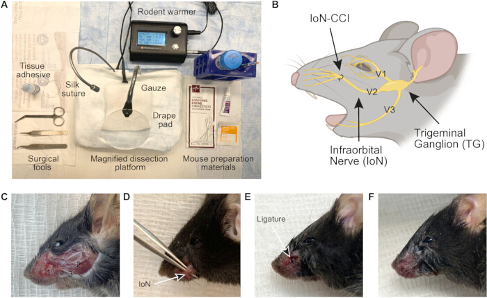
Figure 1. Surgical setup and anatomical overview of infraorbital nerve chronic constriction injury (IoN-CCI) in mice. (A) Surgical setup for infraorbital nerve exposure and ligation. (B) Diagram illustrates the trigeminal sensory pathway, including the trigeminal ganglion (TG), the ophthalmic (V1), maxillary (V2), and mandibular (V3) divisions of the trigeminal nerve. The infraorbital nerve (IoN), a major branch of the maxillary division (V2), is shown with the ligation site used for infraorbital nerve chronic constriction injury (IoN-CCI). Created in BioRender.com. (C) Representative anatomical image of the surgical field showing the infraorbital nerve and adjacent facial nerve branches in a C57BL/6J mouse. Labels indicate 1, infraorbital nerve; 2, buccal nerve; and 3, marginal mandibular nerve. (D) Representative image of the exposed infraorbital nerve (IoN) prior to ligation. (E) Representative appearance of the ligated infraorbital nerve showing an estimated approximately 25% reduction in apparent nerve diameter, with the nerve remaining grossly intact and no blanching, discoloration, complete occlusion, or visible transection. The degree of constriction was estimated visually and was not measured quantitatively. (F) Representative appearance of the surgical site following wound closure with tissue adhesive. Proper apposition of the wound edges is maintained without evidence of dehiscence or excessive tissue disruption. Please click here to view a larger version of this figure.
דיון
The IoN-CCI model provides a reproducible approach for inducing trigeminal neuropathic pain while preserving the gross anatomical continuity of the nerve, allowing investigation of persistent changes in sensory processing3,4,5,14. A critical step in this protocol is the placement and tightening of the ligature around the infraorbital nerve. The degree of constriction must be carefully controlled to achieve a controlled constrictive injury without complete transection, as excessive tightening can produce axotomy-like damage, whereas insufficient constriction may fail to generate consistent hypersensitivity3,14. Clear identification of the infraorbital nerve and careful handling of surrounding tissues are equally important, as misidentification can lead to inconsistent injury severity and increased variability in experimental outcomes. In practice, performing the procedure under magnification and using a consistent anatomical landmark for nerve exposure reduces inter-operator variability and improves reproducibility.
Postoperative recovery conditions have a substantial impact on experimental outcomes. Maintenance of body temperature and provision of supportive care, including hydration and soft diet, improve recovery quality and reduce perioperative complications. Variability in perioperative conditions can directly influence downstream behavioral outcomes, particularly in assays measuring mechanical hypersensitivity. Differences in anesthetic response and recovery dynamics across animals can also introduce variability if not carefully controlled, highlighting the importance of consistent dosing, monitoring, and recovery support. Although postoperative ketoprofen is required for animal welfare and institutional compliance, its administration may influence early behavioral measurements and should be considered when designing experimental timelines and interpreting postoperative outcomes. Because vibrissae contribute to exploratory behavior and whisker-pad sensory function, the extent of trimming and the interval between trimming and behavioral testing should be standardized across experimental groups. Behavioral assays such as von Frey stimulation of the whisker pad remain a standard readout, but differences in testing paradigms and handling can affect sensitivity measurements12,13. Complementary approaches, including assessment of spontaneous grooming or operant-based assays, may provide additional insight into ongoing pain-like states12.
The present procedure represents an adaptation and detailed standardization of established distal IoN-CCI approaches and is not intended to constitute an entirely distinct nerve-injury model. Several variations of infraorbital nerve injury models have been described, including distal IoN constriction and trigeminal inflammatory compression (TIC), each with distinct technical and biological characteristics8,9,10,11. Deseure and Hans described a distal extraorbital IoN-CCI approach in mice in which the main infraorbital nerve is exposed between its exit from the skull and its branching toward the whisker pad8. The present protocol uses a similar general anatomical region but differs in several technical details, including a 7-0 silk ligature instead of 6-0 chromic gut, a two-instrument square-knot technique in place of a sliding slip knot, explicit exclusion of blanching or discoloration during tightening, and a defined perioperative analgesia and postoperative monitoring regimen. Unlike the original sciatic CCI model described by Bennett and Xie, which uses multiple loose ligatures, trigeminal IoN-CCI models commonly employ a single circumferential ligature because of the smaller diameter and anatomical constraints of the infraorbital nerve. IoN-CCI models preserve the gross anatomical continuity of the nerve while producing sustained hypersensitivity and transcriptional remodeling, making them well suited for studying mechanisms that support chronic pain states3,4,5,6. Alternative surgical approaches to the infraorbital nerve, including peripheral or intraoral access, have also been reported, each with tradeoffs in technical difficulty and potential tissue disruption. The approach described here prioritizes consistent nerve exposure while minimizing additional injury to surrounding structures. In addition to evoked hypersensitivity, trigeminal IoN-CCI models can also capture aspects of ongoing or spontaneous pain-like behavior, which more closely reflect patient-reported symptoms4,5,12. For experimental design, unilateral CCI with contralateral sham surgery allows within-animal comparisons in molecular studies, although this configuration may not be appropriate for all behavioral assays due to potential bilateral or central effects.
There are several limitations to consider. Surgical variability remains a primary source of inconsistency, especially with respect to ligature tension and precise anatomical targeting. Small differences at this step can lead to substantial variation in both behavioral and molecular outcomes. Because the silk ligature remains permanently in place, local foreign-body inflammation may contribute to molecular or behavioral outcomes and should be considered when interpreting effects attributed to nerve constriction alone15. Behavioral readouts themselves introduce additional variability and are indirect measures of pain, requiring careful interpretation and, in some cases, repeated measurements to achieve sufficient statistical power. In addition, the intraoperative visual endpoint described in this protocol standardizes ligature placement and constriction but does not independently confirm the extent of microscopic nerve injury or the resulting behavioral phenotype. Because this article is intended as a technique-focused protocol, quantitative behavioral and histological validation were not included. Investigators should therefore confirm successful model induction using behavioral, histological, or molecular outcome measures appropriate for their experimental objectives. In addition, while IoN-CCI captures key features of trigeminal neuropathic pain, it does not fully recapitulate the complexity of human conditions such as trigeminal neuralgia, which involve contributions from central circuits and broader systemic factors2,4,6.
This protocol is well-suited for studies focused on molecular and cellular mechanisms of pain chronification. The model can be readily integrated with approaches such as single-cell or single-nucleus RNA sequencing, imaging, and targeted genetic manipulation to examine cell type-specific responses within the trigeminal ganglion. It is also compatible with pharmacological studies aimed at evaluating interventions that target peripheral or central drivers of neuropathic pain6,7. Because the gross anatomical continuity of the nerve is preserved after constriction, this model can be used to examine molecular and cellular changes that persist beyond the initial injury period. Future studies could pair this procedure with longitudinal behavioral testing and cell type-specific molecular analyses to determine how trigeminal ganglion responses change from the acute to chronic stages of injury.
גילויים
The authors declare that they have no competing interests.
No generative artificial intelligence was used for the manuscript.
תודות
We thank Dr. Lauren Davidson and the staff of the Veterinary Research Core at the National Institute of Dental and Craniofacial Research (NIDCR) for guidance on animal care and perioperative support. We thank Advait Limaye of the Gene Targeting Core for assistance with protocol development. This work was supported by the Intramural Research Program of the NIDCR, National Institutes of Health.
חומרים
| שם | חברה | מספר קטלוג | הערות |
|---|---|---|---|
| Autoclave | Getinge Lancer | 433/533HC-E | Initial instrument sterilization |
| C57BL/6J mice (8–10 weeks) | The Jackson Laboratory (JAX) | 000664 | Experimental subjects |
| Curved forceps | Fine Science Tools | 11274-20 | Passing ligature around nerve |
| Depilatory cream | Nair | N/A | ~30 sec application; hair removal |
| Electric razor | N/A | N/A | Optional vibrissae trimming |
| Ethanol wipes (70%) | Webcol | 6818- | Skin disinfection |
| Fine forceps | Fine Science Tools (FST) | 11251-10 | Dumont #5; nerve isolation |
| Fine scissors | Fine Science Tools (FST) | 14058-11 | Blunt dissection |
| Glass bead sterilizer | CellPoint Scientific | GS-500 | 250 °C, ~15 sec; between-animal sterilization |
| Hydrogel packs | ClearH2O | 70-01-5022 | Hydration support |
| Insulin syringes | BD (Becton Dickinson) | 329461 | |
| Ketoprofen | Zoetis Inc., Kalamazoo, MI | N/A | 5 mg/kg, subcutaneous; administered preoperatively and at approximately 24, 48, and 72 h after the initial dose |
| Magnified dissection platform | AIMS | ASS7 | Illuminated magnifier; Visualization of nerve |
| Needles (27-30G) | BD (Becton Dickinson) | 305109 (27G) or similar | |
| Ophthalmic ointment | BAUSCH & LOMB | 53131610 | Prevent corneal drying |
| Povidone-iodine | Medline | MDS093902 | Skin disinfection |
| Recovery cages | N/A | N/A | Postoperative housing |
| Rodent warmer | Stoelting | 53800M | 37 °C; intraoperative warming |
| Silk suture | Ethicon / Fine Science Tools | K872H | 7-0 silk suture |
| Slide warmer | Fisher Scientific | N/A | Recovery warming |
| Soft diet | Bio-Serv | N/A | Facilitates feeding |
| Sterile gauze | Fisher Scientific | 22-415-506 | Maintain aseptic field |
| Sterile gloves | Fisher Scientific | 19-168-803 | Maintain sterile technique |
| Sterile saline | Hospira, Inc., Lake Forest, IL | 0409-4888-10 | Drug dilution if needed |
| Surgical drape | Fisher Scientific | 19-160-140 | Maintain sterile field |
| Tissue adhesive | 3M | 1469SB | Vetbond skin closure |
| Xylazine | Dechra Veterinary Products | 17033-099-05 | 10 mg/kg, intraperitoneal; combined with ketamine |
| Zetamine (Ketamine Hydrochloride) 100 mg/ml injection | VetOne | 13985-584-10 | 100 mg/kg, intraperitoneal; induction anesthesia |
מקורות
- Bennett GJ, Xie YK. A peripheral mononeuropathy in rat that produces disorders of pain sensation like those seen in man. Pain. 1988;33(1):87-107.
- Castro A, et al. Neuropathic pain after chronic nerve constriction may not correlate with chloride dysregulation in mouse trigeminal nucleus caudalis neurons. Pain. 2017;158(7):1366-72.
- Deseure K, Hans GH. Chronic Constriction Injury of the Rat's Infraorbital Nerve (IoN-CCI) to Study Trigeminal Neuropathic Pain. J Vis Exp. 2015(103).
- Vos BP, Strassman AM, Maciewicz RJ. Behavioral evidence of trigeminal neuropathic pain following chronic constriction injury to the rat's infraorbital nerve. J Neurosci. 1994;14(5 Pt 1):2708-23.
- Xu M, Aita M, Chavkin C. Partial infraorbital nerve ligation as a model of trigeminal nerve injury in the mouse: behavioral, neural, and glial reactions. J Pain. 2008;9(11):1036-48.
- Cui X, et al. Transcriptome-wide analysis of trigeminal ganglion and subnucleus caudalis in a mouse model of chronic constriction injury-induced trigeminal neuralgia. Front Pharmacol. 2023;14:1230633.
- Trevisan G, et al. TRPA1 mediates trigeminal neuropathic pain in mice downstream of monocytes/macrophages and oxidative stress. Brain. 2016;139(Pt 5):1361-77.
- Deseure KR, Hans GH. Chronic Constriction Injury of the Distal Infraorbital Nerve (DIoN-CCI) in Mice to Study Trigeminal Neuropathic Pain. J Vis Exp. 2024(205).
- Ding W, et al. An Improved Rodent Model of Trigeminal Neuropathic Pain by Unilateral Chronic Constriction Injury of Distal Infraorbital Nerve. J Pain. 2017;18(8):899-907.
- Lyons DN, et al. Trigeminal Inflammatory Compression (TIC) injury induces chronic facial pain and susceptibility to anxiety-related behaviors. Neuroscience. 2015;295:126-38.
- Ma F, Zhang L, Lyons D, Westlund KN. Orofacial neuropathic pain mouse model induced by Trigeminal Inflammatory Compression (TIC) of the infraorbital nerve. Mol Brain. 2012;5:44.
- Krzyzanowska A, Avendano C. Behavioral testing in rodent models of orofacial neuropathic and inflammatory pain. Brain Behav. 2012;2(5):678-97.
- Krzyzanowska A, et al. Assessing nociceptive sensitivity in mouse models of inflammatory and neuropathic trigeminal pain. J Neurosci Methods. 2011;201(1):46-54.
- Kernisant M, Gear RW, Jasmin L, Vit JP, Ohara PT. Chronic constriction injury of the infraorbital nerve in the rat using modified syringe needle. J Neurosci Methods. 2008;172(1):43-7.
- Lindenlaub T, Sommer C. Partial sciatic nerve transection as a model of neuropathic pain: a qualitative and quantitative neuropathological study. Pain. 2000;89(1):97-106.
הדפסות חוזרות והרשאות
מאמר זה פורסם
הסרטון יגיע בקרוב