This video demonstrates live confocal imaging of HIV-1-infected human macrophages to visualize and quantify phagosome migration using GFP fluorescence and bright field tracking, providing insights into how HIV alters intracellular trafficking during Fc receptor–mediated phagocytosis.
È richiesta un abbonamento a JoVE per visualizzare questo contenuto. Accedi o inizia la tua prova gratuita.
Articolo metodologico
Live Imaging of Phagosome Migration in HIV-1-Infected Human Macrophages
206 visualizzazioni
⸱
1 luglio 2026
In questo articolo
Abstract
Protocollo
1. Live Cell Video Microscopy Phagocytosis Assay
- Use a confocal imaging system such as a spinning disk system equipped with a heating chamber at 37°C with CO2 passing through a bottle with water for humidification.
- Turn on the heating chamber prior to the experiment to have the microscope stage at 37 °C before the beginning of the phagocytosis assay. Turn on the microscope and computer, and load the imaging software.
- Optimize the imaging settings, such as scanning speed, magnification, resolution, etc., to have at least one cell per field and to image one frame every minute between 60 and 120 min.
NOTE: Here, the sample is imaged with one frame every minute for 60 minutes with a 63X lens. - Place the imaging dish on the microscope stage. Adjust the focus and the location to find just one whole HIV (human immunodeficiency virus)-1-infected macrophage in the field. Use appropriate excitation/emission settings based on the used imaging system and probe. Include a bright field (BF) channel to observe phagosomes (Figure 1ii). Optimize the appearance of the different channel images by adjusting the percentage of transmitted light and exposure time.
NOTE: Here, NLR4.3 HIV-1 Gag-iGFP (green fluorescent protein) was excited using a 491 nm laser with 50 msec of time exposure with 20% of the laser (Figure 1i). - Remove the imaging dish and add 1 ml of SRBC (sheep red blood cells) suspension at 7 x 10⁶ SRBCs/ml to the dish (Figure 1).
- Centrifuge at 500 x g for 2 min at RT to synchronize phagocytosis, record the time at the end of this centrifugation, and return the dish to the stage.
- Optimize the focus and capture GFP and BF images in Z-stacks (throughout the thickness of the cell with a step-distance of 0.3 µm – usually 20 planes) every minute for at least 1 hr. Save the time-lapse video in the native file format of the used imaging system.
NOTE: Here, time-lapse movies were saved in the native format, *.stk files.
Accesso limitato. Accedi o avvia una prova gratuita per visualizzare questo contenuto.
Risultati
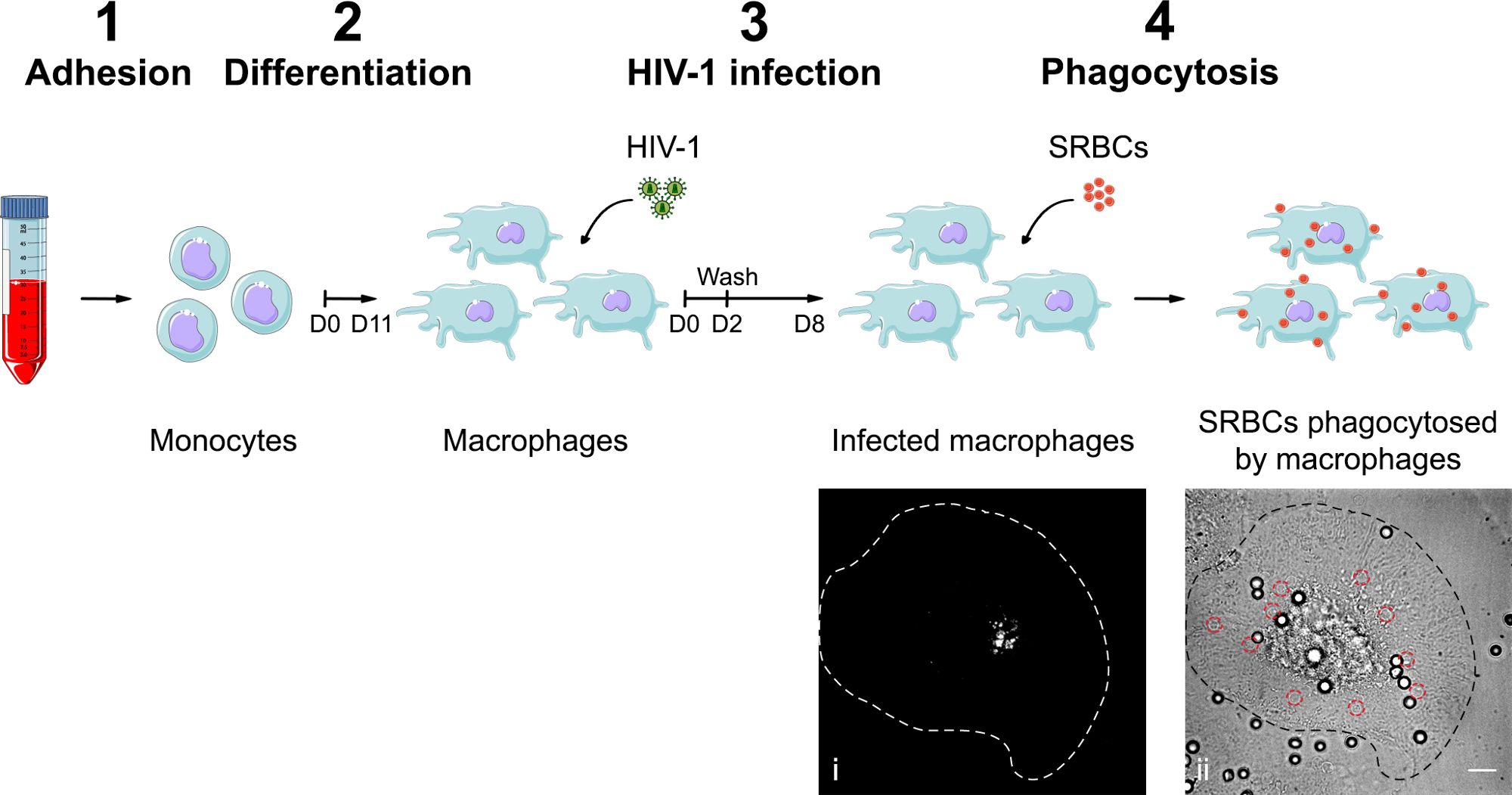
Figure 1. Preparation of control and HIV-infected hMDMs for phagocytosis imaging. Monocytes are purified by adhesion (1) and differentiated into macrophages for 11 days with M-CSF (2). Next, hMDMs are infected with NLR4.3 HIV-1Gag-iGFP i) ...
Accesso limitato. Accedi o avvia una prova gratuita per visualizzare questo contenuto.
Materiali
| Nome | Azienda | Numero di catalogo | Commenti |
|---|---|---|---|
| Glass Bottom Dishes 35 mm uncoated 1.5 | MatTek corporation | P35G-1.5-14-C Case | For acquisition |
| RPMI 1640 medium, no phenol red (10x 500 ml) | Life technologies | 11835-105 | Warm in 37 °C water bath before use ; for phagocytosis assay |
| Sheep red blood cells (SRBCs) | Eurobio | DSGMTN00-0Q | Conserved in Alsever buffer at 4 °C before use |
| Confocal Spinning Disk Unit CSU-X1M1 | Yokogawa | ||
| 491 nm 50 mW laser | COBOLT CALYPSO |
