This video demonstrates the generation of an mCherry-expressing recombinant vaccinia virus using a PKR-based selective replication and visual selection system. The transfection of infected epithelial cells with a recombination vector carrying the gene of interest and the ref fluorescent protein-tagged PKR inhibitor protein cassette enables selective viral replication and visualization of recombinant fluorescent plaques for downstream virological applications.
Articolo metodologico
Generation of a Recombinant Virus Using a Protein Kinase R-Based System
6 visualizzazioni
⸱
29 ottobre 2026
In questo articolo
Abstract
Protocollo
1. Generating the recombination vector
- Design primers to generate the selection cassette. Design each individual amplicon with overlapping sequences with neighboring amplicons and the vector to facilitate isothermal enzymatic assembly of DNA molecules, also called Gibson assembly, using any of several online primer design tools.
NOTE: This protocol can also be completed using traditional restriction endonuclease-based cloning methods. In that case, design primers with the appropriate restriction sites rather than with overlapping sequences. - Using the primers designed in step 1.1, PCR amplify the following elements in order from 5’ to 3’ (Figure 1): ~500 nucleotides of the VACV genomic region 5’ of E3L (5’ arm), EGFP or the gene of interest, ~150 nucleotides from the VACV genomic region immediately 3’ of E3L (short 3’ arm), a synthetic early/late poxvirus promoter, the mCherry-E3L fusion gene, and ~500 nucleotides from the VACV genomic region 3’ of E3L including the short 3’ arm (long 3’ arm).
- In a PCR tube, add the reagents in the following order for each amplicon: 17 μL of DNase free water, 1.2 µL of each primer (initial concentration = 10 μM, final concentration = 0.5 μM), 5 µL of 5x PCR reaction buffer, template DNA (10 ng for amplicons amplified from plasmids: EGFP and E/L promoter-mCherry-E3L cassette; 100 ng for amplicons amplified from viral genomic DNA: 5’ and 3’ arms), and 0.5 µL of DNA polymerase. Adjust the volume of water added for a final reaction volume of 50 μL.
NOTE: The concentration of template DNA should be empirically determined, but we generally start with 10 ng/reaction. - Place the tube(s) in a thermocycler, and melt the DNA at 98 °C for 30 s, and then use 25 rounds of a three-step PCR protocol: 98 °C for 5 s, 55 °C for 10 s, and 72 °C for 1 min.
NOTE: Determine the melting temperature based on the manufacturer’s suggested Tm for each primer set. Determine the appropriate extension time based on the length of each amplicon (1 minute/kb).
- In a PCR tube, add the reagents in the following order for each amplicon: 17 μL of DNase free water, 1.2 µL of each primer (initial concentration = 10 μM, final concentration = 0.5 μM), 5 µL of 5x PCR reaction buffer, template DNA (10 ng for amplicons amplified from plasmids: EGFP and E/L promoter-mCherry-E3L cassette; 100 ng for amplicons amplified from viral genomic DNA: 5’ and 3’ arms), and 0.5 µL of DNA polymerase. Adjust the volume of water added for a final reaction volume of 50 μL.
- Visualize the amplification products on a 1% agarose gel. Add 10 μL of each DNA product and 2 μL of loading buffer to each well, and run at 8 V/cm for 1 h.
- Gel purify each amplicon using a DNA gel extraction kit and manufacturer’s protocol. Elute the amplicons from the column by adding 50 μL of DNase free water and immediately centrifuging.
- Linearize the pUC19 cloning vector using EcoRI endonuclease digestion. To a tube, add 1 μg of pUC19, water to a volume of 17 μL, 2 L of reaction buffer, and 1 μL (20 units) of EcoRI. Incubate at 37 °C for 1 h.
- Visualize the amplification products on a 1% agarose gel run at 8 V/cm for 1 h. Excise the band from the gel, and purify the product using the DNA gel extraction kit as described in step 1.4.
- Ligate all of the individual, gel purified amplicons and the linearized vector using a master mix kit.
- To a PCR tube, add 0.2 pmol of linearized pUC19 and each amplicon (5’ arm, EGFP, short 3’ arm, E/L promoter-mCherry-E3L cassette, 3’arm). Add DNase free water to a final volume of 10 μL, and then add 10 μL of DNA assembly master mix. Incubate samples at 50 °C for 1 h.
- Transform chemically competent E. coli with 2 μL of the assembled product from step 1.6. Plate 100 μL of the transformed cells on LB agarose plates containing 100 μg/mL ampicillin. Incubate the plates overnight at 37 °C.
- Pick well-isolated colonies and transfer individual colonies to tubes containing Luria broth with 100 μg/mL ampicillin. Incubate the tubes overnight at 37 °C while shaking at 225 rpm.
- Isolate the plasmids from the overnight culture using a plasmid miniprep kit. Check the concentration and purity of the DNA using a spectrophotometer. An A260/A280 ratio between 1.8 and 2.0 is acceptable.
- Submit the plasmids for Sanger sequencing to determine whether the desired cloning product is correct. Store the DNA at -20 °C.
2. Generating the recombinant virus
- Infect a confluent monolayer of suitable cells with the virus to be recombined at a multiplicity of infection of 1.0 (MOI = 1.0) in a 6-well plate. Incubate the infected cells at 37 °C and 5% CO2 for 1 h. Then aspirate the medium and replace it with fresh DMEM. Incubate the infected cells at 37 °C and 5% CO2.
NOTE: For replication competent viruses such as a vaccinia virus that lacks K3L22, a cell line such as European rabbit kidney cell line RK13 (ATCC #CCL-37) or BSC-40 is appropriate. However, for replication deficient viruses, such as the virus described in this paper lacking both PKR antagonists E3L and K3L, a complementing cell line expressing these two genes in trans or PKR knock-down or knock-out cells are required. - Transfect the infected cells with 500 ng of the vector generated and validated in step 1.10 using a commercially available transfection reagent following the manufacturer’s protocol. Incubate the cells at 37 °C and 5% CO2 for 48 h.
NOTE: If using a vaccinia virus lacking both E3L and K3L, PKR-mediated selective pressure will drive selection of recombined viruses and maintain expression of the mCherry-E3L fusion protein in these cells. If desired, it should also be possible to PCR amplify only the insert to use for transfection instead of the whole plasmid. - 48 hours post-infection, harvest the infected monolayer. In some cases, the cells can be harvested by pipetting, but if they are still tightly adhered, harvest them with a cell scraper. Freeze-thaw the cells three times, and then sonicate the lysates for 15 s at 50% amplitude. Store this lysate at -80 °C until ready to use.
- Serially 10-fold dilute the lysate harvested in step 2.3 from 10-1 to 10-6 by adding 120 μL of the lysate to 1080 μL of DMEM (10-1), and then adding 120 μL of this dilution to 1080 μL of DMEM (10-2), and repeating this process four more times. Add 1 mL of each dilution to an individual, confluent well of a PKR competent cell line, in this case RK13 cells.
- Incubate the infected cells at 37 °C and 5% CO2 for 1 h. Then aspirate the medium and replace it with fresh DMEM Incubate the infected cells at 37 °C and 5% CO2.
- 24 to 48 hours post-infection, identify recombinant viruses by fluorescence microscopy. Plaques from recombinant viruses express red fluorescence due to integration the mCherry-E3L fusion gene (Figure 2). If a virus devoid of PKR inhibitors was used initially, all plaques will contain recombinant virus.
- Plaque purify recombinant viruses three times on RK13 cells. After the final round of plaque purification, all plaques should express red fluorescence.
- Infect a confluent 6-well plate of RK13 cells expressing the VACV PKR inhibitors E3L and K3L (RK13+E3L+K3L cells) with the plaque-purified red fluorescing virus from step 2.6. Aim for approximately 50-100 plaques per well.
NOTE: These cells provide the VACV PKR antagonists in trans and alleviate the PKR-mediated selective pressure to maintain the mCherry-E3L fusion gene, thus promoting “scarless” generation of the recombinant virus. - Identify collapsed viruses by fluorescence microscopy using an EVOS2 microscope, and a GFP filter cube (Excitation: 470/22, Emission: 525/50) and a RFP filter cube (Excitation: 531/40, Emission: 593/40).
NOTE: The frequency at which the mCherry-E3L fusion gene is lost is approximately 2.5% (Table 1). If EGFP is not included as a marker gene, plaques from mutant viruses that have lost the mCherry-E3L fusion gene will be colorless. - Plaque purify green-only (VC-R4) or colorless plaques (E3L) three times on RK13+E3L+K3L cells. Ensure that no plaques fluoresce red.
- Confirm the loss of mCherry-E3L and the presence of the expected mutation by PCR and Sanger sequencing.
NOTE: If the gene or mutation of interest does not have PKR inhibitory activity, recombinant viruses must be grown on RK13+E3L+K3L cells or an equivalent PKR-inhibited or PKR deficient cell line (Figure 3).
Table 1: Frequency of mCherry-E3L loss from VC-R4+K3L-mCherry-E3L in RK13+E3+K3 cells
| Experiment 1 | Experiment 2 | Experiment 3 | |
| Total plaques (RK13+E3L+K3L) | 115 | 44 | 210 |
| Colorless plaques (RK13+E3L+K3L) | 3 | 1 | 1 |
| Recombination Rate | 2.60% | 2.30% | 0.50% |
Risultati
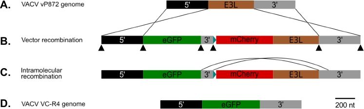
Figure 1: Diagram of p837-GOI-mCherry-E3L as well as the host-range and visual recombination strategy. (A) 5’ arm (black) and 3’ arm (grey) flank the E3L locus (brown) in VACV. (B) In p837-GOI-mCherry-E3L, these arms flank a cassette containing the gene of interest (GOI), in this case EGFP, (green) separated from an mCherry-E3L (red) fusion gene under control of the synthetic early/late poxvirus promoter25 blue) by a short 3’ arm (grey). These external arms drive homologous recombination between VACV and the p837-GOI-mCherry-E3L. Black arrowheads indicate the sites of the overlapping primers used to generate this plasmid by Gibson cloning. (C) When PKR selective pressure is removed, viruses that have undergone intramolecular recombination between the short and long 3’ arms can be selected. (D) Resulting in a virus (VC-R4) containing only the gene of interest in the E3L locus.
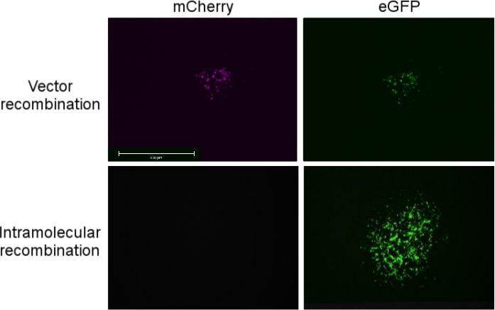
Figure 2: Fluorescent micrographs of (top) a recombinant virus plaque 24 hours after recombination with p837-GOI-mCherry-E3L expressing both mCherry (left) and EGFP (right) in RK13 cells. (Bottom) Micrograph of a recombinant virus plaque 48 hours after PKR-mediated selective pressure has been removed in RK13++ cells, expressing EGFP (right) but not mCherry (left). The scale bar indicates 650 μm for all panels.
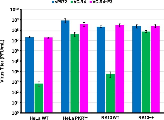
Figure 3: VC-R4 cannot replicate in PKR competent cells. The indicated cell lines were infected with vP872 (blue), VC-R4 (green), or VC-R4+E3L (magenta) at MOI = 0.1. 48 hours post-infection the infected cells were harvested and titered by serial dilution on RK13+E3L+K3L cells. Titers are reported in PFU/mL, errors bars represent the standard deviation of three replicate experiments.
Materiali
| Nome | Azienda | Numero di catalogo | Commenti |
|---|---|---|---|
| 2X-Q5 Master Mix | NEB | M0492L | High-fidelity polymerase used in PCR |
| Ampicillin | ThermoFisher Scientific | 11593027 | Bacterial selective agent |
| Disposable Cell Scrapers | ThermoFisher Scientific | 08-100-242 | Cell scraper to harvest infected cells |
| EVOS FL Auto 2 Cell imaging system | ThermoFisher Scientific | AMAFD2000 | Fluorescent microscope |
| EVOS Light Cube, GFP | ThermoFisher | AMEP4651 | GFP Cube |
| EVOS Light Cube, RFP | ThermoFisher | AMEP4652 | RFP Cube |
| GenJet | SignaGen Laboratories | SL100489 | Transfection reagent |
| Luria Bertani (LB) Broth | Gibco | 10855021 | Bacterial growth medium |
| Monarch DNA gel extraction kit | NEB | T1020L | Gel purification kit used to purify amplicons and linearized vectors |
| Monarch Plasmid Miniprep kit | NEB | T1010L | Miniprep kit ussed to purify plasmids |
| NanoDrop One | ThermoFisher Scientific | ND-ONE-W | Spectrophotometer used to measure RNA and DNA concentration |
| NEBuilder Master Mix | NEB | E2621L | Isothermal enzymatic assembly kit used to generate the recombination vector |
| Q500 Sonicator | Qsonica | Q500-110 | Sonicator for virus lysates |
| RK13 cells | ATCC | CCL-37 | Rabbit kidney cells |
| VWR Multiwell Cell Culture plates | VWR | 10062-892 | Cell culture plates |
