Questo protocollo descrive la generazione e la caratterizzazione fenotipica di un modello murino C57BL/6N-Cracdem1(c.538-83 to c.3352+255 del) mediante l'uso della tecnologia CRISPR/Cas9 per studiare il ruolo di CRACD nel rimodellamento cardiaco successivo all'infarto miocardico.
Articolo metodologico
Generazione e Caratterizzazione Fenotipica di un Modello Murino Deficiente per Cracd, Modificato con CRISPR/Cas9, per Studi sul Rimodellamento Post-Infarto Miocardico
In questo articolo
Sommario
Abstract
Myocardial infarction (MI) remains a major cause of morbidity and mortality worldwide. This protocol describes a method for generating and characterizing a Cracd-deficient mouse line on the C57BL/6N background using CRISPR/Cas9 technology. Zygotes were co-injected with Cas9 mRNA, a gRNA construct, and a donor template designed to generate a 3153-bp genomic deletion. The edited allele was confirmed by PCR genotyping and Sanger sequencing. To assess the functional role of CRACD in post-MI remodeling, Cracd-deficient and wild-type (WT, C57BL/6N) mice underwent MI induced by permanent ligation of the left anterior descending coronary artery. On postoperative day 7, cardiac function and left ventricular wall motion were assessed using transthoracic echocardiography and speckle-tracking strain imaging, followed by histopathological evaluation with H&E and Masson's trichrome staining. Representative results showed that Cracd-deficient mice exhibited reduced ventricular dilation and preserved systolic function compared to WT controls. This protocol provides a reliable experimental platform for mechanistic studies of CRACD in cardiac pathophysiology.
Introduzione
L'infarto miocardico (IM) rimane una delle principali cause di morbilità e mortalità a livello mondiale, contribuendo in modo significativo al carico globale per la salute cardiovascolare1La prognosi a lungo termine dopo infarto miocardico è determinata principalmente dal processo di rimodellamento del ventricolo sinistro (VS).2, che comporta dilatazione ventricolare, assottigliamento della parete e fibrosi interstiziale3,4, che sono fattori chiave nella progressione verso lo scompenso cardiaco (HF). L'incidenza globale di HF derivante dal rimodellamento cardiaco post-infarto miocardico aggrava ulteriormente il carico della malattia cardiovascolare5Nonostante queste osservazioni cliniche ben consolidate, mancano studi sistematici che utilizzino modelli in vivo sensibili e riproducibili per valutare il ruolo di geni specifici nel rimodellamento post-infarto miocardico. Attualmente non esiste un protocollo che combini un modello knock-out ingegnerizzato con CRISPR/Cas9 con tecniche avanzate di imaging della deformazione tissutale per valutare un regolatore citoscheletrico come CRACD. È pertanto urgentemente necessario un metodo che consenta una manipolazione genetica precisa e un fenotipaggio cardiaco ad alta risoluzione.
Il regolatore dell'inibizione della polimerizzazione dell'actina (CRACD), noto anche come KIAA1211, è un regolatore della polimerizzazione dell'actina localizzato principalmente nel citosol che svolge un ruolo fondamentale nella dinamica del citoscheletro6. Regola positivamente la polimerizzazione dell'actina legandosi alle proteine di cappuccio dell'actina (CAPZA1, CAPZA2, CAPZB) e impedendo loro di legarsi alle estremità barbate dei filamenti di actina7. Nei tessuti epiteliali, CRACD svolge un ruolo essenziale nel mantenimento della morfologia cellulare e della trasduzione del segnale preservando l'integrità del citoscheletro di actina8,9. In cellule tumorali, la sua disregolazione è associata a un aumento della metastasi e della proliferazione cellulare6,10. Il ruolo di CRACD nel cuore non è ancora stato completamente chiarito. Studi sul trascrittoma cardiaco e interventi sperimentali mostrano che l'espressione genica di Cracd risponde allo stress patologico e potrebbe regolare geni coinvolti nella dinamica del citoscheletro durante il rimodellamento ventricolare9,11. Inoltre, la sua proteina inibitrice CapZ è strettamente associata alla patologia cardiaca: la fosforilazione di CapZ regola la crescita dei miofibrilli durante l'ipertrofia indotta dalla fenilefrina12. Le nostre ricerche precedenti indicano che una moderata riduzione dell'espressione di CRACD conferisce protezione contro i danni miocardici e preserva la contrattilità dei miofilamenti dopo un'ischemia acuta13. Questi risultati suggeriscono che CRACD potrebbe essere coinvolto nel rimodellamento ventricolare maladattativo dopo infarto miocardico (MI). Tuttavia, mancano studi sistematici in vivo di perdita di funzione che verifichino se la carenza di Cracd moduli il rimodellamento post-MI, principalmente a causa dell'assenza di un protocollo validato passo dopo passo per generare e fenotipizzare un topo carente di Cracd sotto stress ischemico.
Esistono diversi approcci per generare topi knock-out costitutivi. La ricombinazione omologa tradizionale nelle cellule staminali embrionali (ES) è precisa ma costosa, richiede molto tempo (12–18 mesi) ed è limitata a determinati ceppi murini14. La mutagenesi con ENU (N-etil-N-nitrosourea) produce mutazioni puntiformi casuali che richiedono un ampio numero di incroci ripetuti e sequenziamento15. Al contrario, la tecnologia CRISPR/Cas9 offre vantaggi pratici distinti per questo studio: (i) l'iniezione diretta nei zigoti riduce il tempo necessario a 4–6 mesi; (ii) permette l'eliminazione di un ampio frammento genomico (3153 pb) per garantire una perdita completa della funzione; (iii) funziona direttamente sul ceppo C57BL/6N senza necessità di conversione del ceppo; e (iv) evita l'uso di un marcatore selezionabile, minimizzando interferenze trascrizionali indesiderate. Pertanto, CRISPR/Cas9 rappresenta la scelta più efficiente per generare una linea priva di Cracd, adatta a fenotipizzazioni cardiovascolari di routine. Questa tecnologia è stata ampiamente utilizzata per condurre studi precisi in vivo di perdita di funzione16,17.
L'imaging della deformazione con tracciamento del "speckle" è una tecnica di post-elaborazione che segue il movimento dei "speckle" acustici naturali all'interno del miocardio fotogramma per fotogramma, consentendo il calcolo di parametri di deformazione miocardica come la deformazione (variazione frazionaria della lunghezza) e la velocità di deformazione (velocità di variazione della deformazione)18. A differenza dell'ecocardiografia in modo M convenzionale o bidimensionale, che fornisce indici funzionali globali (frazione di eiezione, accorciamento frazionario, diametro interno del ventricolo sinistro) ed è relativamente insensibile alle anomalie regionali del movimento parietale, l'imaging della deformazione può quantificare la funzione contrattile segmentaria. Questo aspetto è particolarmente importante nel periodo immediatamente successivo all'infarto, in cui l'ipercinesia compensatoria dei segmenti non infartuati può mantenere invariata la frazione di eiezione globale nonostante una disfunzione regionale significativa. Il tracciamento del "speckle" offre diversi vantaggi rispetto agli endpoint standard: fornisce valori segmentari di deformazione e velocità di deformazione, spostamento e velocità, consentendo così di rilevare cambiamenti subclinici sottili19; può identificare la disincronia e i deficit contrattili precoci prima che si verifichi un rimodellamento irreversibile; e analizza direttamente la sottosierosa, lo strato più vulnerabile all'ischemia20. In questo studio, CRACD è un regolatore dello scheletro cellulare che potrebbe influenzare la contrattilità delle miofibrille a livello microscopico. Misurazioni basate sulla cavità cardiaca potrebbero facilmente trascurare tali effetti meccanici locali. Pertanto, l'integrazione dell'analisi con tracciamento del "speckle" in questo protocollo migliora la capacità di rilevare precocemente gli effetti protettivi della carenza di Cracd. Di conseguenza, l'inclusione del tracciamento del "speckle" in questo protocollo fornisce uno strumento sensibile e quantitativo per valutare il rimodellamento post-infartuale, in particolare durante la prima settimana dopo l'infarto miocardico, quando emergono la dilatazione precoce e la disfunzione contrattile21,22.
Questo protocollo è stato progettato per ricercatori che studiano l'impatto funzionale della delezione di un gene specifico sul rimodellamento acuto (a 7 giorni) post infarto miocardico, utilizzando una combinazione di knockout costitutivo e imaging ad alta risoluzione. È adatto a studi di generazione di ipotesi in cui la perdita globale del gene bersaglio è accettabile e il principale endpoint riguarda la funzione sistolica precoce e i cambiamenti strutturali. Tuttavia, è necessario considerare diversi limiti. In primo luogo, il topo privo di Cracd è un knockout globale; pertanto, gli effetti cardioprotettivi osservati non possono essere attribuiti esclusivamente alla perdita di CRACD nei cardiomiociti, poiché potrebbero intervenire anche fattori sistemici. Studi futuri che impieghino strategie di knockout condizionale saranno necessari per ottenere una delezione specifica per il tessuto cardiaco. In secondo luogo, il protocollo si concentra su un singolo punto temporale (giorno 7) e non affronta il rimodellamento cronico né la progressione della insufficienza cardiaca. Saranno richiesti studi longitudinali che vadano oltre la fase acuta. In terzo luogo, l'analisi mediante speckle-tracking richiede immagini di alta qualità (bordi endocardici ben definiti) e software dedicato, che potrebbero non essere disponibili in tutte le strutture centralizzate. Nonostante questi limiti, il protocollo fornisce una piattaforma solida e riproducibile per la caratterizzazione funzionale iniziale di geni coinvolti nel rimodellamento cardiaco post infarto miocardico.
Questo studio ha avuto lo scopo di utilizzare il sistema CRISPR/Cas9 per generare e validare un modello murino carente di Cracd (C57BL/6N-Cracdem1 (c.538-83 to c.3352+255 del)) al fine di indagare l'impatto della carenza di Cracd sul rimodellamento cardiaco post-infarto miocardico e sul movimento della parete ventricolare. L'infarto miocardico (MI) è stato indotto sia in topi wild-type (WT, C57BL/6N) che in topi carenti di Cracd, e sono state effettuate valutazioni complete una settimana dopo l'infarto. Le valutazioni hanno incluso parametri ecocardiografici convenzionali, analisi della deformazione mediante speckle-tracking e valutazione istopatologica. Ci siamo concentrati specificamente sulla fase precoce post-MI (giorno 7) per rilevare i primi eventi di rimodellamento e per determinare se la carenza di Cracd influisca sulla dilatazione ventricolare e sulla funzione sistolica durante la fase acuta dell'infarto miocardico. I risultati ottenuti sono attesi per fornire informazioni preziose sul ruolo di CRACD nella modulazione del rimodellamento cardiaco avverso dopo l'infarto miocardico.
Protocollo
All animal experiments were approved by the Institutional Animal Care and Use Committee of the Guangdong Institute of Biotechnology (Approval No. IACUC2025179). The study utilized 20 SPF Cracd-deficient and 20 age- and weight-matched WT mice. All mice were 10–12 weeks old and weighed 20–25 g. All male animals were housed in an AAALAC-accredited facility (License No. SYXK (Yue) 2016-0122) at the Guangdong Institute of Biotechnology (Guangdong Laboratory Animals Monitoring Institute). The housing environment was maintained at 22–25 °C with 50–70% relative humidity under a 12 h light/12 h dark cycle.
1. Generation of Cracd-deficient mice using CRISPR/Cas9
- Use CRISPR/Cas9 gene editing technology to generate Cracd-deficient mice on a C57BL/6N background. Delete the genomic sequence from c.538-83 to c.3352+255 (partial intron 5 to intron 6) of Cracd by homology-directed repair.
NOTE: The mouse Cracd gene (NCBI RefSeq: NM_001163793.1; Ensembl: ENSMUSG00000036377) is located on chromosome 5 and contains 9 exons, with the ATG start codon in exon 2 and the TAA stop codon in exon 9. - Design the sequence of gRNA targeting vector and donor oligonucleotide (with targeting sequence flanked by 120 bp homologous sequences combined on both sides).
- Log in to the NCBI website, select the Primer-BLAST function, and design PCR primers for genotyping.
- Paste the NCBI reference sequence number of Cracd (NM_001163793.1) into the PCR template field, specify the exon range for amplification, and click “Get Primers” to retrieve multiple pairs of candidate primers for PCR-based detection of the deletion and wild-type alleles.
- Design single-guide RNAs (gRNAs) to target sites flanking the intended deletion interval in the Cracd gene.
- Select four gRNA sequences (20 nt upstream of the PAM) using online CRISPR design tools.
- To minimize potential off-target effects, score the candidate gRNAs with CRISPRater and select the two optimal sequences (including the NGG PAM).
NOTE: Example selected sequences:
gRNA-A1: CGGCGCTCTTAACATCCAGAGGG (score: 0.71)
gRNA-A2: CCCTTCAGTCTGGGTAGTAGAGG (score: 0.71)
Accept gRNAs with a CRISPRater score ≥ 0.5. For in vitro validation, accept gRNAs with > 50% cleavage efficiency in a Cas9/gRNA target efficiency detection kit.
- Design a single-stranded donor oligonucleotide to promote efficient joining of the intended breakpoints. Example donor sequence: TTAACCATCAACGGGCCGCTGCAAGGGCTGCTGT
GTCTATA CTCAGGACCACCTGCCCTCTAGAGGCAGGAAGGT
AGAGTTCCAAGCTCTGCTGTGCTAGGTTAAATGCAACTTAACATC. - Co-inject Cas9 mRNA, gRNA and donor oligonucleotide into fertilized eggs to generate Cracd-deficient mice. Prepare Cas9 mRNA and gRNA by in vitro transcription.
- Preparation of gRNA.
- Transcribe gRNAs in vitro from DNA templates using the MEGAshortscript™ T7 transcription kit (see Table of Materials) following the manufacturer's protocol.
- Purify the transcribed gRNAs using an RNA purification kit (see Table of Materials) and elute them in RNase-free water.
- Assess gRNA cleavage activity using an in vitro Cas9/gRNA target efficiency detection kit (see Table of Materials).
- Perform all RNA preparation steps on ice using RNase‑free consumables (pipette tips, tubes, water). Keep RNA samples on ice at all times to prevent degradation.
- To perform the cleavage assay: mix the transcribed gRNA (25 ng/µL) with recombinant Cas9 protein (50 ng/µL) and a PCR-amplified DNA fragment containing the target sequence (50 ng).
- Incubate the reaction at 37 °C for 30 min.
- Run the products on a 2% agarose gel to visualize uncleaved and cleaved bands.
- Calculate cleavage efficiency as the ratio of cleaved band intensity to total (cleaved + uncleaved) band intensity.
- Accept gRNAs with > 50% cleavage efficiency.
- Preparation of Cas9 mRNA.
- Thaw the reagents from the T7 ARCA mRNA Kit (see Table of Materials).
- At room temperature, combine 10 µL of 2× ARCA/NTP mix, 1 µL of template DNA, 2 µL of T7 RNA polymerase mix, and nuclease-free water to 20 µL.
- Mix thoroughly, pulse-spin in a microfuge (5 s at 5000 × g), and incubate at 37 °C for 30 min.
- Remove the template DNA by adding 2 µL of DNase I (1 U/µL), mix well, and incubate at 37 °C for 15 min to obtain Cas9 mRNA. Optionally add 1 µL of RNase inhibitor (40 U/µL) (see Table of Materials) to the reaction to prevent RNA degradation.
- Perform all steps on ice with RNase‑free consumables. Keep the Cas9 mRNA on ice until use.
- Superovulate 4-week-old C57BL/6N female mice by intraperitoneal injection of 5 IU of pregnant mare serum gonadotropin (PMSG), followed 48 h later by 5 IU of human chorionic gonadotropin (hCG).
CAUTION: PMSG and hCG are hormones that may cause skin sensitization. Wear gloves and dispose of needles in a sharps container. - Approximately 14–18 h after hCG injection, euthanize the C57BL/6N female mice and one 8–12-week-old C57BL/6N male mouse by cervical dislocation. Collect oocytes from the oviducts of the females and sperm from the cauda epididymis of the male.
CAUTION: Euthanasia must be performed in accordance with institutional guidelines. Cervical dislocation should only be performed by trained personnel. - Capacitate the sperm in capacitation medium (see Table of Materials).
- Add a small volume of the sperm suspension to the oocytes for in vitro fertilization and co-culture for 3–4 h at 37 °C to obtain fertilized zygotes.
- Dilute the transcribed gRNA and Cas9 mRNA with RNase-free water to working concentrations (typically 25 ng/µL for gRNA and 50 ng/µL for Cas9 mRNA) and donor oligonucleotide at 10 ng/µL.
- Microinject 1–2 pL of the mixture into the cytoplasm of fertilized zygotes using a microinjection system (see Table of Materials).
- Identify healthy zygotes by the presence of two distinct pronuclei and an intact polar body under a stereomicroscope.
- Transplant 300 healthy fertilized zygotes into the oviducts of pseudopregnant female mice that have been mated with vasectomized males.
NOTE: This results in the generation of 2 positive founder mice (F0). - Use RNase‑free water and ice‑cold conditions during dilution to maintain RNA integrity.
- Identify F0 mice by PCR genotyping of tail biopsies (see section 1.6).
- Breed F0 founders with WT mice to generate F1 heterozygous offspring, to obtain germline transmission of the deletion.
- Intercross heterozygous F1 mice to produce F2 homozygous Cracd-deficient mice.
- Preparation of gRNA.
- Genotype by Polymerase Chain Reaction (PCR)
NOTE: The following protocol uses a commercial TIANamp Genomic DNA Kit (see Table of Materials). Buffer GL is the lysis buffer, Buffer GB is the binding buffer, and Buffer WA and Buffer WB are wash buffers.- Place approximately 2–5 mm of mouse tail tissue in a microcentrifuge tube containing 180 µL of Buffer GL, 20 µL of Proteinase K (20 mg/mL stock solution), and 10 µL of RNase A (10 mg/mL stock solution).
- Incubate the sample at 56 °C overnight to digest the tissue.
- Centrifuge the tube at 13800 × g for 2 min at room temperature to pellet tissue debris and transfer the supernatant to a new tube.
- Add 200 µL of Buffer GB and 200 µL of 100% ethanol to the supernatant, mix thoroughly, and apply the mixture to a spin column placed in a collection tube.
- Centrifuge at 13800 × g for 2 min at room temperature and discard the flow-through.
- Add 500 µL of Buffer WA to the column, centrifuge at 13800 × g for 1 min at room temperature and discard the flow-through.
- Carefully add 700 µL of Buffer WB (wash buffer containing ethanol, pre-mixed with 100% ethanol) to the spin column, centrifuge at 13800 × g for 1 min at room temperature and discard the flow-through.
- Apply Buffer WB to the inner wall of the column to wash residual salts.
- Repeat step 1.6.5 once, then transfer the column into a new collection tube and centrifuge at 13800 × g for 2 min at room temperature to dry the membrane and remove residual ethanol.
- Place the column into a new 1.5 mL microcentrifuge tube. Add 50–200 µL of sterilized water or elution buffer (preheated to 65 °C) to the center of the membrane, incubate for 5 min.
- Centrifuge the column at 13800 × g for 2 min at room temperature to elute the DNA.
- To increase DNA yield, either reapply the flow-through to the membrane or add an additional 50–200 µL of sterilized water, incubate for 5 min.
- Perform a final centrifugation at 13800 × g for 2 min.
NOTE: Expect DNA yield of 50–200 ng/µL from tail tissue; A260/A280 ratio of 1.8–2.0 indicates pure DNA.
- Place approximately 2–5 mm of mouse tail tissue in a microcentrifuge tube containing 180 µL of Buffer GL, 20 µL of Proteinase K (20 mg/mL stock solution), and 10 µL of RNase A (10 mg/mL stock solution).
- Genotyping by multiplex PCR using three primers in a single reaction.
- Design three primers to distinguish WT, heterozygous, and homozygous genotypes in one PCR reaction: Forward primer F1 (5’-CAGAAGTCATAGCAGCAAGGCAG-3’, binds upstream of the deletion), Forward primer F2 (5’-GAGGGAATTGAGAGGGAGCC-3’, binds within the deleted region, present only in the WT allele), and Reverse primer R1 (5’-GCCAATTTAGCTGCTTCAAATGTTC-3’, common reverse primer downstream of the deletion).
NOTE: The targeted allele generates a 421 bp amplicon (F1 + R1) only when the deletion is present; the WT allele generates a 357 bp amplicon (F2 + R1) only when the intact allele is present. - Set up the PCR reaction in a 25 µL total volume containing 50–100 ng genomic DNA, 0.2 µM of each primer (F1, F2, and R1), 200 µM dNTPs, 1.5 mM MgCl₂, and 1 U Taq DNA polymerase (see Table of Materials).
- Use the following thermocycling program: initial denaturation at 95 °C for 3 min; 35 cycles of denaturation at 95 °C for 15 s, annealing at 60°C for 15 s, and extension at 72 °C for 25 s; and a final extension at 72 °C for 5 min.
- Separate the PCR products by 1–2% agarose gel electrophoresis (120 V, 30 min) in 1× TAE buffer.
- Genotype interpretation based on band pattern: WT shows only a 357 bp band (F2+R1); Heterozygous shows both 421 bp (F1+R1) and 357 bp (F2+R1) bands; Homozygous shows only a 421 bp band (F1+R1).
- Design three primers to distinguish WT, heterozygous, and homozygous genotypes in one PCR reaction: Forward primer F1 (5’-CAGAAGTCATAGCAGCAAGGCAG-3’, binds upstream of the deletion), Forward primer F2 (5’-GAGGGAATTGAGAGGGAGCC-3’, binds within the deleted region, present only in the WT allele), and Reverse primer R1 (5’-GCCAATTTAGCTGCTTCAAATGTTC-3’, common reverse primer downstream of the deletion).
2. Myocardial infarction model establishment
- Anesthetize the mice via inhalation of 3% isoflurane (delivered in 100% oxygen at 1 L/min), place them in a supine position on a surgical platform, and confirm deep anesthesia by the absence of pedal withdrawal reflex.
CAUTION: Isoflurane is a volatile anesthetic. Use only in a well-ventilated area or under a fume hood to avoid inhalation. - Perform orotracheal intubation using a 20-gauge intravenous catheter sheath. Apply ophthalmic ointment to both eyes to prevent corneal drying during surgery.
- Connect the tube to a mechanical ventilator upon successful intubation to maintain ventilation and anesthesia with 2% isoflurane.
NOTE: Successful intubation is confirmed by visible chest movements synchronized with the ventilator.
- Connect the tube to a mechanical ventilator upon successful intubation to maintain ventilation and anesthesia with 2% isoflurane.
- Remove hair from the anterior chest with depilatory cream. Disinfect the surgical area by applying povidone-iodine and 70% alcohol in a circular motion for multiple times, starting from the center and moving outward.
- After fully exposing the skin, make a 1–1.5 cm incision in the left chest wall between the 2nd and 3rd intercostal spaces to open the thoracic cavity.
- Incise the pericardium to fully expose the heart.
CAUTION: Surgical procedures must be performed under sterile conditions. Use appropriate personal protective equipment (PPE), including gloves and a surgical mask.
- Using the inferior border of the left auricle as a landmark, pass a 7–0 atraumatic suture needle beneath the left anterior descending coronary artery (LAD) approximately 2–3 mm distal to its origin.
- Pass the needle to a depth of approximately 0.5–1 mm and tighten the ligature slowly to avoid myocardial tearing.
- Confirm successful occlusion by observing pallor (whitening) of the left ventricular anterior wall distal to the ligation site within 30 s.
- In sham-operated controls, open the chest and pass the suture beneath the coronary artery without tying the knot.
- After establishing the model, sequentially suture the ribs (4-0 absorbable suture, simple interrupted pattern) and skin (4-0 absorbable suture, continuous pattern) to close the thoracic cavity. Remove the endotracheal tube once the mouse resumes stable spontaneous breathing.
- Place mice on a heating pad (37 °C) until fully conscious.
- Administer postoperative analgesia (e.g., buprenorphine 0.05–0.1 mg/kg, subcutaneously, every 12 h for 48 h) according to institutional IACUC guidelines and closely monitor the postoperative health status for several days.
NOTE: Monitor for signs of distress (hunched posture, ruffled fur, reduced mobility, weight loss > 20%).
- Administer postoperative analgesia (e.g., buprenorphine 0.05–0.1 mg/kg, subcutaneously, every 12 h for 48 h) according to institutional IACUC guidelines and closely monitor the postoperative health status for several days.
- Mortality and exclusion criteria.
- Record the number of mice that die within the first 7 days after MI surgery.
NOTE: In a typical cohort, expect a mortality rate of approximately 10–20% by postoperative day 7, with no significant difference between WT and Cracd‑deficient mice. Common causes of death include extensive infarction leading to acute heart failure, surgical bleeding, or pneumothorax. Exclude any mouse with persistent arrhythmia (heart rate < 350 bpm) or signs of severe distress (e.g., hunched posture, ruffled fur, weight loss > 20%) from subsequent echocardiographic and histological analyses. For sham‑operated mice, mortality should be < 5%.
- Record the number of mice that die within the first 7 days after MI surgery.
3. Echocardiographic assessment
- Animal Preparation and Anesthesia
- Power on the high-resolution small animal ultrasound imaging system (see Table of Materials) and its integrated heated stage.
- Ensure that the appropriate high-frequency transducer is securely connected to the mounting apparatus following the manufacturer’s instructions. Set the temperature of the heated stage to 37 °C and allow it to stabilize.
- Place mice in an induction chamber and anesthetize them with 3% isoflurane in oxygen (1–2 L/min) until the pedal withdrawal reflex is absent. Transfer anesthetized mice to the heated platform in a supine position.
- Maintain anesthesia with 1–1.5% isoflurane delivered via a nose cone, adjusting to maintain heart rate between 450–550 beats per min.
- Apply ophthalmic ointment to both eyes to prevent corneal drying.
- Shave the thoracic region, evenly apply depilatory cream for no longer than 3 min, then remove the depilatory cream thoroughly with wet gauze.
- Rinse the skin gently with warm saline if necessary and dry the area. Attach surface ECG electrodes to the limbs using electrode gel at the contact sites and secure them with tape.
NOTE: Confirm stable ECG and heart rate within 450–550 bpm before proceeding to ultrasound imaging.
- Power on the high-resolution small animal ultrasound imaging system (see Table of Materials) and its integrated heated stage.
- Conventional Echocardiography Image Acquisition
- Position the mouse in a horizontal, supine posture on the heated stage, with a slight leftward tilt if needed to optimize cardiac imaging.
- Apply a generous amount of pre-warmed ultrasound coupling gel to the hairless left precordial region, avoiding air bubbles.
- Place the transducer gently on the chest wall at the left parasternal position. Adjust its orientation so that the index mark points toward the mouse’s right shoulder and fine-tune the angle and pressure to optimize image quality.
- Switch to B-mode (two-dimensional) imaging, adjust depth, focus zone, and frame rate to obtain a clear parasternal long-axis view of the left ventricle. Acceptable image quality requires clear visualization of the endocardial border with no dropouts.
- Once the heart rate stabilizes (450–550 bpm), acquire three separate parasternal long-axis B-mode cine loops, each containing at least 3–5 consecutive cardiac cycles.
- Save each loop by pressing “Cine Store”, and store representative still frames using “Frame Store”.
- From the long‑axis view, rotate the transducer 90° clockwise to obtain a short‑axis view at the mid‑papillary level.
- Acquire three separate short‑axis B‑mode cine loops (each with ≥ 3–5 cycles).
- Activate M‑mode, position the cursor perpendicular to the interventricular septum and posterior wall, and record three separate M‑mode tracings.
NOTE: Acceptable M‑mode tracings require clear endocardial borders throughout the cardiac cycle.
- Use the VevoLab 3.2.0 software (see Table of Materials) to measure the following parameters from M‑mode tracings according to the American Society of Echocardiography leading‑edge method: interventricular septum thickness at diastole and systole (IVSd, IVSs), left ventricular internal diameter at diastole and systole (LVIDd, LVIDs), and left ventricular posterior wall thickness at diastole and systole (LVPWd, LVPWs).
- Calculate left ventricular ejection fraction (EF) and fractional shortening (FS) using the Teichholz formula: EF (%) = [(LV Vold - LV Vols) / LV Vold] × 100, LV Vold = ((7 / (2.4 + LVIDd)) × LVIDd3), LV Vol;s = ((7 / (2.4 + LVIDs)) × LVIDs3), FS (%) = [(LVIDd – LVIDs) / LVIDd] × 100.
NOTE: For each mouse, measurements were averaged from three consecutive cardiac cycles per M‑mode recording, and the average was taken across three separate recordings, resulting in a total of 9 measurements per parameter per mouse.
- Calculate left ventricular ejection fraction (EF) and fractional shortening (FS) using the Teichholz formula: EF (%) = [(LV Vold - LV Vols) / LV Vold] × 100, LV Vold = ((7 / (2.4 + LVIDd)) × LVIDd3), LV Vol;s = ((7 / (2.4 + LVIDs)) × LVIDs3), FS (%) = [(LVIDd – LVIDs) / LVIDd] × 100.
- Position the mouse in a horizontal, supine posture on the heated stage, with a slight leftward tilt if needed to optimize cardiac imaging.
- Speckle-Tracking Strain Analysis
- Perform speckle-tracking analysis using the dedicated analysis software (VevoStrain module within VevoLab 3.2.0).
- Open a clear parasternal long-axis B-mode cine loop, select "Vevo Strain" to enter the strain analysis mode, retain only complete cardiac cycles, and choose "Long Axis".
- Manually trace the endocardial border at the end‑diastolic frame. After the endocardial border is drawn, the software automatically generates an epicardial border (adjust as needed). Then start the strain analysis and activate the "toggle contour/vector/orbit line/B mode" function to visualize the motion trajectory.
- Assess tracking quality via the colour‑coded mesh: it should appear green, closely follow the endocardial and epicardial borders without jerking, and move synchronously with myocardial contraction/relaxation. If not, manually refine the borders and re‑analyze.
- Activate "Time‑to‑peak analysis" to divide the left ventricle into six segments (basal anterior, mid anterior, apical anterior, apical posterior, mid posterior, basal posterior), with each colour representing an individual segment. Once satisfactory tracking is achieved, export data for wall motion parameters: velocity (cm/s), displacement (cm), strain (%), strain rate (1/s) for both longitudinal and radial.
NOTE: If tracking quality remains poor after three acquisition attempts (e.g., erratic strain curves, or inability to visualise clear motion trajectories), exclude the mouse from strain analysis.
- Perform speckle-tracking analysis using the dedicated analysis software (VevoStrain module within VevoLab 3.2.0).
4. Histopathological analysis
- After echocardiography, administer a lethal dose of isoflurane to deeply anesthetized mice and apply a secondary physical method (e.g., cervical dislocation) according to institutional guidelines.
- Open the thoracic cavity, rapidly harvest the hearts and rinse the hearts gently in cold phosphate-buffered saline (PBS).
CAUTION: Euthanasia must be performed according to institutionally approved protocols.
- Open the thoracic cavity, rapidly harvest the hearts and rinse the hearts gently in cold phosphate-buffered saline (PBS).
- Trim the hearts, remove the great vessels and cut transversely from apex to base into 2–3 slices (approximately 2–3 mm thick).
- Immerse the tissue slices in 4% paraformaldehyde in PBS and fix them at 4 °C for 12–24 h.
NOTE: Use a volume ratio of fixative to tissue of at least 10:1.
CAUTION: Paraformaldehyde is toxic and a suspected carcinogen. Handle in a fume hood and wear appropriate PPE (gloves, lab coat, safety goggles). Dispose of paraformaldehyde waste as hazardous chemical waste according to institutional guidelines.
- Immerse the tissue slices in 4% paraformaldehyde in PBS and fix them at 4 °C for 12–24 h.
- Dehydrate tissue through a graded ethanol series (50%, 70%, 80%, 95%, 100% ethanol, each for 1 h), clear them in xylene (2 changes, 30 min each), embed them in paraffin, and section them at 4–5 µm thickness using a microtome.
NOTE: Sections should be free of folds and tears.
CAUTION: Xylene is flammable and toxic. Use only in a fume hood and avoid skin contact. Collect xylene waste in a designated container and dispose of as hazardous chemical waste. - Deparaffinize the sections in xylene (2 changes, 5–10 min each), rehydrate them through descending ethanol series (100%, 95%, 80%, 70%, each for 3 min) to water.
- Stain them using standard Hematoxylin and Eosin (H&E) (see Table of Materials) and Masson’s trichrome protocols (see Table of Materials).
CAUTION: Xylene is toxic. Use only in a fume hood and avoid skin contact. Dispose of xylene waste as hazardous material.
- Stain them using standard Hematoxylin and Eosin (H&E) (see Table of Materials) and Masson’s trichrome protocols (see Table of Materials).
- Examine the stained sections under a light microscope (see Table of Materials).
- For quantitative analysis, capture digital images at 4×, 20×, and 40× magnification using a digital camera attached to the microscope. Measure infarct size and fibrotic area using ImageJ.
NOTE: Infarct size (%) = (infarct area / total LV area) × 100; collagen content (%) = (blue-stained area / total myocardial area) × 100.
- For quantitative analysis, capture digital images at 4×, 20×, and 40× magnification using a digital camera attached to the microscope. Measure infarct size and fibrotic area using ImageJ.
Risultati
Generation of Cracd-deficient mice
A heritable mouse model carrying a targeted deficiency in the Cracd gene was generated using CRISPR/Cas9-mediated genome editing (Figure 1A, Figure 1B). To distinguish WT, heterozygous, and homozygous genotypes, a multiplex PCR was performed using three primers (F1, F2, R1) in a single reaction. Mice homozygous for the deletion (Cracd‑deficient) displayed only the 421 bp band, WT mice showed only the 357 bp band (Figure 1C). Sanger sequencing of genomic DNA from Cracd-deficient mice confirmed the predicted deletion in the target gene compared to WT controls (Figure 1D). Western blot analysis further demonstrated a marked reduction in CRACD protein levels in cardiac tissues from Cracd-deficient mice compared with WT controls (Figure 1E). Together, these data confirm the successful establishment of the Cracd-deficient mouse line.
Echocardiography
Echocardiographic assessment on postoperative day 7 revealed pronounced pathological cardiac remodeling in WT mice after MI (WT-MI) compared with sham-operated controls (Figure 2A). The WT-MI group showed thinning of the interventricular septum (Figure 2B, C), significant left ventricular (LV) dilation as reflected by increased LVIDd (Figure 2D) and LVIDs (Figure 2E), and thinning of the posterior wall (Figure 2F, G). These structural alterations were associated with a pronounced decline in global systolic function, as demonstrated by significantly reduced EF (Figure 2H) and FS (Figure 2I). In contrast, Cracd-deficient mice displayed a cardioprotective phenotype following MI. LV dilation was significantly attenuated, with notably smaller LVIDd and LVIDs compared with the WT-MI group. Furthermore, these mice exhibited improved systolic thickening of the posterior wall and maintained significantly higher EF and FS values. These findings indicate that Cracd deficiency effectively attenuates post-infarction LV dilation and preserves global systolic function.
Speckle-tracking strain analysis
Left ventricular myocardial motion and deformation were further evaluated by speckle‑tracking echocardiography using only parasternal long‑axis views (Figure 3A). To ensure data reliability, we defined pragmatic criteria for acceptable output based on the capabilities of the VevoStrain software. Acceptable strain analysis required: (i) a clear parasternal long‑axis view with endocardial borders visible throughout the cardiac cycle; (ii) a stable heart rate between 450–550 bpm during acquisition; (iii) after automatic tracking, the colour‑coded mesh (activated via “toggle contour/vector/orbit line/B mode”) had to closely follow the endocardial and epicardial borders without jerking or losing contact; and (iv) strain curves had to be smooth, show consistent waveforms across segments, and lack sudden spikes or arrhythmic interruptions. Suboptimal output—most commonly erratic strain curves or inability to visualise clear motion trajectories—led to manual adjustment of endocardial or epicardial contours and re‑analysis. If quality remained poor after three attempts, the mouse was excluded. Using these criteria, we obtained reliable strain data in WT‑MI mice and Cracd‑deficient MI mice.
Compared with the sham group, WT-MI mice exhibited significantly reduced radial velocity (Figure 3B) and longitudinal velocity (Figure 3C), as well as radial displacement (Figure 3D) and longitudinal displacement (Figure 3E), indicating impaired global myocardial motion. The absolute values of radial strain (Figure 3F), radial strain rate (Figure 3G), longitudinal strain (Figure 3H), and longitudinal strain rate (Figure 3I) were also markedly decreased, reflecting severely compromised regional myocardial deformation and function. In contrast, Cracd-deficient mice showed significant improvement in these parameters. Compared with the WT-MI group, they displayed better recovery of radial and longitudinal displacements. Furthermore, radial velocity, radial strain, and radial strain rate were maintained at higher levels, while longitudinal strain and longitudinal strain rate were significantly improved. These results suggest that Cracd deficiency not only preserves global systolic function but also enhances regional LV myocardial deformation after MI.
The speckle‑tracking imaging analysis also included visualization of segmental strain distribution and cardiac motion trajectories. Parasternal long‑axis B‑mode images with segmented six regions (BA, MA, AA, AP, MP, BP) and color gradient mapping are displayed in Figure 4A. Cardiac motion trajectories, time‑to‑peak curves for radial and longitudinal strain, parametric distribution maps of strain synchrony, and three‑dimensional reconstructions are presented in Figure 4B. These data further support the functional improvements observed in Cracd‑deficient mice.
Histological examination
To assess the protective effect of Cracd deficiency against MI, histopathological examination was performed on heart sections from WT and Cracd-deficient mice at 7 days post-MI using H&E and Masson's trichrome staining. Whole‑heart longitudinal sections are shown in Figure 5A, with quantitative analysis of infarct size and collagen content presented in Figure 5B and Figure 5C, respectively. High‑magnification views of the infarct zone, border zone, and remote zone are displayed in Figure 5D. In WT-MI hearts, H&E staining showed pronounced left ventricular wall thinning and a well-demarcated infarct zone. Within the infarct region, extensive inflammatory cell infiltration, myocardial necrosis, and marked fibrosis with scar formation were observed. In the border zone, inflammatory infiltration, myocardial necrosis, fibrosis and cardiomyocyte hypertrophy were evident, while the remote zone displayed prominent hypertrophic changes. Corresponding Masson's trichrome staining further demonstrated extensive interstitial fibrosis within both the infarct and border zones of WT hearts following MI. In contrast, Cracd-deficient mice exhibited substantially attenuated pathological alterations, including reduced wall thinning, diminished inflammatory infiltration and myocardial necrosis, less pronounced cardiomyocyte hypertrophy, and markedly decreased interstitial fibrosis compared with WT-MI hearts. These histological findings indicate that Cracd deficiency mitigates post-infarction myocardial damage and adverse remodeling.
Successful protocol execution should yield: (i) a well‑demarcated infarct zone with extensive collagen deposition in WT-MI hearts; (ii) clear cellular morphology without staining artifacts; and (iii) significantly attenuated pathology in Cracd‑deficient MI hearts. Although infarct size may vary with ligation precision, the genotype‑dependent difference is a reliable indicator of successful performance.
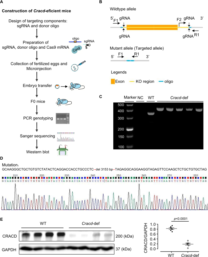
Figure 1: Generation of Cracd-deficient mice. (A) Construction of Cracd-deficient mice. (B) Schematic of the Cracd gene modification strategy. (C) PCR genotyping of tail genomic DNA. WT mice show a single 357 bp band, while homozygous (Cracd‑deficient) mice show a single 421 bp band. (D) Sanger sequencing of genomic DNA confirmed the predicted deletion in the Cracd locus of deficient mice compared to WT controls. (E) Western blot analysis showed significantly reduced CRACD protein levels in the hearts of Cracd-deficient mice compared to WT controls. n = 6 mice per group. Data are presented as mean ± SEM. Statistical significance was calculated with a two-tailed unpaired Student's t-test. Please click here to view a larger version of this figure.
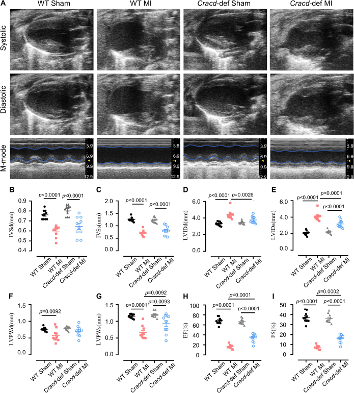
Figure 2: Echocardiographic assessment of cardiac function. (A) Representative B-mode and M-mode echocardiographic images of mouse hearts at diastole and systole. Quantitative analysis of (B) interventricular septum thickness at diastole (IVSd) and (C) systole (IVSs), (D) left ventricular internal diameter at diastole (LVIDd) and (E) systole (LVIDs), (F) left ventricular posterior wall thickness at diastole (LVPWd) and (G) systole (LVPWs), (H) ejection fraction (EF), and (I) fractional shortening (FS). n = 10 mice per group. Data are presented as mean ± SEM. Statistical significance was determined by one-way ANOVA followed by Tukey's multiple comparisons test. Please click here to view a larger version of this figure.
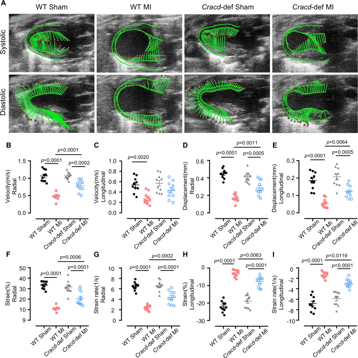
Figure 3: Speckle-tracking strain analysis. (A) Representative images of left ventricular wall motion and deformation at diastole and systole. Quantitative analyses of the following parameters: (B) radial velocity, (C) longitudinal velocity, (D) radial displacement, (E) longitudinal displacement, (F) radial strain, (G) radial strain rate, (H) longitudinal strain, and (I) longitudinal strain rate. n = 10 mice per group. Data are presented as mean ± SEM. Statistical significance was determined by one-way ANOVA followed by Tukey's multiple comparisons test. Please click here to view a larger version of this figure.
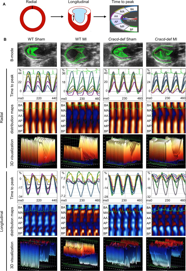
Figure 4: Speckle-tracking imaging in WT-Sham, WT-MI, Cracd-deficient Sham, and Cracd-deficient MI groups at 7 days post-MI. (A) Parasternal long‑axis B‑mode images. Left ventricular wall was segmented into six regions: BA (basal anterior), MA (mid anterior), AA (apical anterior), AP (apical posterior), MP (mid posterior), and BP (basal posterior). Color gradient from blue to red represents increasing strain magnitude. (B) Cardiac motion trajectories; time-to-peak curves for radial and longitudinal strain values along with corresponding peak times. Parametric distribution maps of strain synchrony (blue/red indicate direction: radial – away from/toward center; longitudinal – away from/toward apex). White dots = highest velocity regions, black dots = lowest velocity. Three-dimensional reconstructions (X: time, Y: spatial position, Z: strain magnitude). Please click here to view a larger version of this figure.
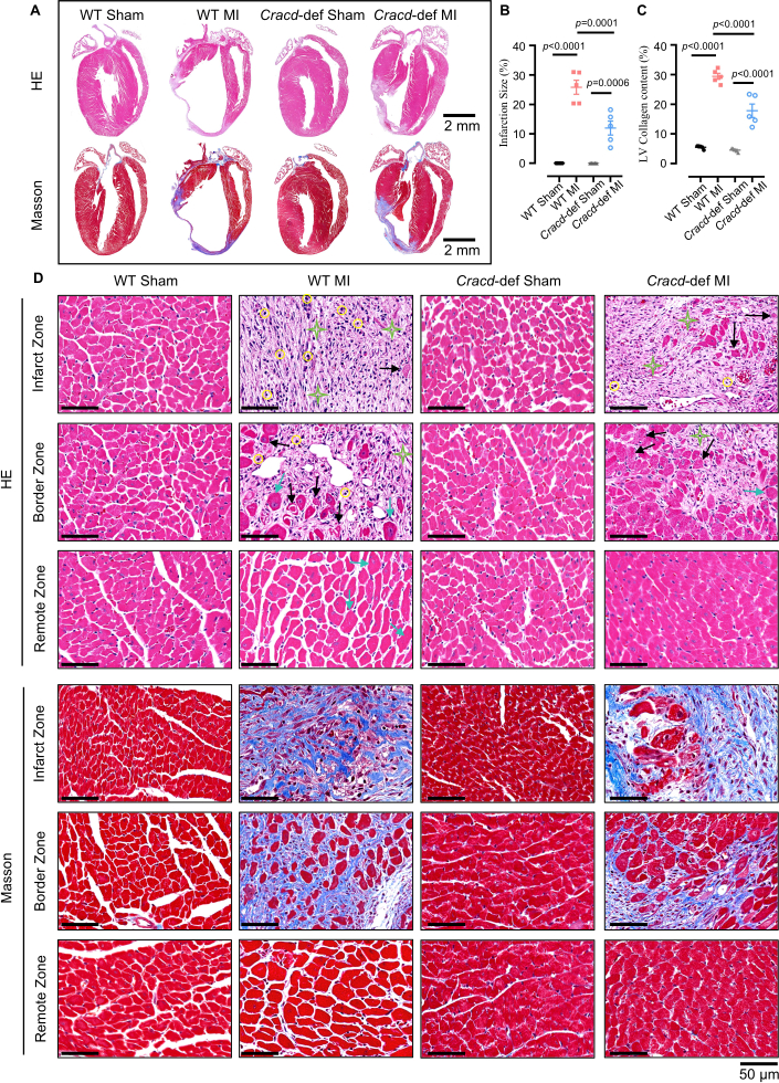
Figure 5: Histopathological analysis of cardiac remodeling after MI. (A) Whole-heart longitudinal sections with H&E and Masson's trichrome staining. Scale bars: 2 mm. (B) Quantitative analysis of infarct size and (C) collagen content. (D) High‑magnification views of infarct zone (IZ), border zone (BZ), and remote zone (RZ). H&E: yellow circles = inflammatory infiltration; black arrows = myocardial necrosis; green stars = fibrosis; blue arrows = hypertrophy. Masson’s trichrome: blue indicates collagen deposition. Scale bar: 50 μm. n = 5 mice per group. Data are presented as mean ± SEM. Statistical significance was determined by one-way ANOVA followed by Tukey's multiple comparisons test. Please click here to view a larger version of this figure.
Discussione
In questo studio, abbiamo generato un topo privo del gene Cracd mediante tecnologia CRISPR/Cas9 e abbiamo messo a punto un protocollo per fenotipizzarlo dopo infarto miocardico (MI) utilizzando ecocardiografia convenzionale, imaging ecocardiografico con speckle-tracking strain e istologia. Il protocollo ha prodotto topi knock-out con perdita confermata della proteina CRACD. I topi deficienti di Cracd hanno mostrato una minore dilatazione ventricolare, una migliore frazione di eiezione (EF) e una ridotta fibrosi rispetto ai controlli wild-type (WT) dopo l'infarto miocardico, confermando l'utilità del modello nello studio del rimodellamento cardiaco.
Il successo di questo modello dipende da diversi passaggi critici. La progettazione dell'gRNA e la relativa validazione in vitro influenzano direttamente l'efficienza della modifica nei zigoti23. Utilizziamo CRISPick o Benchling per selezionare gRNA con punteggi CRISPRater ≥ 0,5 e successivamente testiamo l'efficienza di scissione mediante un saggio Cas9/gRNA in vitro. Solo gli gRNA che raggiungono una scissione > 50% vengono microiniettati. Anche la legatura corretta dell'arteria discendente anteriore (LAD) è fondamentale: il punto di sutura con filo 7‑0 deve essere posizionato a 2–3 mm dall'origine della LAD e a una profondità di 0,5–1 mm, per evitare di perforare il ventricolo destro (troppo profondo) o di non occludere adeguatamente il vaso (troppo superficiale); la comparsa immediata di un'area bianca sulla parete anteriore conferma l'occlusione. Anche il controllo della frequenza cardiaca durante l'ecocardiografia è essenziale24: mantenere la frequenza tra 450 e 550 bpm regolando l'isoflurano (tipicamente 1–2%) migliora la riproducibilità delle misurazioni convenzionali e delle misure di deformazione (strain). Infine, per l'analisi mediante speckle-tracking, la valutazione visiva della mesh codificata a colori è fondamentale25. La mesh deve seguire i bordi miocardici senza movimenti irregolari; in caso contrario, è necessario un aggiustamento manuale; se la qualità rimane scadente dopo tre tentativi, il topo viene escluso.
Anche quando il protocollo viene seguito meticolosamente, possono insorgere alcune difficoltà tecniche. La risoluzione sistematica dei problemi può risolvere la maggior parte di essi. Bassi tassi di trasmissione germinale (inferiori al 10%) indicano solitamente un'integrazione scadente dell'oligonucleotide donatore o un'attività debole della gRNA26. Di conseguenza, aumentare l'oligo donatore a 20 ng/µL, estendere le braccia di omologia a 120–200 bp e verificare nuovamente il taglio della gRNA (> 50%) può migliorare la trasmissione. Un'elevata mortalità post-IM (superiore al 30%) è spesso causata da pneumotorace, emorragia e un'ampia infarto27. Pertanto, mantenere i topi sotto ossigeno continuo fino al risveglio e utilizzare un tappetino riscaldante per mantenere la temperatura corporea riduce in modo evidente i tassi di mortalità. Se la parete anteriore non diventa pallida dopo la legatura, probabilmente la CAD è stata identificata in modo errato o il punto di sutura è troppo superficiale. In questo caso, ridefinire la posizione della CAD (un vaso diagonale di colore rosso brillante) e ripassare l'ago a una profondità di 0,5–1 mm, talvolta con l'ausilio di un microscopio stereoscopico, risolve il problema. Per una scarsa qualità del tracciamento del contrasto, i sospetti abituali sono una bassa frequenza di acquisizione, bordi sfocati o artefatti dovuti al movimento28. Di conseguenza, aumentare la frequenza di acquisizione, regolare finemente il guadagno, correggere manualmente il tracciato endocardico ed escludere i cicli con aritmie aiutano a migliorare i risultati.
Oltre a queste considerazioni operative, il metodo presenta chiare limitazioni. Il knockout di Cracd è globale, quindi CRACD manca in tutti i tessuti. Di conseguenza, la protezione osservata non può essere attribuita esclusivamente ai cardiomiociti, poiché potrebbero intervenire anche fattori sistemici. Sarebbe necessario un knockout condizionale per distinguere i ruoli specifici per tipo cellulare. Un'altra limitazione è rappresentata dall'unico punto temporale analizzato al giorno 7; in tal modo non si ottiene alcuna informazione sul rimodellamento cronico. Pertanto, per studi a lungo termine dovrebbero essere inclusi punti temporali successivi (4 o 8 settimane). Anche la riproducibilità risente della dipendenza dalla qualità delle immagini: non tutti i sistemi ecografici sono in grado di fornire contorni endocardici nitidi, e l'esperienza dell'operatore è determinante. Per questo motivo, si raccomanda che tutte le chirurgie e le misurazioni siano eseguite da personale professionalmente addestrato.
Alla luce di questi limiti, è utile confrontare il nostro approccio con strategie alternative per lo studio della funzione genica nel rimodellamento post-infarto miocardico. La manipolazione genica mediata da AAV e gli oligonucleotidi antisenso (ASO) rappresentano due alternative comuni29,30. L'AAV9 può indurre un'overespressione o un silenziamento transitorio specifico per il tessuto cardiaco senza necessità di incroci, ma presenta svantaggi come il limite di confezionamento (~4,7 kb), un'efficienza di trasduzione variabile e effetti fuori bersaglio31. Al contrario, il nostro modello di knockout mediante CRISPR/Cas9 offre una perdita di funzione permanente e completa, vantaggio utile per studi a lungo termine. Gli ASO consentono un silenziamento reversibile, dipendente dalla dose e con insorgenza rapida, risultando adatti a interventi acuti; tuttavia, richiedono somministrazioni ripetute e mancano di specificità tissutale32. Pertanto, il knockout costitutivo descritto in questo studio è il più indicato per la scoperta iniziale e la fenotipizzazione cronica.
Infine, il protocollo può essere esteso ben oltre il CRACD. Lo stesso flusso di lavoro — la generazione di topi knock-out mediante CRISPR/Cas9 combinata con ecocardiografia, imaging della deformazione e istologia — può valutare qualsiasi gene candidato nel rimodellamento post-infarto miocardico. Il quadro fenotipico si adatta anche ad altri modelli di malattie cardiovascolari, come la costrizione aortica trasversa (sovraccarico da pressione) o il danno da ischemia-riperfusione. Inoltre, i campioni di tessuto ottenuti con questo protocollo possono essere utilizzati per analisi multi-omiche (transcrittomica, proteomica, metabolomica) al fine di individuare i meccanismi molecolari. Il modello deficitario per Cracd può fungere da strumento per la sperimentazione di farmaci: composti il cui meccanismo d'azione è sospettato di coinvolgere la via CRACD possono essere valutati. Pertanto, questo protocollo fornisce una piattaforma adattabile a un'ampia gamma di studi meccanicistici e traslazionali nella ricerca cardiovascolare.
Dichiarazioni
Gli autori dichiarano di non avere interessi concorrenti.
Ringraziamenti
Questo lavoro è stato sostenuto dalla Guangdong Basic and Applied Basic Research Foundation (2023A1515011278), dal Progetto autofinanziato dell'Istituto di Ricerca in Biotecnologia della Provincia di Guangdong (GDBRI-ZL202602) e dall'Interscambio e Cooperazione Accademica Cina-Canada sui Modelli e la Patogenesi delle Malattie Cardiovascolari (MS202500058).
Materiali
| Nome | Azienda | Numero di catalogo | Commenti |
|---|---|---|---|
| ACRISPR/Cas9 | |||
| Soluzione di Trizma Cloridrato | Sigma-Aldrich, USA | T2663 | Utilizzata per la preparazione del buffer di estrazione del DNA |
| Proteinasi K | Sigma-Aldrich, USA | 539480 | Utilizzata per la digestione della coda del topo |
| Green Taq Mix | Vazyme, Cina | P131 | Utilizzata per l'amplificazione PCR |
| Agarosio | BioFroxx, Germania | 1110GR500 | Utilizzato per l'elettroforesi su gel del DNA a concentrazione 1–2% |
| Marcatore di DNA | Thermo Scientific, USA | SM0242 | Utilizzato come scala di DNA per determinare la dimensione dei prodotti PCR |
| TIANamp Genomic DNA Kit | Tiangen Biotech, Cina | DP304 | Utilizzato per l'estrazione del DNA genomico dal tessuto della coda del topo |
| MEGAshortscript™ T7 transcription kit | Thermo Scientific, USA | AM1354 | Utilizzato per la trascrizione in vitro dell'gRNA |
| Kit di purificazione dell'RNA | Zymo Research, USA | R1016 | Utilizzato per la purificazione dell'gRNA trascritto |
| BeyoCRISPR™ sgRNA Screening Kit | Beyotime, Cina | D8412S | Utilizzato per la valutazione in vitro dell'attività di scissione dell'gRNA |
| Inibitore della RNasi | NEB, USA | M0314 | Utilizzato per prevenire la degradazione dell'RNA durante la trascrizione in vitro |
| TaKaRa Taq | Takara Bio, Giappone | R001A | Utilizzato per il genotipaggio PCR del DNA della coda del topo |
| FERTIUP® Mouse Sperm Preincubation Medium: PM | Cosmo Bio, Giappone | KYD-002-EX | Utilizzato per la capacitazione degli spermatozoi prima della fecondazione in vitro |
| T7 ARCA mRNA Kit | NEB, USA | E2060 | Utilizzato per la trascrizione in vitro dell'mRNA di Cas9 |
| Gonadotropina di siero di cavalla gravida | MCE, USA | 9002-70-4 | Utilizzata per la superovulazione, somministrata per iniezione intraperitoneale |
| Gonadotropina corionica umana | ProSpec, Israele | HOR-250 | Utilizzata per indurre l'ovulazione, somministrata per iniezione intraperitoneale |
| Acqua priva di RNasi | Aladdin, Cina | R665526 | Utilizzata per la solubilizzazione dell'RNA e l'allestimento delle reazioni PCR |
| Primer | Sangon Biotech, Cina | / | Utilizzati per il genotipaggio |
| Chirurgia | |||
| Isoflurano | RWD Life Science, Cina | R510-22-10 | Utilizzato per l'induzione e il mantenimento dell'anestesia durante l'intervento chirurgico |
| Crema depilatoria | Veet, Francia | 1000207 | Applicata sulla zona toracica per rimuovere il pelo prima dell'intervento chirurgico e dell'ecografia |
| Chirurgia 7-0 | Lingqiao, Cina | LQ7022380206 | Utilizzato per la legatura permanente dell'arteria coronaria discendente anteriore (LAD) |
| Chirurgia 4-0 | Lingqiao, Cina | LQ4022380412 | Utilizzato per la chiusura dell'incisione cutanea dopo l'intervento chirurgico |
| Respiratore per anestesia | Harvard Apparatus, USA | Tabletop | Utilizzato per somministrare isoflurano e supportare la respirazione |
| Respiratore per piccoli animali | Taimeng, Cina | HX-101E | Utilizzato per fornire ventilazione a pressione positiva durante l'intervento chirurgico a torace aperto |
| Piastra riscaldante a temperatura costante | TigerGene, USA | TG-TP-GS | Utilizzata per mantenere la temperatura corporea del topo durante l'intervento chirurgico e il recupero |
| Retrattore costale | Shenzhen Huayon Biotech, Cina | LN-18-4101 | Utilizzato per separare le costole ed esporre il cuore per la legatura della LAD |
| Forbici | Shenzhen Huayon Biotech, Cina | 18-0551 | Utilizzate per effettuare incisioni cutanee e tagliare i tessuti |
| Microscopio stereoscopico | Nikon, Giappone | SMZ 745 | Utilizzato per ottenere una visione ingrandita durante l'intervento chirurgico di legatura dell'arteria LAD |
| Pinza microscopica per ago | Shenzhen Huayon Biotech, Cina | 18-2211 | Utilizzata per manipolare l'ago sottile da 7-0 sotto il microscopio |
| Prodotti chimici e reagenti | |||
| Agenti accoppianti per ultrasuoni | Tianjin Jinya Technology Development, Cina | TM-100 | Applicati sul torace per l'ecocardiografia, garantiscono un contatto adeguato della sonda |
| Paraformaldeide | Sigma-Aldrich, USA | V900894 | Utilizzata per la fissazione del tessuto cardiaco dopo il prelievo |
| Xilene | Macklin, Cina | 1330-20-7 | Utilizzato per la rimozione della paraffina e la chiarificazione del tessuto prima della colorazione |
| Ematossilina | Sigma-Aldrich, USA | H3136 | Colorante nucleare, utilizzato nella colorazione ematossilina-eosina (HE) |
| Eosina | Sigma-Aldrich, USA | E4009 | Colorante citoplasmatico, utilizzato nella colorazione ematossilina-eosina (HE) |
| Ponceau S | Sigma-Aldrich, USA | P3504 | Utilizzato nella colorazione di Masson per la colorazione del citoplasma |
| Acido fosfomolibdico | Sigma-Aldrich, USA | 221856 | Utilizzato nella colorazione di Masson come mordente e per la differenziazione |
| Soluzione di blu di anilina | Sigma-Aldrich, USA | 415049 | Utilizzata nella colorazione di Masson per la colorazione delle fibre collagene (blu) |
| Anticorpo anti-CRACD | Absea, Cina | KC-35356 | Anticorpo primario per la rilevazione della proteina CRACD mediante Western Blot, diluizione 1:2000 |
| Anticorpo anti-GAPDH | CST, USA | 2118S | Anticorpo di controllo del carico per Western Blot, diluizione 1:5000 |
| Etilene | Macklin, Cina | 64-17-5 | Utilizzato per la disidratazione e la preparazione dei reagenti (gradi 70%, 80%, 95%, 100%) |
| Attrezzature di laboratorio | |||
| Centrifuga Sorvall™ Legend™ Micro 17 | Thermo Scientific, USA | 75002541 | Utilizzata per la preparazione abituale dei campioni |
| Ciclatore termico PCR | Bio-Rad Laboratories, USA | 1861096 | Utilizzato per l'amplificazione del DNA |
| Sistema di microiniezione | Eppendorf, Germania | 5193000020 | Utilizzato per la microiniezione dei reagenti CRISPR/Cas9 negli zigoti |
| Sistema di imaging Vevo 2100 | VisualSonics, Canada | Vevo2100 | Sistema di imaging ad alta frequenza per la valutazione non invasiva della funzione e della struttura cardiaca dopo infarto miocardico (MI) |
| Microscopio ottico | Leica, Germania | DM2500 | Utilizzato per osservare sezioni cardiache colorate con ematossilina-eosina (HE) e Masson |
| Software di analisi | |||
| ImageJ | National Institutes of Health, USA | / | Utilizzato per l'analisi delle immagini, inclusa la misurazione delle aree, la quantificazione del segnale e l'elaborazione delle immagini istologiche (colorazioni HE e Masson) |
| VevoLab 3.2.0 / VevoStrain | VisualSonics, Canada | / | Software VevoLab per l'analisi della funzione cardiaca; modulo VevoStrain utilizzato per l'analisi della deformazione miocardica al fine di valutare le prestazioni cardiache dopo infarto miocardico (MI) |
| GraphPad Prism 8 | GraphPad Software, USA | / | Utilizzato per l'analisi statistica e la preparazione dei grafici |
Riferimenti
- Fauconnier, J., Meli, A. C., Thireau, J., Roberge, S., Shan, J., et al. Ryanodine receptor leak mediated by caspase-8 activation leads to left ventricular injury after myocardial ischemia-reperfusion. Proc Natl Acad Sci U S A. 108 (32), 13258-13263 (2011).
- Cohn, J. N., Ferrari, R., Sharpe, N. Cardiac remodeling-concepts and clinical implications: a consensus paper from an international forum on cardiac remodeling. J Am Coll Cardiol. , (2000).
- Liu, D., Tian, X., Liu, Y., Song, H., Cheng, X., et al. CREG ameliorates the phenotypic switching of cardiac fibroblasts after myocardial infarction via modulation of CDC42. Cell Death Dis. 12 (4), (2021).
- Contessotto, P., Pandit, A. Therapies to prevent post-infarction remodelling: from repair to regeneration. Biomaterials. 275, (2021).
- Zhu, R., Sun, H., Yu, K., Zhong, Y., Shi, H., et al. Interleukin-37 and dendritic cells treated with interleukin-37 plus troponin I ameliorate cardiac remodeling after myocardial infarction. J Am Heart Assoc. 5 (12), (2016).
- Chang, Y., Zhang, J., Huo, X., Qu, X., Xia, C., et al. Substrate rigidity dictates colorectal tumorigenic cell stemness and metastasis via CRAD-dependent mechanotransduction. Cell Rep. 38 (7), (2022).
- Seo, Y., Jang, J., Ko, K. P., Zou, G., Huang, Y., et al. Actin dysregulation induces immune evasion via oxidative stress-activated PD-L1 in gastric cancer. bioRxiv. , (2024).
- Kim, B., Zhang, S., Huang, Y., Ko, K. P., Jung, Y. S., et al. CRACD loss induces neuroendocrine cell plasticity of lung adenocarcinoma. Cell Rep. 43 (6), (2024).
- Jung, Y. S., Wang, W., Jun, S., Zhang, J., Srivastava, M., et al. Deregulation of CRAD-controlled cytoskeleton initiates mucinous colorectal cancer via β-catenin. Nat Cell Biol. 20 (11), 1303-1314 (2018).
- Liu, Z., Cao, H., Shi, Y., Yang, R. KIAA1211 plays an oncogenic role in human non-small cell lung cancer. J Cancer. 10 (26), 6747-6753 (2019).
- Snider, P. L., Snider, E., Simmons, O., Lilly, B., Conway, S. J. Analysis of uncharacterized mKiaa1211 expression during mouse development and cardiovascular morphogenesis. J Cardiovasc Dev Dis. 6 (2), (2019).
- Lin, Y. H., Warren, C. M., Li, J., McKinsey, T. A., Russell, B. Myofibril growth during cardiac hypertrophy is regulated through dual phosphorylation and acetylation of the actin capping protein CapZ. Cell Signal. 28 (8), 1015-1024 (2016).
- Yang, F. H., Pyle, W. G. Reduced cardiac CapZ protein protects hearts against acute ischemia-reperfusion injury and enhances preconditioning. J Mol Cell Cardiol. 52 (3), 761-772 (2012).
- Tong, C., Huang, G., Ashton, C., Wu, H., Yan, H., et al. Rapid and cost-effective gene targeting in rat embryonic stem cells by TALENs. J Genet Genomics. 39 (6), 275-280 (2012).
- Cordes, S. P. N-ethyl-N-nitrosourea mutagenesis: boarding the mouse mutant express. Microbiol Mol Biol Rev. 69 (3), 426-439 (2005).
- Cong, L., Ran, F. A., Cox, D., Lin, S., Barretto, R., et al. Multiplex genome engineering using CRISPR/Cas systems. Science. 339 (6121), 819-823 (2013).
- Kanke, K. L., Rayner, R. E., Bozik, J., Abel, E., Venugopalan, A., et al. Single-stranded DNA with internal base modifications mediates highly efficient knock-in in primary cells using CRISPR-Cas9. Nucleic Acids Res. 52 (22), 13561-13576 (2024).
- Mihos, C. G., Liu, J. E., Anderson, K. M., Pernetz, M. A., O'Driscoll, J. M., et al. Speckle-tracking strain echocardiography for the assessment of left ventricular structure and function: a scientific statement from the American Heart Association. Circulation. 152 (10), (2025).
- Garg, P. K., Biggs, M. L., Kizer, J. R., Shah, S. J., Psaty, B., et al. Glucose dysregulation and subclinical cardiac dysfunction in older adults: the Cardiovascular Health Study. Cardiovasc Diabetol. 21 (1), (2022).
- Asanuma, T., Fukuta, Y., Masuda, K., Hioki, A., Iwasaki, M., et al. Assessment of myocardial ischemic memory using speckle tracking echocardiography. JACC Cardiovasc Imaging. 5 (1), 1-11 (2012).
- Chong, S. Y., Zharkova, O., Yatim, S. M. J. M., Wang, X., Lim, X. C., et al. Tissue factor cytoplasmic domain exacerbates post-infarct left ventricular remodeling via orchestrating cardiac inflammation and angiogenesis. Theranostics. 11 (19), 9243-9261 (2021).
- Hung, C. L., Verma, A., Uno, H., Shin, S. H., Bourgoun, M., et al. Longitudinal and circumferential strain rate, left ventricular remodeling, and prognosis after myocardial infarction. J Am Coll Cardiol. 56 (22), 1812-1822 (2010).
- Labuhn, M., Adams, F. F., Ng, M., Knoess, S., Schambach, A., et al. Refined sgRNA efficacy prediction improves large- and small-scale CRISPR-Cas9 applications. Nucleic Acids Res. 46 (3), 1375-1385 (2018).
- Wu, J., Bu, L., Gong, H., Jiang, G., Li, L., et al. Effects of heart rate and anesthetic timing on high-resolution echocardiographic assessment under isoflurane anesthesia in mice. J Ultrasound Med. 29 (12), (2010).
- Voigt, J. U., Pedrizzetti, G., Lysyansky, P., Marwick, T. H., Houle, H., et al. Definitions for a common standard for 2D speckle tracking echocardiography: consensus document of the EACVI/ASE/Industry Task Force to standardize deformation imaging. J Am Soc Echocardiogr. 28 (2), 183-193 (2015).
- Port, F., Chen, H. M., Lee, T., Bullock, S. L. Optimized CRISPR/Cas tools for efficient germline and somatic genome engineering in Drosophila. Proc Natl Acad Sci U S A. 111 (29), (2014).
- Gao, E., Lei, Y. H., Shang, X., Huang, Z. M., Zuo, L., et al. A novel and efficient model of coronary artery ligation and myocardial infarction in the mouse. Circ Res. 107 (12), 1445-1453 (2010).
- Rösner, A., Barbosa, D., Aarsæther, E., Kjønås, D., Schirmer, H., et al. The influence of frame rate on two-dimensional speckle-tracking strain measurements: a study on silico-simulated models and images recorded in patients. Eur Heart J Cardiovasc Imaging. 16 (10), 1137-1147 (2015).
- Argiro, A., Bui, Q., Hong, K. N., Ammirati, E., Olivotto, I., et al. Applications of gene therapy in cardiomyopathies. JACC Heart Fail. 12 (2), 248-260 (2024).
- Micheletti, R., Plaisance, I., Abraham, B. J., Sarre, A., Ting, C. C., et al. The long noncoding RNA Wisper controls cardiac fibrosis and remodeling. Sci Transl Med. 9 (395), (2017).
- Wu, Z., Yang, H., Colosi, P. Effect of genome size on AAV vector packaging. Mol Ther. 18 (1), 80-86 (2010).
- Juliano, R. L. The delivery of therapeutic oligonucleotides. Nucleic Acids Res. 44 (14), 6518-6548 (2016).
Ristampe e permessi
