このプロトコルでは、C57BL/6N-の作製および表現型解析について説明します。Cracdem1(c.538-83からc.3352+255欠損) 心筋梗塞後の心室再構築におけるCRACDの役割を研究するための、CRISPR/Cas9技術を用いたマウスモデル。
この記事について
サマリー
要約
Myocardial infarction (MI) remains a major cause of morbidity and mortality worldwide. This protocol describes a method for generating and characterizing a Cracd-deficient mouse line on the C57BL/6N background using CRISPR/Cas9 technology. Zygotes were co-injected with Cas9 mRNA, a gRNA construct, and a donor template designed to generate a 3153-bp genomic deletion. The edited allele was confirmed by PCR genotyping and Sanger sequencing. To assess the functional role of CRACD in post-MI remodeling, Cracd-deficient and wild-type (WT, C57BL/6N) mice underwent MI induced by permanent ligation of the left anterior descending coronary artery. On postoperative day 7, cardiac function and left ventricular wall motion were assessed using transthoracic echocardiography and speckle-tracking strain imaging, followed by histopathological evaluation with H&E and Masson's trichrome staining. Representative results showed that Cracd-deficient mice exhibited reduced ventricular dilation and preserved systolic function compared to WT controls. This protocol provides a reliable experimental platform for mechanistic studies of CRACD in cardiac pathophysiology.
概要
心筋梗塞(MI)は、世界的に見て罹患率および死亡率の主要な原因となっており、世界の心血管健康に大きな負担を与えています1。MI後の長期予後は、主に左室(LV)リモデリングのプロセスによって決定されます2。これには心室の拡大、壁の菲薄化、および間質性線維化が含まれ3,4、これらが心不全(HF)への進行における主要な要因となります。MI後の心リモデリングに起因するHFの世界的な発生率は、心血管疾患の負担をさらに悪化させています5。これらの確立された臨床的観察にもかかわらず、感度が高く再現性のあるin vivoモデルを用いて、MI後のリモデリングにおける特定遺伝子の役割を評価する系統的な研究は不足しています。現在、CRACDのような細胞骨格調節因子を評価するために、CRISPR/Cas9で構築されたノックアウトモデルと高度なストレインイメージングを組み合わせたプロトコルは存在しません。したがって、精密な遺伝子操作と高解像度の心機能表現型解析を可能にする手法が急務となっています。
Capping protein inhibiting regulator of actin dynamics (CRACD)(別名KIAA1211)は、主に細胞質に局在するアクチン重合の調節因子であり、細胞骨格のダイナミクスにおいて重要な役割を果たしている6。CRACDは、アクチンキャッピングタンパク質(CAPZA1、CAPZA2、CAPZB)に結合し、それらがアクチンフィラメントのバーブド端をキャッピングすることを妨げることで、アクチン重合を正に制御する7。上皮組織において、CRACDはアクチン細胞骨格の完全性を維持することで、細胞形態の維持および信号伝達において重要な役割を担っている8,9。腫瘍細胞では、その調節不全が細胞の転移および増殖の亢進に関連している6,10。心臓におけるCRACDの役割は完全には解明されていない。心臓のトランスクリプトーム解析および実験的介入研究により、Cracd遺伝子の発現は病理学的ストレスに応答し、心室リモデリング中の細胞骨格ダイナミクスに関連する遺伝子を調節している可能性が示されている9,11。さらに、その抑制タンパク質であるCapZは心疾患と密接に関連しており、CapZのリン酸化はフェニレフリン誘発性の肥大における筋原線維の成長を調節する12。我々の初期研究では、CRACDの緩やかなダウンレギュレーションが心筋損傷に対する保護効果をもたらし、急性虚血後の筋フィラメント収縮能を維持することが示唆されている13。これらの知見は、心筋梗塞(MI)後の不適応的な心室リモデリングにおいてCRACDが役割を果たしている可能性を提起している。しかしながら、虚血ストレス下でのCracd欠損マウスの作製および表現型解析に関する検証済みのステップバイステップのプロトコルが欠如していたため、Cracd欠損がMI後のリモデリングを調節するかどうかを検証する体系的なin vivo機能喪失研究は行われていなかった。
構成的ノックアウトマウスを作製する方法はいくつか存在する。胚性幹細胞(ES細胞)を用いた伝統的な相同組換えは精密であるが、コストが高く、時間がかかり(12〜18ヶ月)、また特定のマウス系統に限定される14。ENU(N-エチル-N-ニトロソ尿素)変異導入法はランダムな点変異を誘導するため、広範なバッククロスとシーケンシングが必要となる15。対照的に、CRISPR/Cas9は本研究において明確な実用的利点を持つ。(i) 接合子への直接注入により、期間を4〜6ヶ月に短縮できる。(ii) 大きなゲノム断片(3153 bp)を欠失させることができ、完全な機能喪失を確実にできる。(iii) 系統変換を行うことなく、C57BL/6Nバックグラウンドで直接的に作用する。(iv) 選抜マーカーを回避でき、意図しない転写干渉を最小限に抑えられる。したがって、日常的な心血管表現型解析に適したCracd欠損ラインを作製するには、CRISPR/Cas9が最も効率的な選択肢である。この技術は、精密なin vivoでの機能喪失研究を遂行するために広く利用されてきた16,17。
スペックルトラッキング心筋ストレインイメージングは、心筋内の自然な音響スペックルの動きをフレームごとに追跡する後処理手法であり、これによりストレイン(長さの分率変化)やストレインレート(変形速度)などの心筋変形パラメータを算出することが可能です18。全般的機能指標(駆出率、短縮率、左室内部径)を提供し、局所的な壁運動異常に対しては比較的感度が低い従来のMモードや二次元心エコー検査とは異なり、ストレインイメージングでは分節的な収縮機能を定量化できます。これは、特に心筋梗塞後の早期において重要であり、重大な局所機能不全があるにもかかわらず、非梗塞部位の代償性過剰収縮によって全般的な駆出率が維持される場合があるためです。スペックルトラッキングは、標準的なエンドポイントと比較していくつかの利点があります。まず、分節的なストレイン、ストレインレート、変位、および速度を算出できるため、微細な亜臨床的変化を検出できます19。また、不可逆的なリモデリングが起こる前に、同期不全や早期の収縮不全を特定できます。さらに、虚血に最も脆弱な層である心内膜下を直接的に評価できます20。本研究において、CRACDは微視的なレベルで心筋細胞の収縮性に影響を与える可能性のある細胞骨格調節因子です。従来の心腔ベースの測定では、このような局所的な機械的影響を見落とす可能性があります。したがって、本プロトコルにスペックルトラッキング解析を組み込むことで、Cracd欠損による早期の保護効果を検出する能力を高めることができます。以上のことから、本プロトコルにスペックルトラッキングを統合することは、心筋梗塞後のリモデリング、特に早期の拡大と収縮不全が出現する心筋梗塞後1週目の評価において、感度の高い定量的なツールを提供することになります21,22。
本プロトコルは、構成的ノックアウトと高解像度イメージングを組み合わせ、心筋梗塞(MI)後急性期(7日目)のリモデリングに対する特定遺伝子欠損の機能的影響を調査する研究者向けに設計されています。本手法は、標的遺伝子の全身的な喪失が許容され、主要エンドポイントが早期の収縮機能および構造的変化である、仮説生成研究に適しています。ただし、いくつかの制限を考慮する必要があります。第一に、Cracd欠損マウスは全身性ノックアウトであるため、観察された心保護効果を心筋細胞におけるCRACDの喪失のみに帰属させることはできず、全身的な寄与が関与している可能性があります。心臓特異的な欠損を実現するには、条件付きノックアウト戦略を用いた今後の研究が必要です。第二に、本プロトコルは単一の時点(7日目)に焦点を当てており、慢性的なリモデリングや心不全(HF)の進行については扱っていません。急性期以降の縦断的研究が必要となります。第三に、スペックルトラッキング解析には高品質な画像(明瞭な心内膜境界)と専用ソフトウェアが必要であり、すべてのコアファシリティで利用可能とは限りません。これらの制限はあるものの、本プロトコルは、MI後の心リモデリングに関与する遺伝子の初期機能特性を評価するための、堅牢で再現性の高いプラットフォームを提供します。
本研究は、CRISPR/Cas9システムを利用して、~を生成および検証することを目的とした。 クラックド欠損マウスモデル(C57BL/6N-Cracdem1 (c.538-83 から c.3352+255 欠失))〜の影響を調査するための Cracd 心筋梗塞(MI)後の心リモデリングおよび心室壁運動における欠損。野生型(WT, C57BL/6N)および Cracd欠損マウスを用い、心筋梗塞(MI)の1週間後に包括的な評価を行った。評価項目には、従来の心エコー図パラメータ、スペックルトラッキング心筋ストレイン解析、および組織病理学的評価が含まれた。特に、初期のリモデリング事象を捉え、〜かどうかを判定するため、MI後の早期段階(7日目)に焦点を当てた。 Cracd 欠損は、心筋梗塞(MI)の急性期における心室拡大および収縮機能に影響を及ぼします。これらの知見は、MI後の悪性心室リモデリングの調節におけるCRACDの役割について、有用な洞察を提供することが期待されます。
プロトコル
All animal experiments were approved by the Institutional Animal Care and Use Committee of the Guangdong Institute of Biotechnology (Approval No. IACUC2025179). The study utilized 20 SPF Cracd-deficient and 20 age- and weight-matched WT mice. All mice were 10–12 weeks old and weighed 20–25 g. All male animals were housed in an AAALAC-accredited facility (License No. SYXK (Yue) 2016-0122) at the Guangdong Institute of Biotechnology (Guangdong Laboratory Animals Monitoring Institute). The housing environment was maintained at 22–25 °C with 50–70% relative humidity under a 12 h light/12 h dark cycle.
1. Generation of Cracd-deficient mice using CRISPR/Cas9
- Use CRISPR/Cas9 gene editing technology to generate Cracd-deficient mice on a C57BL/6N background. Delete the genomic sequence from c.538-83 to c.3352+255 (partial intron 5 to intron 6) of Cracd by homology-directed repair.
NOTE: The mouse Cracd gene (NCBI RefSeq: NM_001163793.1; Ensembl: ENSMUSG00000036377) is located on chromosome 5 and contains 9 exons, with the ATG start codon in exon 2 and the TAA stop codon in exon 9. - Design the sequence of gRNA targeting vector and donor oligonucleotide (with targeting sequence flanked by 120 bp homologous sequences combined on both sides).
- Log in to the NCBI website, select the Primer-BLAST function, and design PCR primers for genotyping.
- Paste the NCBI reference sequence number of Cracd (NM_001163793.1) into the PCR template field, specify the exon range for amplification, and click “Get Primers” to retrieve multiple pairs of candidate primers for PCR-based detection of the deletion and wild-type alleles.
- Design single-guide RNAs (gRNAs) to target sites flanking the intended deletion interval in the Cracd gene.
- Select four gRNA sequences (20 nt upstream of the PAM) using online CRISPR design tools.
- To minimize potential off-target effects, score the candidate gRNAs with CRISPRater and select the two optimal sequences (including the NGG PAM).
NOTE: Example selected sequences:
gRNA-A1: CGGCGCTCTTAACATCCAGAGGG (score: 0.71)
gRNA-A2: CCCTTCAGTCTGGGTAGTAGAGG (score: 0.71)
Accept gRNAs with a CRISPRater score ≥ 0.5. For in vitro validation, accept gRNAs with > 50% cleavage efficiency in a Cas9/gRNA target efficiency detection kit.
- Design a single-stranded donor oligonucleotide to promote efficient joining of the intended breakpoints. Example donor sequence: TTAACCATCAACGGGCCGCTGCAAGGGCTGCTGT
GTCTATA CTCAGGACCACCTGCCCTCTAGAGGCAGGAAGGT
AGAGTTCCAAGCTCTGCTGTGCTAGGTTAAATGCAACTTAACATC. - Co-inject Cas9 mRNA, gRNA and donor oligonucleotide into fertilized eggs to generate Cracd-deficient mice. Prepare Cas9 mRNA and gRNA by in vitro transcription.
- Preparation of gRNA.
- Transcribe gRNAs in vitro from DNA templates using the MEGAshortscript™ T7 transcription kit (see Table of Materials) following the manufacturer's protocol.
- Purify the transcribed gRNAs using an RNA purification kit (see Table of Materials) and elute them in RNase-free water.
- Assess gRNA cleavage activity using an in vitro Cas9/gRNA target efficiency detection kit (see Table of Materials).
- Perform all RNA preparation steps on ice using RNase‑free consumables (pipette tips, tubes, water). Keep RNA samples on ice at all times to prevent degradation.
- To perform the cleavage assay: mix the transcribed gRNA (25 ng/µL) with recombinant Cas9 protein (50 ng/µL) and a PCR-amplified DNA fragment containing the target sequence (50 ng).
- Incubate the reaction at 37 °C for 30 min.
- Run the products on a 2% agarose gel to visualize uncleaved and cleaved bands.
- Calculate cleavage efficiency as the ratio of cleaved band intensity to total (cleaved + uncleaved) band intensity.
- Accept gRNAs with > 50% cleavage efficiency.
- Preparation of Cas9 mRNA.
- Thaw the reagents from the T7 ARCA mRNA Kit (see Table of Materials).
- At room temperature, combine 10 µL of 2× ARCA/NTP mix, 1 µL of template DNA, 2 µL of T7 RNA polymerase mix, and nuclease-free water to 20 µL.
- Mix thoroughly, pulse-spin in a microfuge (5 s at 5000 × g), and incubate at 37 °C for 30 min.
- Remove the template DNA by adding 2 µL of DNase I (1 U/µL), mix well, and incubate at 37 °C for 15 min to obtain Cas9 mRNA. Optionally add 1 µL of RNase inhibitor (40 U/µL) (see Table of Materials) to the reaction to prevent RNA degradation.
- Perform all steps on ice with RNase‑free consumables. Keep the Cas9 mRNA on ice until use.
- Superovulate 4-week-old C57BL/6N female mice by intraperitoneal injection of 5 IU of pregnant mare serum gonadotropin (PMSG), followed 48 h later by 5 IU of human chorionic gonadotropin (hCG).
CAUTION: PMSG and hCG are hormones that may cause skin sensitization. Wear gloves and dispose of needles in a sharps container. - Approximately 14–18 h after hCG injection, euthanize the C57BL/6N female mice and one 8–12-week-old C57BL/6N male mouse by cervical dislocation. Collect oocytes from the oviducts of the females and sperm from the cauda epididymis of the male.
CAUTION: Euthanasia must be performed in accordance with institutional guidelines. Cervical dislocation should only be performed by trained personnel. - Capacitate the sperm in capacitation medium (see Table of Materials).
- Add a small volume of the sperm suspension to the oocytes for in vitro fertilization and co-culture for 3–4 h at 37 °C to obtain fertilized zygotes.
- Dilute the transcribed gRNA and Cas9 mRNA with RNase-free water to working concentrations (typically 25 ng/µL for gRNA and 50 ng/µL for Cas9 mRNA) and donor oligonucleotide at 10 ng/µL.
- Microinject 1–2 pL of the mixture into the cytoplasm of fertilized zygotes using a microinjection system (see Table of Materials).
- Identify healthy zygotes by the presence of two distinct pronuclei and an intact polar body under a stereomicroscope.
- Transplant 300 healthy fertilized zygotes into the oviducts of pseudopregnant female mice that have been mated with vasectomized males.
NOTE: This results in the generation of 2 positive founder mice (F0). - Use RNase‑free water and ice‑cold conditions during dilution to maintain RNA integrity.
- Identify F0 mice by PCR genotyping of tail biopsies (see section 1.6).
- Breed F0 founders with WT mice to generate F1 heterozygous offspring, to obtain germline transmission of the deletion.
- Intercross heterozygous F1 mice to produce F2 homozygous Cracd-deficient mice.
- Preparation of gRNA.
- Genotype by Polymerase Chain Reaction (PCR)
NOTE: The following protocol uses a commercial TIANamp Genomic DNA Kit (see Table of Materials). Buffer GL is the lysis buffer, Buffer GB is the binding buffer, and Buffer WA and Buffer WB are wash buffers.- Place approximately 2–5 mm of mouse tail tissue in a microcentrifuge tube containing 180 µL of Buffer GL, 20 µL of Proteinase K (20 mg/mL stock solution), and 10 µL of RNase A (10 mg/mL stock solution).
- Incubate the sample at 56 °C overnight to digest the tissue.
- Centrifuge the tube at 13800 × g for 2 min at room temperature to pellet tissue debris and transfer the supernatant to a new tube.
- Add 200 µL of Buffer GB and 200 µL of 100% ethanol to the supernatant, mix thoroughly, and apply the mixture to a spin column placed in a collection tube.
- Centrifuge at 13800 × g for 2 min at room temperature and discard the flow-through.
- Add 500 µL of Buffer WA to the column, centrifuge at 13800 × g for 1 min at room temperature and discard the flow-through.
- Carefully add 700 µL of Buffer WB (wash buffer containing ethanol, pre-mixed with 100% ethanol) to the spin column, centrifuge at 13800 × g for 1 min at room temperature and discard the flow-through.
- Apply Buffer WB to the inner wall of the column to wash residual salts.
- Repeat step 1.6.5 once, then transfer the column into a new collection tube and centrifuge at 13800 × g for 2 min at room temperature to dry the membrane and remove residual ethanol.
- Place the column into a new 1.5 mL microcentrifuge tube. Add 50–200 µL of sterilized water or elution buffer (preheated to 65 °C) to the center of the membrane, incubate for 5 min.
- Centrifuge the column at 13800 × g for 2 min at room temperature to elute the DNA.
- To increase DNA yield, either reapply the flow-through to the membrane or add an additional 50–200 µL of sterilized water, incubate for 5 min.
- Perform a final centrifugation at 13800 × g for 2 min.
NOTE: Expect DNA yield of 50–200 ng/µL from tail tissue; A260/A280 ratio of 1.8–2.0 indicates pure DNA.
- Place approximately 2–5 mm of mouse tail tissue in a microcentrifuge tube containing 180 µL of Buffer GL, 20 µL of Proteinase K (20 mg/mL stock solution), and 10 µL of RNase A (10 mg/mL stock solution).
- Genotyping by multiplex PCR using three primers in a single reaction.
- Design three primers to distinguish WT, heterozygous, and homozygous genotypes in one PCR reaction: Forward primer F1 (5’-CAGAAGTCATAGCAGCAAGGCAG-3’, binds upstream of the deletion), Forward primer F2 (5’-GAGGGAATTGAGAGGGAGCC-3’, binds within the deleted region, present only in the WT allele), and Reverse primer R1 (5’-GCCAATTTAGCTGCTTCAAATGTTC-3’, common reverse primer downstream of the deletion).
NOTE: The targeted allele generates a 421 bp amplicon (F1 + R1) only when the deletion is present; the WT allele generates a 357 bp amplicon (F2 + R1) only when the intact allele is present. - Set up the PCR reaction in a 25 µL total volume containing 50–100 ng genomic DNA, 0.2 µM of each primer (F1, F2, and R1), 200 µM dNTPs, 1.5 mM MgCl₂, and 1 U Taq DNA polymerase (see Table of Materials).
- Use the following thermocycling program: initial denaturation at 95 °C for 3 min; 35 cycles of denaturation at 95 °C for 15 s, annealing at 60°C for 15 s, and extension at 72 °C for 25 s; and a final extension at 72 °C for 5 min.
- Separate the PCR products by 1–2% agarose gel electrophoresis (120 V, 30 min) in 1× TAE buffer.
- Genotype interpretation based on band pattern: WT shows only a 357 bp band (F2+R1); Heterozygous shows both 421 bp (F1+R1) and 357 bp (F2+R1) bands; Homozygous shows only a 421 bp band (F1+R1).
- Design three primers to distinguish WT, heterozygous, and homozygous genotypes in one PCR reaction: Forward primer F1 (5’-CAGAAGTCATAGCAGCAAGGCAG-3’, binds upstream of the deletion), Forward primer F2 (5’-GAGGGAATTGAGAGGGAGCC-3’, binds within the deleted region, present only in the WT allele), and Reverse primer R1 (5’-GCCAATTTAGCTGCTTCAAATGTTC-3’, common reverse primer downstream of the deletion).
2. Myocardial infarction model establishment
- Anesthetize the mice via inhalation of 3% isoflurane (delivered in 100% oxygen at 1 L/min), place them in a supine position on a surgical platform, and confirm deep anesthesia by the absence of pedal withdrawal reflex.
CAUTION: Isoflurane is a volatile anesthetic. Use only in a well-ventilated area or under a fume hood to avoid inhalation. - Perform orotracheal intubation using a 20-gauge intravenous catheter sheath. Apply ophthalmic ointment to both eyes to prevent corneal drying during surgery.
- Connect the tube to a mechanical ventilator upon successful intubation to maintain ventilation and anesthesia with 2% isoflurane.
NOTE: Successful intubation is confirmed by visible chest movements synchronized with the ventilator.
- Connect the tube to a mechanical ventilator upon successful intubation to maintain ventilation and anesthesia with 2% isoflurane.
- Remove hair from the anterior chest with depilatory cream. Disinfect the surgical area by applying povidone-iodine and 70% alcohol in a circular motion for multiple times, starting from the center and moving outward.
- After fully exposing the skin, make a 1–1.5 cm incision in the left chest wall between the 2nd and 3rd intercostal spaces to open the thoracic cavity.
- Incise the pericardium to fully expose the heart.
CAUTION: Surgical procedures must be performed under sterile conditions. Use appropriate personal protective equipment (PPE), including gloves and a surgical mask.
- Using the inferior border of the left auricle as a landmark, pass a 7–0 atraumatic suture needle beneath the left anterior descending coronary artery (LAD) approximately 2–3 mm distal to its origin.
- Pass the needle to a depth of approximately 0.5–1 mm and tighten the ligature slowly to avoid myocardial tearing.
- Confirm successful occlusion by observing pallor (whitening) of the left ventricular anterior wall distal to the ligation site within 30 s.
- In sham-operated controls, open the chest and pass the suture beneath the coronary artery without tying the knot.
- After establishing the model, sequentially suture the ribs (4-0 absorbable suture, simple interrupted pattern) and skin (4-0 absorbable suture, continuous pattern) to close the thoracic cavity. Remove the endotracheal tube once the mouse resumes stable spontaneous breathing.
- Place mice on a heating pad (37 °C) until fully conscious.
- Administer postoperative analgesia (e.g., buprenorphine 0.05–0.1 mg/kg, subcutaneously, every 12 h for 48 h) according to institutional IACUC guidelines and closely monitor the postoperative health status for several days.
NOTE: Monitor for signs of distress (hunched posture, ruffled fur, reduced mobility, weight loss > 20%).
- Administer postoperative analgesia (e.g., buprenorphine 0.05–0.1 mg/kg, subcutaneously, every 12 h for 48 h) according to institutional IACUC guidelines and closely monitor the postoperative health status for several days.
- Mortality and exclusion criteria.
- Record the number of mice that die within the first 7 days after MI surgery.
NOTE: In a typical cohort, expect a mortality rate of approximately 10–20% by postoperative day 7, with no significant difference between WT and Cracd‑deficient mice. Common causes of death include extensive infarction leading to acute heart failure, surgical bleeding, or pneumothorax. Exclude any mouse with persistent arrhythmia (heart rate < 350 bpm) or signs of severe distress (e.g., hunched posture, ruffled fur, weight loss > 20%) from subsequent echocardiographic and histological analyses. For sham‑operated mice, mortality should be < 5%.
- Record the number of mice that die within the first 7 days after MI surgery.
3. Echocardiographic assessment
- Animal Preparation and Anesthesia
- Power on the high-resolution small animal ultrasound imaging system (see Table of Materials) and its integrated heated stage.
- Ensure that the appropriate high-frequency transducer is securely connected to the mounting apparatus following the manufacturer’s instructions. Set the temperature of the heated stage to 37 °C and allow it to stabilize.
- Place mice in an induction chamber and anesthetize them with 3% isoflurane in oxygen (1–2 L/min) until the pedal withdrawal reflex is absent. Transfer anesthetized mice to the heated platform in a supine position.
- Maintain anesthesia with 1–1.5% isoflurane delivered via a nose cone, adjusting to maintain heart rate between 450–550 beats per min.
- Apply ophthalmic ointment to both eyes to prevent corneal drying.
- Shave the thoracic region, evenly apply depilatory cream for no longer than 3 min, then remove the depilatory cream thoroughly with wet gauze.
- Rinse the skin gently with warm saline if necessary and dry the area. Attach surface ECG electrodes to the limbs using electrode gel at the contact sites and secure them with tape.
NOTE: Confirm stable ECG and heart rate within 450–550 bpm before proceeding to ultrasound imaging.
- Power on the high-resolution small animal ultrasound imaging system (see Table of Materials) and its integrated heated stage.
- Conventional Echocardiography Image Acquisition
- Position the mouse in a horizontal, supine posture on the heated stage, with a slight leftward tilt if needed to optimize cardiac imaging.
- Apply a generous amount of pre-warmed ultrasound coupling gel to the hairless left precordial region, avoiding air bubbles.
- Place the transducer gently on the chest wall at the left parasternal position. Adjust its orientation so that the index mark points toward the mouse’s right shoulder and fine-tune the angle and pressure to optimize image quality.
- Switch to B-mode (two-dimensional) imaging, adjust depth, focus zone, and frame rate to obtain a clear parasternal long-axis view of the left ventricle. Acceptable image quality requires clear visualization of the endocardial border with no dropouts.
- Once the heart rate stabilizes (450–550 bpm), acquire three separate parasternal long-axis B-mode cine loops, each containing at least 3–5 consecutive cardiac cycles.
- Save each loop by pressing “Cine Store”, and store representative still frames using “Frame Store”.
- From the long‑axis view, rotate the transducer 90° clockwise to obtain a short‑axis view at the mid‑papillary level.
- Acquire three separate short‑axis B‑mode cine loops (each with ≥ 3–5 cycles).
- Activate M‑mode, position the cursor perpendicular to the interventricular septum and posterior wall, and record three separate M‑mode tracings.
NOTE: Acceptable M‑mode tracings require clear endocardial borders throughout the cardiac cycle.
- Use the VevoLab 3.2.0 software (see Table of Materials) to measure the following parameters from M‑mode tracings according to the American Society of Echocardiography leading‑edge method: interventricular septum thickness at diastole and systole (IVSd, IVSs), left ventricular internal diameter at diastole and systole (LVIDd, LVIDs), and left ventricular posterior wall thickness at diastole and systole (LVPWd, LVPWs).
- Calculate left ventricular ejection fraction (EF) and fractional shortening (FS) using the Teichholz formula: EF (%) = [(LV Vold - LV Vols) / LV Vold] × 100, LV Vold = ((7 / (2.4 + LVIDd)) × LVIDd3), LV Vol;s = ((7 / (2.4 + LVIDs)) × LVIDs3), FS (%) = [(LVIDd – LVIDs) / LVIDd] × 100.
NOTE: For each mouse, measurements were averaged from three consecutive cardiac cycles per M‑mode recording, and the average was taken across three separate recordings, resulting in a total of 9 measurements per parameter per mouse.
- Calculate left ventricular ejection fraction (EF) and fractional shortening (FS) using the Teichholz formula: EF (%) = [(LV Vold - LV Vols) / LV Vold] × 100, LV Vold = ((7 / (2.4 + LVIDd)) × LVIDd3), LV Vol;s = ((7 / (2.4 + LVIDs)) × LVIDs3), FS (%) = [(LVIDd – LVIDs) / LVIDd] × 100.
- Position the mouse in a horizontal, supine posture on the heated stage, with a slight leftward tilt if needed to optimize cardiac imaging.
- Speckle-Tracking Strain Analysis
- Perform speckle-tracking analysis using the dedicated analysis software (VevoStrain module within VevoLab 3.2.0).
- Open a clear parasternal long-axis B-mode cine loop, select "Vevo Strain" to enter the strain analysis mode, retain only complete cardiac cycles, and choose "Long Axis".
- Manually trace the endocardial border at the end‑diastolic frame. After the endocardial border is drawn, the software automatically generates an epicardial border (adjust as needed). Then start the strain analysis and activate the "toggle contour/vector/orbit line/B mode" function to visualize the motion trajectory.
- Assess tracking quality via the colour‑coded mesh: it should appear green, closely follow the endocardial and epicardial borders without jerking, and move synchronously with myocardial contraction/relaxation. If not, manually refine the borders and re‑analyze.
- Activate "Time‑to‑peak analysis" to divide the left ventricle into six segments (basal anterior, mid anterior, apical anterior, apical posterior, mid posterior, basal posterior), with each colour representing an individual segment. Once satisfactory tracking is achieved, export data for wall motion parameters: velocity (cm/s), displacement (cm), strain (%), strain rate (1/s) for both longitudinal and radial.
NOTE: If tracking quality remains poor after three acquisition attempts (e.g., erratic strain curves, or inability to visualise clear motion trajectories), exclude the mouse from strain analysis.
- Perform speckle-tracking analysis using the dedicated analysis software (VevoStrain module within VevoLab 3.2.0).
4. Histopathological analysis
- After echocardiography, administer a lethal dose of isoflurane to deeply anesthetized mice and apply a secondary physical method (e.g., cervical dislocation) according to institutional guidelines.
- Open the thoracic cavity, rapidly harvest the hearts and rinse the hearts gently in cold phosphate-buffered saline (PBS).
CAUTION: Euthanasia must be performed according to institutionally approved protocols.
- Open the thoracic cavity, rapidly harvest the hearts and rinse the hearts gently in cold phosphate-buffered saline (PBS).
- Trim the hearts, remove the great vessels and cut transversely from apex to base into 2–3 slices (approximately 2–3 mm thick).
- Immerse the tissue slices in 4% paraformaldehyde in PBS and fix them at 4 °C for 12–24 h.
NOTE: Use a volume ratio of fixative to tissue of at least 10:1.
CAUTION: Paraformaldehyde is toxic and a suspected carcinogen. Handle in a fume hood and wear appropriate PPE (gloves, lab coat, safety goggles). Dispose of paraformaldehyde waste as hazardous chemical waste according to institutional guidelines.
- Immerse the tissue slices in 4% paraformaldehyde in PBS and fix them at 4 °C for 12–24 h.
- Dehydrate tissue through a graded ethanol series (50%, 70%, 80%, 95%, 100% ethanol, each for 1 h), clear them in xylene (2 changes, 30 min each), embed them in paraffin, and section them at 4–5 µm thickness using a microtome.
NOTE: Sections should be free of folds and tears.
CAUTION: Xylene is flammable and toxic. Use only in a fume hood and avoid skin contact. Collect xylene waste in a designated container and dispose of as hazardous chemical waste. - Deparaffinize the sections in xylene (2 changes, 5–10 min each), rehydrate them through descending ethanol series (100%, 95%, 80%, 70%, each for 3 min) to water.
- Stain them using standard Hematoxylin and Eosin (H&E) (see Table of Materials) and Masson’s trichrome protocols (see Table of Materials).
CAUTION: Xylene is toxic. Use only in a fume hood and avoid skin contact. Dispose of xylene waste as hazardous material.
- Stain them using standard Hematoxylin and Eosin (H&E) (see Table of Materials) and Masson’s trichrome protocols (see Table of Materials).
- Examine the stained sections under a light microscope (see Table of Materials).
- For quantitative analysis, capture digital images at 4×, 20×, and 40× magnification using a digital camera attached to the microscope. Measure infarct size and fibrotic area using ImageJ.
NOTE: Infarct size (%) = (infarct area / total LV area) × 100; collagen content (%) = (blue-stained area / total myocardial area) × 100.
- For quantitative analysis, capture digital images at 4×, 20×, and 40× magnification using a digital camera attached to the microscope. Measure infarct size and fibrotic area using ImageJ.
結果
Generation of Cracd-deficient mice
A heritable mouse model carrying a targeted deficiency in the Cracd gene was generated using CRISPR/Cas9-mediated genome editing (Figure 1A, Figure 1B). To distinguish WT, heterozygous, and homozygous genotypes, a multiplex PCR was performed using three primers (F1, F2, R1) in a single reaction. Mice homozygous for the deletion (Cracd‑deficient) displayed only the 421 bp band, WT mice showed only the 357 bp band (Figure 1C). Sanger sequencing of genomic DNA from Cracd-deficient mice confirmed the predicted deletion in the target gene compared to WT controls (Figure 1D). Western blot analysis further demonstrated a marked reduction in CRACD protein levels in cardiac tissues from Cracd-deficient mice compared with WT controls (Figure 1E). Together, these data confirm the successful establishment of the Cracd-deficient mouse line.
Echocardiography
Echocardiographic assessment on postoperative day 7 revealed pronounced pathological cardiac remodeling in WT mice after MI (WT-MI) compared with sham-operated controls (Figure 2A). The WT-MI group showed thinning of the interventricular septum (Figure 2B, C), significant left ventricular (LV) dilation as reflected by increased LVIDd (Figure 2D) and LVIDs (Figure 2E), and thinning of the posterior wall (Figure 2F, G). These structural alterations were associated with a pronounced decline in global systolic function, as demonstrated by significantly reduced EF (Figure 2H) and FS (Figure 2I). In contrast, Cracd-deficient mice displayed a cardioprotective phenotype following MI. LV dilation was significantly attenuated, with notably smaller LVIDd and LVIDs compared with the WT-MI group. Furthermore, these mice exhibited improved systolic thickening of the posterior wall and maintained significantly higher EF and FS values. These findings indicate that Cracd deficiency effectively attenuates post-infarction LV dilation and preserves global systolic function.
Speckle-tracking strain analysis
Left ventricular myocardial motion and deformation were further evaluated by speckle‑tracking echocardiography using only parasternal long‑axis views (Figure 3A). To ensure data reliability, we defined pragmatic criteria for acceptable output based on the capabilities of the VevoStrain software. Acceptable strain analysis required: (i) a clear parasternal long‑axis view with endocardial borders visible throughout the cardiac cycle; (ii) a stable heart rate between 450–550 bpm during acquisition; (iii) after automatic tracking, the colour‑coded mesh (activated via “toggle contour/vector/orbit line/B mode”) had to closely follow the endocardial and epicardial borders without jerking or losing contact; and (iv) strain curves had to be smooth, show consistent waveforms across segments, and lack sudden spikes or arrhythmic interruptions. Suboptimal output—most commonly erratic strain curves or inability to visualise clear motion trajectories—led to manual adjustment of endocardial or epicardial contours and re‑analysis. If quality remained poor after three attempts, the mouse was excluded. Using these criteria, we obtained reliable strain data in WT‑MI mice and Cracd‑deficient MI mice.
Compared with the sham group, WT-MI mice exhibited significantly reduced radial velocity (Figure 3B) and longitudinal velocity (Figure 3C), as well as radial displacement (Figure 3D) and longitudinal displacement (Figure 3E), indicating impaired global myocardial motion. The absolute values of radial strain (Figure 3F), radial strain rate (Figure 3G), longitudinal strain (Figure 3H), and longitudinal strain rate (Figure 3I) were also markedly decreased, reflecting severely compromised regional myocardial deformation and function. In contrast, Cracd-deficient mice showed significant improvement in these parameters. Compared with the WT-MI group, they displayed better recovery of radial and longitudinal displacements. Furthermore, radial velocity, radial strain, and radial strain rate were maintained at higher levels, while longitudinal strain and longitudinal strain rate were significantly improved. These results suggest that Cracd deficiency not only preserves global systolic function but also enhances regional LV myocardial deformation after MI.
The speckle‑tracking imaging analysis also included visualization of segmental strain distribution and cardiac motion trajectories. Parasternal long‑axis B‑mode images with segmented six regions (BA, MA, AA, AP, MP, BP) and color gradient mapping are displayed in Figure 4A. Cardiac motion trajectories, time‑to‑peak curves for radial and longitudinal strain, parametric distribution maps of strain synchrony, and three‑dimensional reconstructions are presented in Figure 4B. These data further support the functional improvements observed in Cracd‑deficient mice.
Histological examination
To assess the protective effect of Cracd deficiency against MI, histopathological examination was performed on heart sections from WT and Cracd-deficient mice at 7 days post-MI using H&E and Masson's trichrome staining. Whole‑heart longitudinal sections are shown in Figure 5A, with quantitative analysis of infarct size and collagen content presented in Figure 5B and Figure 5C, respectively. High‑magnification views of the infarct zone, border zone, and remote zone are displayed in Figure 5D. In WT-MI hearts, H&E staining showed pronounced left ventricular wall thinning and a well-demarcated infarct zone. Within the infarct region, extensive inflammatory cell infiltration, myocardial necrosis, and marked fibrosis with scar formation were observed. In the border zone, inflammatory infiltration, myocardial necrosis, fibrosis and cardiomyocyte hypertrophy were evident, while the remote zone displayed prominent hypertrophic changes. Corresponding Masson's trichrome staining further demonstrated extensive interstitial fibrosis within both the infarct and border zones of WT hearts following MI. In contrast, Cracd-deficient mice exhibited substantially attenuated pathological alterations, including reduced wall thinning, diminished inflammatory infiltration and myocardial necrosis, less pronounced cardiomyocyte hypertrophy, and markedly decreased interstitial fibrosis compared with WT-MI hearts. These histological findings indicate that Cracd deficiency mitigates post-infarction myocardial damage and adverse remodeling.
Successful protocol execution should yield: (i) a well‑demarcated infarct zone with extensive collagen deposition in WT-MI hearts; (ii) clear cellular morphology without staining artifacts; and (iii) significantly attenuated pathology in Cracd‑deficient MI hearts. Although infarct size may vary with ligation precision, the genotype‑dependent difference is a reliable indicator of successful performance.
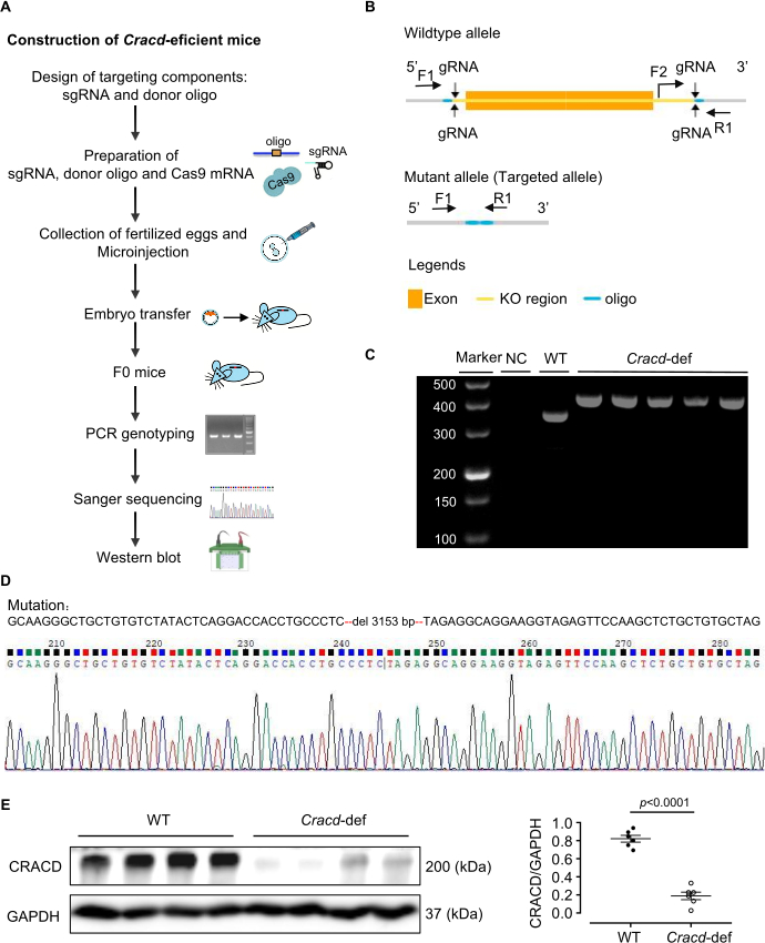
Figure 1: Generation of Cracd-deficient mice. (A) Construction of Cracd-deficient mice. (B) Schematic of the Cracd gene modification strategy. (C) PCR genotyping of tail genomic DNA. WT mice show a single 357 bp band, while homozygous (Cracd‑deficient) mice show a single 421 bp band. (D) Sanger sequencing of genomic DNA confirmed the predicted deletion in the Cracd locus of deficient mice compared to WT controls. (E) Western blot analysis showed significantly reduced CRACD protein levels in the hearts of Cracd-deficient mice compared to WT controls. n = 6 mice per group. Data are presented as mean ± SEM. Statistical significance was calculated with a two-tailed unpaired Student's t-test. Please click here to view a larger version of this figure.
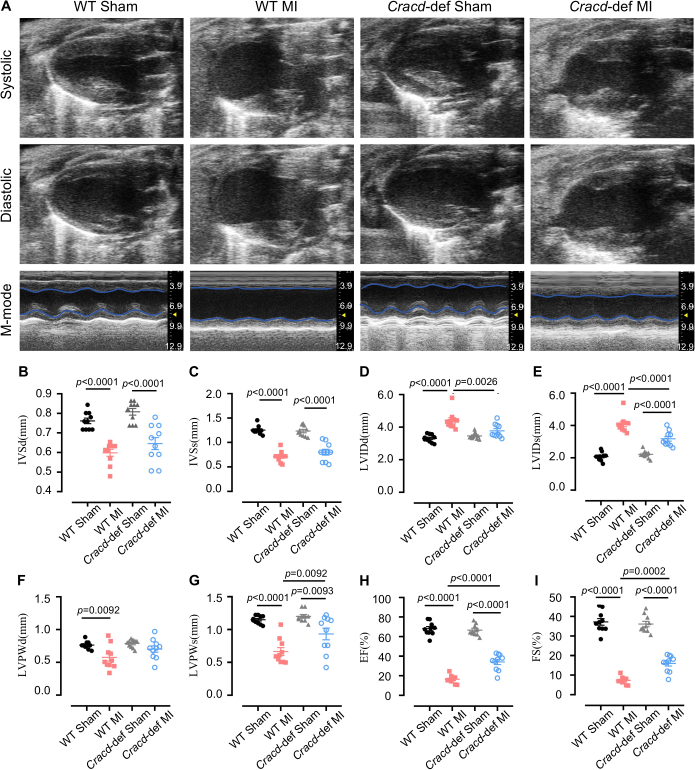
Figure 2: Echocardiographic assessment of cardiac function. (A) Representative B-mode and M-mode echocardiographic images of mouse hearts at diastole and systole. Quantitative analysis of (B) interventricular septum thickness at diastole (IVSd) and (C) systole (IVSs), (D) left ventricular internal diameter at diastole (LVIDd) and (E) systole (LVIDs), (F) left ventricular posterior wall thickness at diastole (LVPWd) and (G) systole (LVPWs), (H) ejection fraction (EF), and (I) fractional shortening (FS). n = 10 mice per group. Data are presented as mean ± SEM. Statistical significance was determined by one-way ANOVA followed by Tukey's multiple comparisons test. Please click here to view a larger version of this figure.
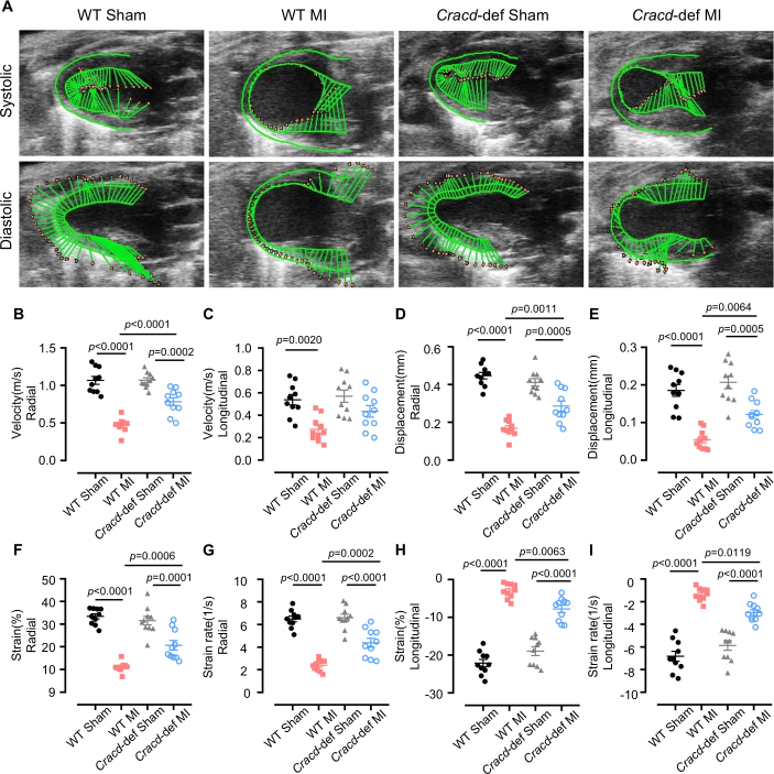
Figure 3: Speckle-tracking strain analysis. (A) Representative images of left ventricular wall motion and deformation at diastole and systole. Quantitative analyses of the following parameters: (B) radial velocity, (C) longitudinal velocity, (D) radial displacement, (E) longitudinal displacement, (F) radial strain, (G) radial strain rate, (H) longitudinal strain, and (I) longitudinal strain rate. n = 10 mice per group. Data are presented as mean ± SEM. Statistical significance was determined by one-way ANOVA followed by Tukey's multiple comparisons test. Please click here to view a larger version of this figure.
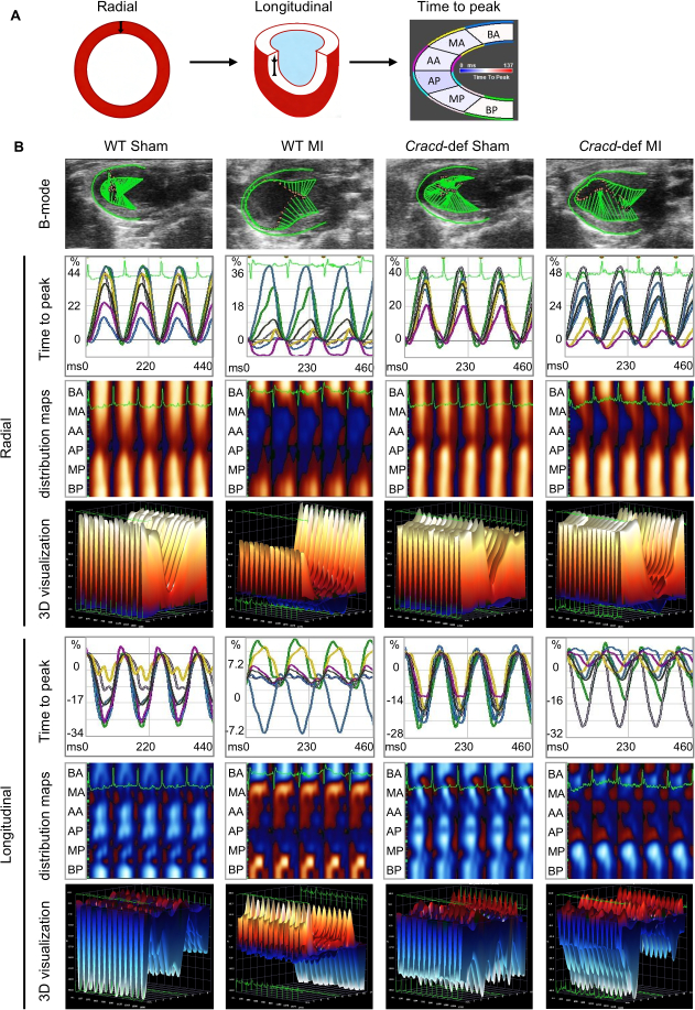
Figure 4: Speckle-tracking imaging in WT-Sham, WT-MI, Cracd-deficient Sham, and Cracd-deficient MI groups at 7 days post-MI. (A) Parasternal long‑axis B‑mode images. Left ventricular wall was segmented into six regions: BA (basal anterior), MA (mid anterior), AA (apical anterior), AP (apical posterior), MP (mid posterior), and BP (basal posterior). Color gradient from blue to red represents increasing strain magnitude. (B) Cardiac motion trajectories; time-to-peak curves for radial and longitudinal strain values along with corresponding peak times. Parametric distribution maps of strain synchrony (blue/red indicate direction: radial – away from/toward center; longitudinal – away from/toward apex). White dots = highest velocity regions, black dots = lowest velocity. Three-dimensional reconstructions (X: time, Y: spatial position, Z: strain magnitude). Please click here to view a larger version of this figure.
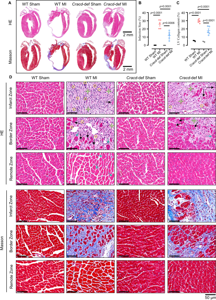
Figure 5: Histopathological analysis of cardiac remodeling after MI. (A) Whole-heart longitudinal sections with H&E and Masson's trichrome staining. Scale bars: 2 mm. (B) Quantitative analysis of infarct size and (C) collagen content. (D) High‑magnification views of infarct zone (IZ), border zone (BZ), and remote zone (RZ). H&E: yellow circles = inflammatory infiltration; black arrows = myocardial necrosis; green stars = fibrosis; blue arrows = hypertrophy. Masson’s trichrome: blue indicates collagen deposition. Scale bar: 50 μm. n = 5 mice per group. Data are presented as mean ± SEM. Statistical significance was determined by one-way ANOVA followed by Tukey's multiple comparisons test. Please click here to view a larger version of this figure.
ディスカッション
本研究では、CRISPR/Cas9を用いてCracd欠損マウスを作製し、心筋梗塞(MI)後の表現型解析を行うために、従来の心エコー検査、スペックル追跡ストレインイメージング、および組織学的解析からなるワークフローを構築しました。このプロトコルにより、CRACDタンパク質の消失が確認されたノックアウトマウスを作製しました。Cracd欠損マウスは、MI後に野生型(WT)コントロールと比較して、心室拡張の抑制、EFの改善、および線維化の減少を示し、心リモデリングの研究における本モデルの有用性が確認されました。
このモデルの成功は、いくつかの重要なステップに依存しています。gRNAの設計およびインビトロでの検証は、受精卵編集の効率に直接的に影響します。23CRISPickまたはBenchlingを用いてCRISPRaterスコアが0.5以上のgRNAを選定し、その後、in vitro Cas9/gRNAアッセイで切断効率を検証します。以下のgRNAのみを > 50%の切片をマイクロインジェクションします。また、左前下行枝(LAD)の適切な結紮も同様に極めて重要です。右心室への穿刺(深すぎる場合)や血管の閉塞不全(浅すぎる場合)を避けるため、7-0縫合糸をLAD起始部から2〜3 mm離れた、深さ0.5〜1 mmの位置に通す必要があります。前壁が即座に白くなることで閉塞が確認されます。さらに、心エコー検査中の心拍数制御も不可欠です。24イソフルラン(通常1~2%)を調整して450~550 bpmに維持することで、従来の測定および歪み(ストレイン)測定の再現性が向上します。最後に、スペックルトラッキング解析においては、カラーコード化されたメッシュの視覚的評価が不可欠です。25メッシュは、急激な変動なく心筋の境界に沿っている必要があります。そうでない場合は手動での修正が必要です。3回試行しても品質が低いままである場合、そのマウスは除外されます。
プロトコルを厳密に遵守しても、特定の技術的な問題が発生することがあります。体系的なトラブルシューティングによって、そのほとんどを解決できます。生殖細胞系列への伝達率が低い(10%未満)場合は、通常、ドナーオリゴヌクレオチドの組み込み効率が低いか、gRNA活性が弱いことを示しています26。したがって、ドナーオリゴの濃度を20 ng/µLに上げ、ホモロジーアームを120–200 bpに延長し、gRNAの切断効率(> 50%)を再確認することで、伝達率を向上させることができます。心筋梗塞(MI)後の死亡率が高い(30%超)場合、多くは気胸、出血、または広範囲の梗塞に起因します27。そのため、マウスが覚醒するまで持続的に酸素を投与し、加熱パッドを用いて体温を維持することで、死亡率を著しく低下させられます。結紮後に前壁が蒼白にならない場合は、LADの同定ミスか、縫合が浅すぎる可能性があります。この場合は、LAD(鮮やかな赤色の対角線上の血管)を再確認し、時には解剖顕微鏡を用いて、0.5–1 mmの深さに針を通し直すことで解決します。スペックルトラッキングの品質が低い場合、通常は低フレームレート、境界の不鮮明さ、またはモーションアーチファクトが原因となります28。したがって、フレームレートを上げ、ゲインを微調整し、心内膜トレースを手動で補正し、不整脈のあるサイクルを除外することが有効です。
これらの運用上の考慮事項に加えて、本手法には明確な限界があります。Cracdノックアウトは全身性であるため、すべての組織でCRACDが欠損しています。その結果、全身的な寄与が関与している可能性があるため、観察された保護作用を心筋細胞のみに起因するものと断定することはできません。細胞型特異的な役割を切り分けるには、条件付きノックアウトが必要となります。もう一つの限界は、7日目という単一の時点での評価であることです。これにより、慢性的なリモデリングについては何も分かっていません。したがって、長期的な研究には、より遅い時点(4週または8週)を追加する必要があります。また、再現性は画像品質への依存性という課題があります。すべての超音波診断装置で鮮明な心内膜境界が得られるわけではなく、操作者の習熟度も影響します。そのため、専門的な訓練を受けた者がすべての手術および測定を行うことが推奨されます。
これらの制限を考慮すると、心筋梗塞(MI)後のリモデリングにおける遺伝子機能を研究するための代替戦略と、我々のアプローチを比較することは有用である。AAVを介した遺伝子操作とアンチセンスオリゴヌクレオチド(ASO)は、一般的な2つの代替手段である29,30。AAV9は、交配を行うことなく心臓特異的な一過性の過剰発現またはノックダウンを導入できるが、パッケージング容量の制限(約4.7 kb)、導入効率のばらつき、およびオフターゲット効果が欠点となる31。対照的に、本研究のCRISPR/Cas9ノックアウトモデルは、永続的かつ完全な機能喪失を可能にし、長期的な研究において有利である。ASOは、迅速に発現し、用量依存的な可逆的ノックダウンを提供するため、急性期の介入に適しているが、繰り返しの投与が必要であり、組織特異性に欠ける32。したがって、本稿で述べる構成的ノックアウトは、初期の発見および慢性期の表現型解析に最適である。
最終的に、本プロトコルはCRACD以外にも幅広く応用可能です。CRISPR/Cas9によるノックアウトマウスの作製に、心エコー検査、ストレインイメージング、および組織学的解析を組み合わせた同一のワークフローを用いることで、心筋梗塞(MI)後のリモデリングにおけるあらゆる候補遺伝子の評価が行えます。この表現型解析の枠組みは、横大動脈狭窄(圧力負荷)や虚血再灌流傷害などの他の心血管疾患モデルにも適合します。さらに、本プロトコルで得られた組織サンプルは、分子メカニズムを解明するためのマルチオミクス解析(トランスクリプトミクス、プロテオミクス、メタボロミクス)に使用できます。Cracd欠損モデルは薬剤試験のツールとしても活用でき、CRACD経路を介して作用すると考えられる化合物を評価することが可能です。したがって、本プロトコルは、心血管研究における幅広いメカニズム研究およびトランスレーショナル研究に適応可能なプラットフォームを提供します。
開示事項
著者は、競合する利益がないことを宣言します。
謝辞
本研究は、広東省基礎・応用基礎研究財団(2023A1515011278)、広東省生物技術研究院自費プロジェクト(GDBRI-ZL202602)、および心血管疾患モデルと病因に関する中加学術交流・協力(MS202500058)の支援を受けて行われました。
材料
| 名前 | 会社 | カタログ番号 | コメント |
|---|---|---|---|
| CRISPR/Cas9 | |||
| トリスマ塩酸塩溶液 | Sigma-Aldrich, USA | T2663 | DNA抽出バッファーの調製に使用 |
| プロテイナーゼK | Sigma-Aldrich, USA | 539480 | マウス尾部の消化に使用 |
| Green Taq Mix | Vazyme, China | P131 | PCR増幅に使用 |
| アガロース | BioFroxx, Germany | 1110GR500 | 1–2%濃度のDNAゲル電気泳動に使用 |
| DNAマーカー | Thermo Scientific, USA | SM0242 | PCR産物のサイズ決定用DNAラダーとして使用 |
| TIANamp Genomic DNA Kit | Tiangen Biotech, China | DP304 | マウス尾部組織からのゲノムDNA抽出に使用 |
| MEGAshortscript™ T7転写キット | Thermo Scientific, USA | AM1354 | gRNAのインビトロ転写に使用 |
| RNA精製キット | Zymo Research, USA | R1016 | 転写されたgRNAの精製に使用 |
| BeyoCRISPR™ sgRNAスクリーニングキット | Beyotime, China | D8412S | gRNA切断活性のインビトロ評価に使用 |
| RNaseインヒビター | NEB, USA | M0314 | インビトロ転写中のRNA分解防止に使用 |
| TaKaRa Taq | Takara Bio, Japan | R001A | マウス尾部DNAのPCRジェノタイピングに使用 |
| FERTIUP® マウス精子前培養培地: PM | Cosmo Bio, Japan | KYD-002-EX | 体外受精前の精子キャパシテーションに使用 |
| T7 ARCA mRNAキット | NEB, USA | E2060 | Cas9 mRNAのインビトロ転写に使用 |
| 妊娠馬血清性ゴナドトロピン | MCE, USA | 9002-70-4 | 超排卵に使用。腹腔内投与 |
| ヒト絨毛性ゴナドトロピン | ProSpec, Israel | HOR-250 | 排卵誘導に使用。腹腔内投与 |
| RNaseフリー水 | Aladdin, China | R665526 | RNAの溶解およびPCRセットアップに使用 |
| プライマー | Sangon Biotech, China | / | ジェノタイピングに使用 |
| 手術 | |||
| イソフルラン | RWD Life Science, China | R510-22-10 | 手術中の麻酔導入および維持に使用 |
| 除毛クリーム | Veet, France | 1000207 | 手術および超音波検査前に胸部の被毛を除去するために塗布 |
| 7-0 縫合糸 | Lingqiao, China | LQ7022380206 | 左前下降(LAD)冠動脈の永久結紮に使用 |
| 4-0 縫合糸 | Lingqiao, China | LQ4022380412 | 手術後の皮膚切開部の閉鎖に使用 |
| 麻酔用ベンチレーター | Harvard Apparatus, USA | Tabletop | イソフルランの投与および呼吸維持に使用 |
| 小動物用ベンチレーター | Taimeng, China | HX-101E | 開胸手術中の陽圧換気に使用 |
| 恒温加熱パッド | TigerGene, USA | TG-TP-GS | 手術中および回復中のマウス体温維持に使用 |
| 肋骨開展器 | Shenzhen Huayon Biotech, China | LN-18-4101 | 肋骨を分離し、LAD結紮のために心臓を露出させるために使用 |
| はさみ | Shenzhen Huayon Biotech, China | 18-0551 | 皮膚切開および組織切断に使用 |
| 実体顕微鏡 | Nikon, Japan | SMZ 745 | LAD冠動脈結紮手術中に拡大視野を提供するために使用 |
| 微細針保持器 | Shenzhen Huayon Biotech, China | 18-2211 | 顕微鏡下で小型の7-0縫合針を操作するために使用 |
| 化学薬品および試薬 | |||
| 超音波カップリング剤 | Tianjin Jinya Technology Development, China | TM-100 | 心エコー検査時に胸部に塗布し、プローブの適切な接触を確保 |
| パラホルムアルデヒド | Sigma-Aldrich, USA | V900894 | 摘出後の心臓組織の固定に使用 |
| キシレン | Macklin, China | 1330-20-7 | 染色の前のパラフィン除去および組織透明化に使用 |
| ヘマトキシリン | Sigma-Aldrich, USA | H3136 | 核染色。HE染色に使用 |
| エオシン | Sigma-Aldrich, USA | E4009 | 細胞質染色。HE染色に使用 |
| ポンソーS | Sigma-Aldrich, USA | P3504 | マッソン染色における細胞質染色に使用 |
| ホスホモリブデン酸 | Sigma-Aldrich, USA | 221856 | マッソン染色における媒染および分化に使用 |
| アニリンブルー溶液 | Sigma-Aldrich, USA | 415049 | マッソン染色におけるコラーゲン繊維(青)の染色に使用 |
| CRACD抗体 | Absea, China | KC-35356 | ウェスタンブロットによるCRACDタンパク質検出用の一次抗体、希釈倍率 1:2000 |
| GAPDH抗体 | CST, USA | 2118S | ウェスタンブロット用のローディングコントロール抗体、希釈倍率 1:5000 |
| エタノール | Macklin, China | 64-17-5 | 脱水および試薬調製に使用(70%、80%、95%、100%グレード) |
| 実験機器 | |||
| Sorvall™ Legend™ Micro 17遠心機 | Thermo Scientific, USA | 75002541 | 日常的なサンプル調製に使用 |
| PCRサーマルサイクラー | Bio-Rad Laboratories, USA | 1861096 | DNA増幅に使用 |
| マイクロインジェクションシステム | Eppendorf, Germany | 5193000020 | 接合子へのCRISPR/Cas9試薬のマイクロインジェクションに使用 |
| Vevo 2100 イメージングシステム | VisualSonics, Canada | Vevo2100 | 心筋梗塞(MI)後の心機能および構造を非侵襲的に評価するための高周波イメージングシステム |
| 光学顕微鏡 | Leica, Germany | DM2500 | HE染色およびマッソン染色を施した心臓切片の観察に使用 |
| 解析ソフトウェア | |||
| ImageJ | National Institutes of Health, USA | / | 面積測定、シグナル定量化、組織学的画像(HEおよびマッソン染色)の処理を含む画像解析に使用 |
| VevoLab 3.2.0 / VevoStrain | VisualSonics, Canada | / | 心機能解析用VevoLabソフトウェア。VevoStrainモジュールは、MI後の心機能を評価するための心筋ストレイン解析に使用 |
| GraphPad Prism 8 | GraphPad Software, USA | / | 統計解析およびグラフ作成に使用 |
参考文献
- Fauconnier, J., Meli, A. C., Thireau, J., Roberge, S., Shan, J., et al. Ryanodine receptor leak mediated by caspase-8 activation leads to left ventricular injury after myocardial ischemia-reperfusion. Proc Natl Acad Sci U S A. 108 (32), 13258-13263 (2011).
- Cohn, J. N., Ferrari, R., Sharpe, N. Cardiac remodeling-concepts and clinical implications: a consensus paper from an international forum on cardiac remodeling. J Am Coll Cardiol. , (2000).
- Liu, D., Tian, X., Liu, Y., Song, H., Cheng, X., et al. CREG ameliorates the phenotypic switching of cardiac fibroblasts after myocardial infarction via modulation of CDC42. Cell Death Dis. 12 (4), (2021).
- Contessotto, P., Pandit, A. Therapies to prevent post-infarction remodelling: from repair to regeneration. Biomaterials. 275, (2021).
- Zhu, R., Sun, H., Yu, K., Zhong, Y., Shi, H., et al. Interleukin-37 and dendritic cells treated with interleukin-37 plus troponin I ameliorate cardiac remodeling after myocardial infarction. J Am Heart Assoc. 5 (12), (2016).
- Chang, Y., Zhang, J., Huo, X., Qu, X., Xia, C., et al. Substrate rigidity dictates colorectal tumorigenic cell stemness and metastasis via CRAD-dependent mechanotransduction. Cell Rep. 38 (7), (2022).
- Seo, Y., Jang, J., Ko, K. P., Zou, G., Huang, Y., et al. Actin dysregulation induces immune evasion via oxidative stress-activated PD-L1 in gastric cancer. bioRxiv. , (2024).
- Kim, B., Zhang, S., Huang, Y., Ko, K. P., Jung, Y. S., et al. CRACD loss induces neuroendocrine cell plasticity of lung adenocarcinoma. Cell Rep. 43 (6), (2024).
- Jung, Y. S., Wang, W., Jun, S., Zhang, J., Srivastava, M., et al. Deregulation of CRAD-controlled cytoskeleton initiates mucinous colorectal cancer via β-catenin. Nat Cell Biol. 20 (11), 1303-1314 (2018).
- Liu, Z., Cao, H., Shi, Y., Yang, R. KIAA1211 plays an oncogenic role in human non-small cell lung cancer. J Cancer. 10 (26), 6747-6753 (2019).
- Snider, P. L., Snider, E., Simmons, O., Lilly, B., Conway, S. J. Analysis of uncharacterized mKiaa1211 expression during mouse development and cardiovascular morphogenesis. J Cardiovasc Dev Dis. 6 (2), (2019).
- Lin, Y. H., Warren, C. M., Li, J., McKinsey, T. A., Russell, B. Myofibril growth during cardiac hypertrophy is regulated through dual phosphorylation and acetylation of the actin capping protein CapZ. Cell Signal. 28 (8), 1015-1024 (2016).
- Yang, F. H., Pyle, W. G. Reduced cardiac CapZ protein protects hearts against acute ischemia-reperfusion injury and enhances preconditioning. J Mol Cell Cardiol. 52 (3), 761-772 (2012).
- Tong, C., Huang, G., Ashton, C., Wu, H., Yan, H., et al. Rapid and cost-effective gene targeting in rat embryonic stem cells by TALENs. J Genet Genomics. 39 (6), 275-280 (2012).
- Cordes, S. P. N-ethyl-N-nitrosourea mutagenesis: boarding the mouse mutant express. Microbiol Mol Biol Rev. 69 (3), 426-439 (2005).
- Cong, L., Ran, F. A., Cox, D., Lin, S., Barretto, R., et al. Multiplex genome engineering using CRISPR/Cas systems. Science. 339 (6121), 819-823 (2013).
- Kanke, K. L., Rayner, R. E., Bozik, J., Abel, E., Venugopalan, A., et al. Single-stranded DNA with internal base modifications mediates highly efficient knock-in in primary cells using CRISPR-Cas9. Nucleic Acids Res. 52 (22), 13561-13576 (2024).
- Mihos, C. G., Liu, J. E., Anderson, K. M., Pernetz, M. A., O'Driscoll, J. M., et al. Speckle-tracking strain echocardiography for the assessment of left ventricular structure and function: a scientific statement from the American Heart Association. Circulation. 152 (10), (2025).
- Garg, P. K., Biggs, M. L., Kizer, J. R., Shah, S. J., Psaty, B., et al. Glucose dysregulation and subclinical cardiac dysfunction in older adults: the Cardiovascular Health Study. Cardiovasc Diabetol. 21 (1), (2022).
- Asanuma, T., Fukuta, Y., Masuda, K., Hioki, A., Iwasaki, M., et al. Assessment of myocardial ischemic memory using speckle tracking echocardiography. JACC Cardiovasc Imaging. 5 (1), 1-11 (2012).
- Chong, S. Y., Zharkova, O., Yatim, S. M. J. M., Wang, X., Lim, X. C., et al. Tissue factor cytoplasmic domain exacerbates post-infarct left ventricular remodeling via orchestrating cardiac inflammation and angiogenesis. Theranostics. 11 (19), 9243-9261 (2021).
- Hung, C. L., Verma, A., Uno, H., Shin, S. H., Bourgoun, M., et al. Longitudinal and circumferential strain rate, left ventricular remodeling, and prognosis after myocardial infarction. J Am Coll Cardiol. 56 (22), 1812-1822 (2010).
- Labuhn, M., Adams, F. F., Ng, M., Knoess, S., Schambach, A., et al. Refined sgRNA efficacy prediction improves large- and small-scale CRISPR-Cas9 applications. Nucleic Acids Res. 46 (3), 1375-1385 (2018).
- Wu, J., Bu, L., Gong, H., Jiang, G., Li, L., et al. Effects of heart rate and anesthetic timing on high-resolution echocardiographic assessment under isoflurane anesthesia in mice. J Ultrasound Med. 29 (12), (2010).
- Voigt, J. U., Pedrizzetti, G., Lysyansky, P., Marwick, T. H., Houle, H., et al. Definitions for a common standard for 2D speckle tracking echocardiography: consensus document of the EACVI/ASE/Industry Task Force to standardize deformation imaging. J Am Soc Echocardiogr. 28 (2), 183-193 (2015).
- Port, F., Chen, H. M., Lee, T., Bullock, S. L. Optimized CRISPR/Cas tools for efficient germline and somatic genome engineering in Drosophila. Proc Natl Acad Sci U S A. 111 (29), (2014).
- Gao, E., Lei, Y. H., Shang, X., Huang, Z. M., Zuo, L., et al. A novel and efficient model of coronary artery ligation and myocardial infarction in the mouse. Circ Res. 107 (12), 1445-1453 (2010).
- Rösner, A., Barbosa, D., Aarsæther, E., Kjønås, D., Schirmer, H., et al. The influence of frame rate on two-dimensional speckle-tracking strain measurements: a study on silico-simulated models and images recorded in patients. Eur Heart J Cardiovasc Imaging. 16 (10), 1137-1147 (2015).
- Argiro, A., Bui, Q., Hong, K. N., Ammirati, E., Olivotto, I., et al. Applications of gene therapy in cardiomyopathies. JACC Heart Fail. 12 (2), 248-260 (2024).
- Micheletti, R., Plaisance, I., Abraham, B. J., Sarre, A., Ting, C. C., et al. The long noncoding RNA Wisper controls cardiac fibrosis and remodeling. Sci Transl Med. 9 (395), (2017).
- Wu, Z., Yang, H., Colosi, P. Effect of genome size on AAV vector packaging. Mol Ther. 18 (1), 80-86 (2010).
- Juliano, R. L. The delivery of therapeutic oligonucleotides. Nucleic Acids Res. 44 (14), 6518-6548 (2016).
再版と許可
