Guest Editor

Collection Overview
Cardiovascular diseases encompass a range of heart and blood vessel disorders and are the leading cause of death globally. Developing animal models of these diseases is a primary strategy for understanding the underlying pathogenic mechanisms and testing new treatments.
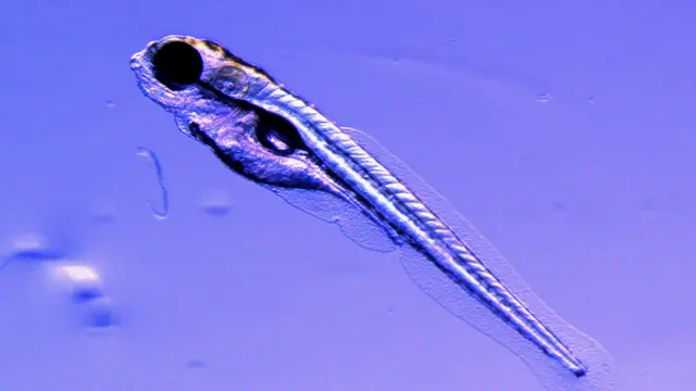
Zebrafish, native to the freshwaters of South Asia, offers a unique model for cardiovascular research. Their transparent embryos develop outside the womb, with the heart as the first organ to form. This allows researchers to observe heart development and function in real time using a simple light microscope. The zebrafish genome is fully sequenced.
Remarkably, zebrafish can survive without a functional heart for a week after fertilization. Unlike humans, they can regenerate heart tissue entirely within 4 to 6 weeks following injury. These numerous advantages make zebrafish an invaluable model for studying cardiac valve development, hemodynamics, and severe cardiovascular phenotypes.
This Methods Collection aims to compile essential techniques used in zebrafish heart research, including video imaging of heart function in zebrafish embryos and adults and adult zebrafish cardiomyocyte cell culture.